Changing stroke rehab and research worldwide now.Time is Brain! trillions and trillions of neurons that DIE each day because there are NO effective hyperacute therapies besides tPA(only 12% effective). I have 523 posts on hyperacute therapy, enough for researchers to spend decades proving them out. These are my personal ideas and blog on stroke rehabilitation and stroke research. Do not attempt any of these without checking with your medical provider. Unless you join me in agitating, when you need these therapies they won't be there.

What this blog is for:

My blog is not to help survivors recover, it is to have the 10 million yearly stroke survivors light fires underneath their doctors, stroke hospitals and stroke researchers to get stroke solved. 100% recovery. The stroke medical world is completely failing at that goal, they don't even have it as a goal. Shortly after getting out of the hospital and getting NO information on the process or protocols of stroke rehabilitation and recovery I started searching on the internet and found that no other survivor received useful information. This is an attempt to cover all stroke rehabilitation information that should be readily available to survivors so they can talk with informed knowledge to their medical staff. It lays out what needs to be done to get stroke survivors closer to 100% recovery. It's quite disgusting that this information is not available from every stroke association and doctors group.

Showing posts with label proteostasis. Show all posts

Friday, March 20, 2026

Blood protein structure changes may enable earlier detection of Alzheimer’s

Your competent? doctor figured out what to do about proteostasis years ago, right.

Do you prefer your doctor, hospital and board of director's incompetence NOT KNOWING? OR NOT DOING? Your choice; let them be incompetent or demand action!

Blood protein structure changes may enable earlier detection of Alzheimer’s

A recent study in Nature Aging combined mass spectrometry-based structural proteomics with machine learning to establish a minimally invasive, reliable, and potentially scalable research strategy for early detection and classification of Alzheimer’s disease (AD) and related cognitive conditions.

Protein homeostasis disruption and structural biomarkers in Alzheimer’s disease

Proteostasis, or protein homeostasis, refers to the cellular processes that maintain proper protein folding, stability, and degradation. These mechanisms are crucial because a substantial proportion of newly synthesized proteins can misfold, disrupting normal cell function if not managed by cellular quality control systems.

In AD, the machinery responsible for proteostasis becomes less effective, allowing misfolded proteins and damaged cellular components to build up over time. This impaired clearance supports the early accumulation of amyloid-β aggregates, abnormal protein clumps that can form in the brain years before the first signs of Alzheimer’s symptoms appear. A comprehensive understanding of protein conformational changes and interactions, beyond the traditional focus on amyloid plaques and tau tangles, could uncover disease mechanisms and plasma-based structural biomarkers.

Apolipoprotein E (APOE) is a polymorphic plasma protein with three major isoforms (ε2, ε3, ε4) differing by one or two amino acids, leading to altered binding properties. The ε4 allele is strongly associated with increased AD risk, while ε2 confers protection. Despite extensive characterization of APOE genotype expression profiles and network effects, the impact of APOE variants on the structure of ApoE-interacting proteins remains underexplored.

Neuropsychiatric symptoms (NPSs) are prevalent in AD, with sex differences noted in progression and symptomatology. Women tend to experience more rapid cognitive decline and higher rates of delusion, while men exhibit increased apathy and agitation. Despite growing efforts to define molecular correlates of NPSs, the relationship between sex and NPSs remains unclear due to clinical heterogeneity in AD.

Assessing protein structure alterations in Alzheimer’s disease

Blood samples were collected from participants at the University of California, San Diego (UCSD) and the University of Southern California Alzheimer’s Disease Research Centers. Alzheimer’s pathology in the UCSD cohort was supported by cerebrospinal fluid (CSF) measurements of amyloid-β and tau, while clinical status across cohorts was evaluated using established diagnostic criteria. Participants were assessed biannually for cognitive function and categorized using standard criteria, including the Clinical Dementia Rating (CDR) and neuropsychological testing.

Peptide samples were analyzed by Liquid Chromatography–Tandem Mass Spectrometry (LC–MS/MS) coupled to a timsTOF Pro mass spectrometer. A machine learning framework was used to classify mass spectrometry data, with a deep neural network selected after benchmarking against 17 additional machine learning algorithms.

Identification of structural blood biomarkers for early AD detection

A total of 520 blood samples were obtained from two large cohorts. By combining blood assessment findings with detailed clinical and biomarker data, including cognitive tests and cerebrospinal fluid (CSF) measures, where available, researchers classified Alzheimer’s disease (AD) status and progression.


Wednesday, August 20, 2025

Reshaping Nuclear Speckles Could Halt Alzheimer’s

 Hopefully your competent? doctor get further research going to prevent Alzheimers.

Proteostasis
also known as protein homeostasis, proteostasis is the maintenance of physiological balance in the proteome, and includes protein synthesis, folding, trafficking, and degradation.

Reshaping Nuclear Speckles Could Halt Alzheimer’s

eveals that reshaping nuclear speckles — tiny structures inside cell nuclei that regulate protein maintenance — could be a novel way to treat proteinopathies like Alzheimer’s, Parkinson’s, and prion diseases. Researchers found that pyrvinium pamoate, an existing FDA-approved drug, alters the surface tension of nuclear speckles, making them less round and more effective at supporting proteostasis.

In neurons and animal models of tauopathy, the drug dramatically reduced toxic protein accumulation and improved movement and cellular health. The work represents a new frontier in neurodegenerative research, with plans to move toward human clinical trials.Key Facts: Pyrvinium pamoate works by lowering nuclear speckle surface tension, improving protein maintenance across hundreds of genes.Strong Results: The drug reduced tau protein buildup by ~70% and improved symptoms in fly and retinal disease models.Broad Potential: Could target multiple protein-related disorders, from Alzheimer’s to retinitis pigmentosa.Source: Universityof Pittsburgh Targeting cellular structures called nuclear speckles could be a completely new approach for treating proteinopathies — diseases driven by abnormal accumulation of misfolded proteins — such as Alzheimer’s, Parkinson’s and prion diseases, according to new research publishing in Nature Communications led by the University of Pittsburgh. “Our research is painting a picture where dysregulation of nuclear speckles is important for neuron degeneration in the context of many diseases,” said senior author Bokai Zhu, Ph.D., assistant professor in Pitt’s Department of Medicine and the Aging Institute.
This shows a cell.
Adding pyrvinium pamoate greatly improved climbing prowess in both larvae and adult flies — important evidence of the drug’s effectiveness in living creatures. Credit: Neuroscience News

“The concept of rejuvenating nuclear speckles to treat these diseases is completely novel, but I believe it’s the next frontier of neurodegenerative research.” 

Nuclear speckles are structures within cell nuclei that regulate proper protein production, folding and degradation — a balance known as proteostasis. Zhu’s previous research found that nuclear speckle shape affects function: more spherical speckles were linked with worse proteostasis than irregularly shaped ones. 

Hypothesizing that drugs that make nuclear speckles less round could treat proteinopathies, Zhu’s team screened hundreds of FDA-approved drugs. When they measured the effects on nuclear speckle sphericity, one stood out: pyrvinium pamoate, originally approved for treating pinworm infections. 

And sure enough, pyrvinium pamoate improved proteostasis within cells in a dish, found first author William Dion, Ph.D., a former graduate student in Zhu’s lab. 

“We were thrilled that our hypothesis was correct,” said Zhu. “This led us to ask: Does this drug work in disease models, and how does it work?” 

Promising Disease Model Results 

Zhu collaborated with Xu Chen, Ph.D., at UC San Diego, who studies tauopathies — diseases driven by tau protein buildup in the brain, causing memory, cognitive and locomotive deficits.  

In mouse neurons expressing human tau, pyrvinium pamoate reduced the pathological protein by about 70%. 

“Tau is a really difficult protein to degrade, so I didn’t think this experiment was going to work,” said Zhu. “But a week after starting my collaboration with Dr. Chen, I got a text message from her saying that she had collected some of the best data in her career.” 

Human neurons carrying a frontotemporal dementia-associated tau mutation had abnormally shaped nuclear speckles and elevated tau levels. Led by Yuren Tao, a graduate student in Dr. Xu Chen’s lab, the researchers showed that low doses of the drug restored nuclear speckle shape and dramatically reduced tau levels without causing cellular stress or toxicity. 

In fly models of tauopathy, locomotive symptoms can be measured by assessing climbing ability. Adding pyrvinium pamoate greatly improved climbing prowess in both larvae and adult flies — important evidence of the drug’s effectiveness in living creatures. 

In experiments led by Yuanyuan Chen, Ph.D., assistant professor of ophthalmology at Pitt, the researchers used mouse retinas cultured in a dish to show that the drug held promise for treating retinitis pigmentosa, a disease caused by a faulty gene that leads to misfolding of the retinal protein rhodopsin, which clogs up the rod cells of the eye and causes progressive vision loss. 

Unique Mechanism of Action 

To understand how the drug works, Bennett Van Houten, Ph.D., professor in the Pitt Department of Pharmacology & Chemical Biology, led experiments with optical tweezers, which use lasers to precisely manipulate microscopic structures. Nuclear speckles were typically difficult to stretch because of high surface tension, but adding the drug dramatically lowered surface tension, making speckles easy to stretch and rupture. 

According to Zhu, when nuclear speckles have lower surface tension, they become less round and spread out to make better contact with chromosomes, leading to greater production of genes regulating proteostasis. 

“This was the killer experiment,” said Zhu. “Unlike most drugs, which target a specific receptor, pyrvinium pamoate acts by changing the surface tension of nuclear speckles. This is a totally new idea. Because nuclear speckles act globally on chromosomes, the drug can potentially alter the expression of hundreds of genes — which may be why it is so effective.” 

Zhu hopes to move this research into clinical trials soon to test whether pyrvinium pamoate could effectively treat proteinopathies in humans. 

Other authors on the study were Shanshan Zhao, Ph.D., Yuhang Nie, M.S., all of UC San Diego; and Maci Chambers, Riley K. Arbuckle, Michelle Sun, Syeda Kubra, Ph.D., Matthew A. Schaich, Ph.D., Megan Ye, Imran Jamal, Ph.D., Mads B. Larsen, Ph.D., Daniel Camarco, M.S., Eleanor Ickes, Haokun H. Wang, M.S., C. DuPont, Bingjie Wang, Ph.D., Silvia Liu, Ph.D., Shaohua Pi, Ph.D., Bill B. Chen, Ph.D., all of Pitt and UPMC. 

About this neurodegeneration research news

Author: Anastasia Gorelova
Source: University of Pittsburgh
Contact: Anastasia Gorelova – University of Pittsburgh
Image: The image is credited to Neuroscience News

Original Research: Open access.
SON-dependent nuclear speckle rehabilitation alleviates proteinopathies” by Bokai Zhu et al. Nature Communications

Saturday, May 3, 2025

In Vivo Visualization and Quantification of Brain Heat Shock Protein 90 with [11C]HSP990 in Healthy Aging and Neurodegeneration

 I'm sure your competent? doctor already has protocols on heat and cold shock proteins for your recovery. Oh no, that's not the case!

  • cold shock proteins (2 posts to October 2023)
  • heat shock proteins (3 posts to April 2013)
  • In Vivo Visualization and Quantification of Brain Heat Shock Protein 90 with [11C]HSP990 in Healthy Aging and Neurodegeneration

    Romy Cools, Koen Vermeulen, Eline Vonck, Veerle Baekelandt, Cassis Varlow, Valeria Narykina, Christopher Cawthorne, Koen Van Laere, Wim Vanduffel, Neil Vasdev and Guy Bormans

    Abstract

    Heat shock protein 90 (Hsp90) is essential for maintaining cellular proteostasis and may play an important role in the development of neurodegenerative proteinopathies. Therefore, we aimed to develop an Hsp90-specific PET brain tracer to quantify Hsp90 expression in the brain in vivo in order to explore its potential as a biomarker for neurodegenerative disease characterization and to support Hsp90-targeted drug development. Methods: We developed the radiosynthesis of (R)-2-amino-7-(4-fluoro-2-(6-(methoxy-11C)pyridin-2-yl)phenyl)-4-methyl-7,8-dihydropyrido[4,3-d]pyrimidin-5(6H)-one, [11C]HSP990, and validated the tracer using in vitro autoradiography, in vitro brain homogenate saturation binding, ex vivo biodistribution, and in vivo PET imaging in rodent models of Alzheimer disease (AD) and Parkinson disease versus healthy age-matched and young controls. Human brain samples from AD patients and healthy subjects were included in our in vitro binding studies. A nonhuman primate PET brain study with arterial blood sampling was conducted under baseline and blocking conditions. Results: In vitro and in vivo [11C]HSP990 studies in rodents and a nonhuman primate revealed saturable Hsp90 binding pools in natural killer lymphocytes, bone marrow, and notably the brain, where the highest binding was observed, particularly in gray matter. Blocking studies indicated that saturable Hsp90 in natural killer lymphocytes considerably influences the pharmacokinetics of Hsp90-targeting probes, which is critical for Hsp90 drug development. In vitro [3H]HSP990 brain homogenate saturation binding assays suggested that the tracer binds a distinct subfraction of the total Hsp90 pool, which is significantly diminished in both rodent and human AD brain tissue compared with age-matched controls. In vivo PET imaging confirmed reduced [11C]HSP990 brain binding on aging and an even stronger decrease in AD mice, suggesting that Hsp90 depletion may impair protein quality control and accelerate proteinopathies. Conclusion: [11C]HSP990 is a promising Hsp90-specific tracer and reveals strong Hsp90 binding in the brain. Uniformly reduced tracer binding was observed in AD brain tissue compared with age-matched controls. [11C]HSP990 holds potential as a biomarker for neurodegenerative disease characterization and progression, and it may aid in patient stratification and therapy monitoring. Human [11C]HSP990 PET neuroimaging studies are under way to investigate whether these findings translate to humans.

    Heat shock protein 90 (Hsp90), present in 4 isoforms, is a key chaperone in the protein quality control system, maintaining cellular proteostasis by stabilizing, folding and refolding, and regulating many client proteins (1). Accordingly, Hsp90 function is implicated in diseases associated with proteotoxic stress, such as neurodegenerative disorders and cancer (2,3). The role of Hsp90 in neurodegenerative diseases, including Alzheimer disease (AD), Parkinson disease (PD), amyotrophic lateral sclerosis, and Huntington disease, remains controversial in the literature and has been linked to both protective functions and pathologic roles.

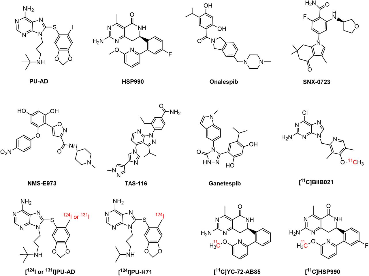
    Proteins involved in aggregation, such as β-amyloid, phosphorylated tau, and α-synuclein (αSyn), are reported Hsp90 clients (46). Some studies have shown that upregulated Hsp90 colocalized with these aggregates in a neurodegenerative brain, potentially contributing to disease pathology by exacerbating aggregate accumulation and hindering misfolded protein degradation (2,6). This has sparked interest in Hsp90 inhibitors, to suppress its aberrant neuronal activity, as potential treatments for neurodegenerative disorders (7). Conversely, reduced Hsp90 levels have been linked to neuronal cell death. In this context, induction of the heat shock response through Hsp90 inhibition has been explored to upregulate chaperone function, thereby reducing protein aggregation and supporting cytoprotection (1,711). For instance, (S)-2-fluoro-6-((tetrahydrofuran-3-yl)amino)-4-(3,6,6-trimethyl-4-oxo-4,5,6,7-tetrahydro-1H-indol-1-yl)benzamide) (SNX-0723) and 9-(3-(tert-butylamino)propyl)-8-((6-iodobenzo[d][1,3]dioxol-5-yl)thio)-9H-purin-6-amine (PU-AD) (Fig. 1) have shown promise in preclinical studies by preventing αSyn oligomerization and rescuing striatal dopamine levels in a PD rat model and by inducing the degradation of misfolded proteins and restoring memory in an AD mouse model, respectively (2,12). PU-AD was evaluated in clinical trials for AD, amyotrophic lateral sclerosis, and glioblastoma, but these studies were withdrawn or terminated and results have yet to be published (1315). The compound (R)-2-amino-7-(4-fluoro-2-(6-methoxypyridin-2-yl)phenyl)-4-methyl-7,8-dihydropyrido[4,3-d]pyrimidin-5(6H)-one (HSP990) (Fig. 1) has also shown therapeutic potential, improving cognitive function in AD mouse models and reducing huntingtin aggregation in Huntington disease, but exhibited neurotoxicity in a phase I trial for solid tumors (1618).

    FIGURE 1.

    Chemical structure of Hsp90 inhibitors and PET brain tracers.

    The potential clinical efficacy of Hsp90 inhibitors depends on their ability to exert therapeutic effects at safe doses. Accurate measurement of Hsp90 occupancy in the brain using PET can significantly contribute to determining optimal dosing regimens and avoiding toxic effects (19). Furthermore, Hsp90 PET brain imaging may provide insights into the role of Hsp90 in neurologic diseases.

    Only a few Hsp90-targeting PET probes that can permeate the blood–brain barrier have been developed so far (2,20,21). [124I]PU-AD (Fig. 1) showed higher hippocampal retention in human AD brain than in controls on static delayed (3 h after injection) PET. However, absolute brain [124I]PU-AD concentrations were very low, and 124I produces low-quality PET images and exposes patients to high radiation because of its long half-life (2,22). More recently, 6-chloro-9-((4-(methoxy-11C)-3,5-dimethyl-2-pyridinyl)methyl]-9H-purin-2-amine ([11C]BIIB021) (Fig. 1) was developed, exhibiting Hsp90-specific binding in the rat brain but showing the presence of brain radiometabolites, which may complicate PET image quantification (21). Our group previously developed (R)-2-amino-7-(2-(6-(methoxy-11C)pyridin-2-yl)phenyl)-4-methyl-7,8-dihydropyrido[4,3-d]pyrimidin-5(6H)-one ([11C]YC-72-AB85) (Fig. 1), demonstrating reversible Hsp90-specific brain binding in healthy rodents and a nonhuman primate (NHP) (20), but it has not been explored in neurodegeneration. Given that HSP990 has already been used in a clinical trial, this study sought to develop and evaluate [11C]HSP990 (Fig. 1) as a Hsp90 PET tracer to elucidate saturable Hsp90 binding and its role in health and neurodegenerative disease and to advance clinical translation.

    More at link.

    Wednesday, July 26, 2023

    Proteomics analysis of plasma from middle-aged adults identifies protein markers of dementia risk in later life

    I can't imagine any hospital using this to test for dementia risk; so useless.  And with NO protocols to prevent dementia, doubly useless.

    What is the method of proteomics analysis?

    The techniques that are most often used are electrospray ionization (ESI) and matrix-assisted laser desorption/ionization (MALDI). Both of them are representatives of so-called soft ionization techniques in which ions are created with low internal energies and thus undergo little fragmentation.

    Proteomics analysis of plasma from middle-aged adults identifies protein markers of dementia risk in later life

    Science Translational Medicine
    19 Jul 2023
    Vol 15, Issue 705

    Abstract

    A diverse set of biological processes have been implicated in the pathophysiology of Alzheimer’s disease (AD) and related dementias. However, there is limited understanding of the peripheral biological mechanisms relevant in the earliest phases of the disease. Here, we used a large-scale proteomics platform to examine the association of 4877 plasma proteins with 25-year dementia risk in 10,981 middle-aged adults. We found 32 dementia-associated plasma proteins that were involved in proteostasis, immunity, synaptic function, and extracellular matrix organization. We then replicated the association between 15 of these proteins and clinically relevant neurocognitive outcomes in two independent cohorts. We demonstrated that 12 of these 32 dementia-associated proteins were associated with cerebrospinal fluid (CSF) biomarkers of AD, neurodegeneration, or neuroinflammation. We found that eight of these candidate protein markers were abnormally expressed in human postmortem brain tissue from patients with AD, although some of the proteins that were most strongly associated with dementia risk, such as GDF15, were not detected in these brain tissue samples. Using network analyses, we found a protein signature for dementia risk that was characterized by dysregulation of specific immune and proteostasis/autophagy pathways in adults in midlife ~20 years before dementia onset, as well as abnormal coagulation and complement signaling ~10 years before dementia onset. Bidirectional two-sample Mendelian randomization genetically validated nine of our candidate proteins as markers of AD in midlife and inferred causality of SERPINA3 in AD pathogenesis. Last, we prioritized a set of candidate markers for AD and dementia risk prediction in midlife.

    Editor’s summary

    Pathological changes involved in Alzheimer’s disease (AD) occur decades before the onset of cognitive deficits but are not well understood. Here, Walker and colleagues used proteomics and genomics on a cohort of middle-aged adults followed longitudinally and identified pathway-specific plasma proteins that increased dementia risk up to 25 years later. The pathway signature of these proteins was characterized by dysregulated immune signaling and proteostasis in the earliest preclinical stages and abnormal coagulation and complement signaling around 10 years before dementia onset. The study indicates that distinct biological mechanisms may be relevant in earlier and later preclinical stages of AD. –Daniela Neuhofer

    Get full access to this article

    Monday, March 27, 2023

    How does dietary intervention affect aging?

    Well you lost 5 cognitive years from your stroke.

    So your doctor has to have protocols to recover that for you also, so ask them to include these anti-aging protocols in your rehab interventions. If they have nothing you don't have a functioning stroke doctor. 

    RUN AWAY!

    (Or more likely, walk as fast as you can).

    How does dietary intervention affect aging?

    Aging is a process that includes multiple interwoven mechanisms that interact with lifestyle factors. A new review seeks to encapsulate the available information on the effect of diet on aging at the cellular level in humans.

    Review: The role of dietary strategies in the modulation of hallmarks of aging. Image Credit: UfaBizPhoto / ShutterstockReview: The role of dietary strategies in the modulation of hallmarks of aging. Image Credit: UfaBizPhoto / Shutterstock

    Introduction

    Age is a universal factor that contributes to most chronic diseases. However, it is not inevitable at a specific age and may be considered malleable.

    The hallmarks of aging occur at the systemic, cellular, and molecular levels. They include genomic instability, attrition of telomeres, epigenetic modification, reduced proteostasis, loss of nutrient sensing regulation, mitochondrial dysfunction, cellular senescence, stem cell exhaustion, and changes in intercellular communication. Furthermore, three new hallmarks have been identified: impaired macroautophagy, chronic inflammation, and dysbiosis associated with aging.

    Dietary restriction (DR) and several dietary patterns have been studied for their ability to improve multiple health outcomes associated with aging, such as changes in body fat, insulin sensitivity, blood sugar levels, blood pressure, and inflammation.

    The present question is whether it also can slow down the speed of aging in the cells. Earlier research shows that DR can stabilize the genome from undesirable changes that occur naturally but promote aging, preserves protein integrity, helps maintain the regulation of nutrient-sensing pathways that are important in energy homeostasis, and slow cellular aging processes. It also sustains beneficial intercellular communication.

    Diets such as the Mediterranean diet (MD), the ketogenic diet (KD), and other plant-based diets have also been evaluated for their effectiveness in this field. Similar benefits have been reported. The researchers in the current study, published in the journal Ageing Research Reviews, sum up the potential for nutritional interventions to increase the healthy lifespan, exploring facets such as adherence levels, adverse effects, and broad-spectrum applicability.

    What does the evidence show?

    In general, DR shows reduced nuclear damage with a higher rate of DNA repair under calorie restriction (CR) conditions, at 30% to 55% of normal caloric intake, in rodents. Possible mechanisms include lowering the production of oxidants like hydrogen peroxide in the mitochondria or the leakage of free radicals in mitochondrial DNA (mtDNA).

    Encouragingly, CR late in life still promotes the healing of oxidative damage and keeps the genome functioning by inducing DNA repair and increased debris removal by autophagy. This has not been found in non-rodent species, however.

    CR did reduce DNA damage in humans in just six months while fasting between sunrise and sunset for two days a week (Muslims call this the Sunnah fast) produced similar results within six weeks, lasting for up to 36 months, despite CR being mostly below a fifth of normal.

    As for epigenetic modifications that promote aging, preclinical but not human models indicate that 30% CR can reduce and reverse these changes. However, much more extensive studies are required in this area. In preclinical studies, DR has been observed to ensure proper disposal of toxic proteins, which is essential for proteostasis.

    40% CR and intermittent fasting are associated with improved protein homeostasis, even with a high-fat diet. This could help prevent aging-related changes, especially in neurodegeneration.  

    The MD model also promoted genomic health, as shown by improvements in various markers of DNA oxidation in humans. It also prevented telomere shortening, a sign of age-related disease in many conditions. Dietary energy restriction by 30% is also potentially beneficial in producing this effect, especially when fats, meats, and sweets are reduced.

    Epigenetic modifications favorable to health were observed with MD-like diets but not with corn oil. MD could also activate autophagy to remove toxic proteins within the visceral white fat in obese adult women. This is perhaps due to the presence of polyphenols from olive oil and wine in this diet.

    Protein restriction and a plant-based KD are potential interventions to increase autophagy and maintain proteostasis. Fasting, CR, and the MD seem to mediate better responses to nutrient sensing, extending the lifespan in preclinical studies, but further human studies are essential to establish the clinical utility of this response.

    Replicative senescence is a circadian phenomenon in cells whereby the telomere progressively shortens during successive cell divisions, ending in growth arrest. However, senescence also follows cell damage through various mechanisms, whether mediated by oxidative or DNA damage or external stressors.  

    The buildup of senescent cells can lead to deterioration in functional tissue, promoting faster aging and degenerative disease. However, 30% CR for a mean of ten years is reduced with reduced senescence in adults above 60. This may be reflected in reduced senescence-related markers when following the MD.

    MD adherence is also associated with reduced inflammaging, a low-grade chronic inflammatory state that leads to tissue senescence, dysfunction, degeneration, and aging caused by altered cell communication. Interestingly, this is less obvious with DR.

    What are the implications?

    While CR is the nutritional intervention most often used in dietary experiments, it is not broadly applicable to humans, both because it is unappealing to most people and because it may cause psychological stress when followed over the long term. The selective restriction of some amino acids is still a research area.

    Most research evidence points to the use of plant-based foods and the MD as helpful in slowing down aging-associated changes, but the KD appears, from the available studies, to be more specific in its effects.

    Stem cell exhaustion is significantly related to tissue aging, hindering regeneration. Unfortunately, little evidence is available on the effects of CR or MD/KD on stem cell exhaustion, and this appears to be a wide-open field for future work.

    Overall, “dietary restriction and certain dietary patterns, such as the Mediterranean dietary pattern, are nutritional strategies that can impact aging rates and the development of age-related clinical conditions.”

    However, this cannot be applied with confidence or precision until more evidence is available from human studies using standardized methods in more significant numbers of people.

    Journal reference: