WHOM is going to do the intervention research that prevents this from happening?
If your doctor and hospital aren't involved in getting this research to occur, YOU DON'T HAVE A FUNCTIONING STROKE DOCTOR OR HOSPITAL!
Approximately 30% of stroke patients develop dementia within 1 year of stroke onset, so it is imperative your doctor solves this problem.
Cognitive Decline May Be Triggered by Astrocyte Dysfunction
Source: Weill Cornell Medicine
People with dementia have protein build-up in astrocytes that may trigger abnormal antiviral activity and memory loss, according to a preclinical study by a team of Weill Cornell Medicine investigators.
Dysfunction in cells called neurons, which transmit messages throughout the brain, has long been the prime suspect in dementia-related cognitive deficits. But a new study, published in Science Advances on April 19, suggests that abnormal immune activity in non-neuronal brain cells called astrocytes is sufficient to cause cognitive deficits in dementia.
The discovery could lead to new treatments that reduce excess immune activity in astrocytes and their detrimental effects on other brain cells and cognition.
“Astrocyte dysfunction alone can drive memory loss, even when neurons and other cells are otherwise healthy,” said co-senior author Dr. Anna Orr, the Nan and Stephen Swid Assistant Professor of Frontotemporal Dementia Research in the Feil Family Brain and Mind Research Institute and a member of the Helen and Robert Appel Alzheimer’s Disease Research Institute at Weill Cornell Medicine.
“We found, in mice, that astrocytes can cause cognitive decline through their antiviral activities, which can make neurons hyperactive.”
While neurons have been intensively studied in dementia and other diseases, much less research has focused on astrocytes, which many scientists viewed as playing only supporting roles to neurons in brain health.
“We are very interested in the roles of astrocytes in cognitive and behavioral disorders,” she said. “These cells are prevalent in the brain and perform various key functions, but their involvement in neurocognitive disorders like dementia are poorly understood.”
When the investigators, including first author Dr. Avital Licht-Murava, a former postdoctoral associate in the Orr lab, examined tissue samples from deceased individuals who were diagnosed with either Alzheimer’s disease or frontotemporal dementia, they found an accumulation of a protein called TDP-43 in astrocytes within the hippocampus, a brain region crucial for memory.
To understand the effects of this protein build-up, the team conducted a series of experiments in mouse models and brain cells grown in the laboratory. Other senior investigators that contributed to the study include Dr. Robert Schwartz at Weill Cornell Medicine and Dr. Robert Froemke at New York University.
In mice, the build-up of TDP-43 in astrocytes was sufficient to cause progressive memory loss but not other behavioral changes. “Astrocytes in the hippocampus seem to be more vulnerable to this pathology.” she said.
To understand the causes of memory loss at the molecular level, co-senior author Dr. Adam Orr, an assistant professor of research in neuroscience in the Feil Family Brain and Mind Research Institute and a member of the Appel Alzheimer’s Disease Research Institute at Weill Cornell Medicine, analyzed gene expression and found high levels of antiviral gene activities, even though no virus was present in the brain.

Astrocytes produced excessive amounts of immune messengers called chemokines, which can activate CXCR3 chemokine receptors typically found on infiltrating immune cells. To their surprise, the team discovered that CXCR3 receptor levels were elevated in hippocampal neurons, and that excessive CXCR3 receptor activity made neurons “hyperactive,” Dr. Anna Orr said.
“Blocking CXCR3 reduced neuronal firing in individual neurons and eliminating CXCR3 in mice by genetic engineering alleviated cognitive deficits caused by astrocytic TDP-43 build-up,” Dr. Adam Orr said. These experiments demonstrate that impaired astrocytes can have a detrimental role in dementia, he said.
Both investigators were excited by the potential clinical implications of their findings.
“For effective therapeutics, we need to consider astrocytes along with neurons,” Dr. Anna Orr said.
Drugs that target the identified immune pathways might help improve cognitive function in people with dementia. She noted that scientists are already testing CXCR3 blockers to treat arthritis and other inflammatory conditions in clinical trials. These drugs could be tested and potentially repurposed for dementia.
This study may also provide insights into how antiviral immune responses can cause cognitive dysfunction. Previous research has linked viral infections to Alzheimer’s disease and to long-term neurocognitive effects such as memory loss and brain fog. Abnormal immune activity in astrocytes might contribute to these cognitive effects as well as increase individuals’ susceptibility to viral infections, which could further worsen brain health and promote some cases of dementia.
The team is currently studying how TDP-43 alters antiviral activities in astrocytes and whether these changes increase brain susceptibility to viral pathogens.
“Astrocytes can promote resilience or vulnerability to brain disease,” Dr. Anna Orr said. “Understanding how they enable cognitive function or cause cognitive decline will be critical to understanding brain health and developing effective therapies.”
About this neuroscience and memory research news
Author: Barbara Prempeh
Source: Weill Cornell University
Contact: Barbara Prempeh – Weill Cornell University
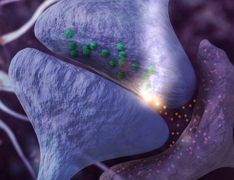
Image: The image is an Original 3D by BROKENGRID
Original Research: Open access.
“Astrocytic TDP-43 dysregulation impairs memory by modulating antiviral pathways and interferon-inducible chemokines” by Anna Orr et al. Science Advances
Abstract
Astrocytic TDP-43 dysregulation impairs memory by modulating antiviral pathways and interferon-inducible chemokines
Transactivating response region DNA binding protein 43 (TDP-43) pathology is prevalent in dementia, but the cell type–specific effects of TDP-43 pathology are not clear, and therapeutic strategies to alleviate TDP-43–linked cognitive decline are lacking.
We found that patients with Alzheimer’s disease or frontotemporal dementia have aberrant TDP-43 accumulation in hippocampal astrocytes.
In mouse models, induction of widespread or hippocampus-targeted accumulation in astrocytic TDP-43 caused progressive memory loss and localized changes in antiviral gene expression. These changes were cell-autonomous and correlated with impaired astrocytic defense against infectious viruses.
Among the changes, astrocytes had elevated levels of interferon-inducible chemokines, and neurons had elevated levels of the corresponding chemokine receptor CXCR3 in presynaptic terminals. CXCR3 stimulation altered presynaptic function and promoted neuronal hyperexcitability, akin to the effects of astrocytic TDP-43 dysregulation, and blockade of CXCR3 reduced this activity. Ablation of CXCR3 also prevented TDP-43–linked memory loss.
Thus, astrocytic TDP-43 dysfunction contributes to cognitive impairment through aberrant chemokine-mediated astrocytic-neuronal interactions.
No comments:
Post a Comment