Changing stroke rehab and research worldwide now.Time is Brain! trillions and trillions of neurons that DIE each day because there are NO effective hyperacute therapies besides tPA(only 12% effective). I have 523 posts on hyperacute therapy, enough for researchers to spend decades proving them out. These are my personal ideas and blog on stroke rehabilitation and stroke research. Do not attempt any of these without checking with your medical provider. Unless you join me in agitating, when you need these therapies they won't be there. Did your doctor somehow miss the recovery protocol that Pedro Bach-y-Rita used to recover fully back in 1958 with only a partial brain?

What this blog is for:

My blog is not to help survivors recover, it is to have the 10 million yearly stroke survivors light fires underneath their doctors, stroke hospitals and stroke researchers to get stroke solved. 100% recovery. The stroke medical world is completely failing at that goal, they don't even have it as a goal. Shortly after getting out of the hospital and getting NO information on the process or protocols of stroke rehabilitation and recovery I started searching on the internet and found that no other survivor received useful information. This is an attempt to cover all stroke rehabilitation information that should be readily available to survivors so they can talk with informed knowledge to their medical staff. It lays out what needs to be done to get stroke survivors closer to 100% recovery. It's quite disgusting that this information is not available from every stroke association and doctors group.

Showing posts with label prostaglandins. Show all posts

Friday, August 16, 2024

Restoring Brain’s Waste-Clearing System Reverses Aging Effects

 Didn't your competent? doctor start creating protocols on this years ago? NO? So you don't have a functioning stroke doctor, do you? Your doctor was incompetent for DOING NOTHING before and will stay incompetent by doing nothing now to get this to human testing! There are NO excuses for such behavior!

Restoring Brain’s Waste-Clearing System Reverses Aging Effects

Summary: New research in mice reveals that aging slows the brain’s ability to clear out harmful waste, contributing to neurological disorders like Alzheimer’s and Parkinson’s. Scientists have found that restoring function in the brain’s waste-clearing system, known as the glymphatic system, can reverse these age-related effects.

Using a clinically approved drug, researchers increased the efficiency of waste removal, offering a potential treatment strategy for age-related brain diseases.

Key facts:

  • Aging slows the brain’s waste-clearing process, increasing the risk of neurological disorders.
  • The glymphatic system, responsible for brain detoxification, can be restored to youthful efficiency.
  • A known drug successfully revived waste-clearing in aged mice, showing promise for future therapies.

Source: University of Rochester

Alzheimer’s, Parkinson’s, and other neurological disorders can be seen as “dirty brain” diseases, where the brain struggles to clear out harmful waste. Aging is a key risk factor because, as we grow older, our brain’s ability to remove toxic buildup slows down.

However, new research in mice demonstrates that it’s possible to reverse age-related effects and restore the brain’s waste-clearing process.
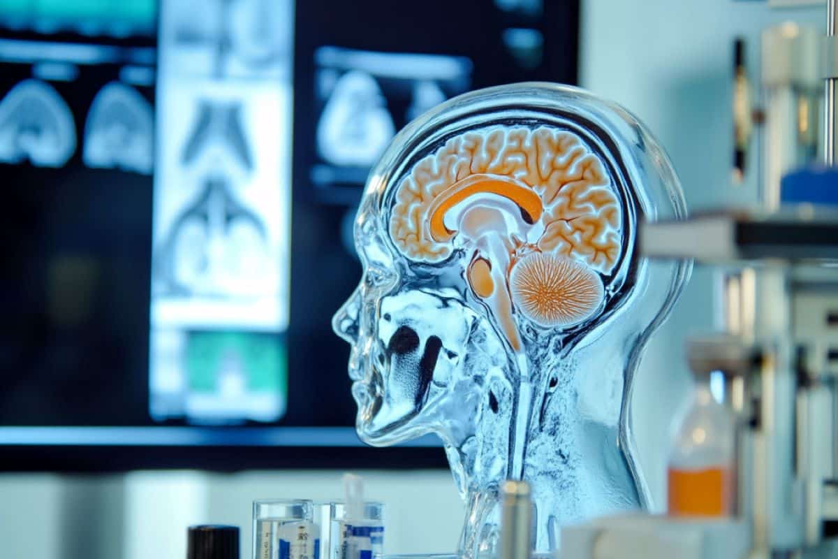
This shows a brain.
The new research combines advanced imaging and particle tracking techniques to describe for the first time in detail the route via the cervical lymph vessels in the neck through which half of dirty CSF exits the brain. Credit: Neuroscience News

“This research shows that restoring cervical lymph vessel function can substantially rescue the slower removal of waste from the brain associated with age,” said Douglas Kelley, PhD, a professor of Mechanical Engineering in the University of Rochester Hajim School of Engineering and Applied Sciences.

“Moreover, this was accomplished with a drug already being used clinically, offering a potential treatment strategy.”  

Kelley is one of the lead authors of the study, which appears in the journal Nature Aging, along with Maiken Nedergaard, MD, DMSc, co-director the University’s Center for Translational Neuromedicine.

First described by Nedergaard and her colleagues in 2012, the glymphatic system is the brain’s unique waste removal process that uses cerebrospinal fluid (CSF) to wash away excess proteins generated by energy hungry neurons and other cells in the brain during normal activity.

This discovery pointed the way for potential new approaches to treat diseases commonly associated with the accumulation of protein waste in the brain, such Alzheimer’s (beta amyloid and tau) and Parkinson’s (alpha-synuclein).

In healthy and young brains, the glymphatic system does a good job of flushing away these toxic proteins, however, as we age, this system slows, setting the stage for these diseases.

A network of tiny pumps draws waste from the brain

Once laden with protein waste, CSF in the skull needs to make its way to the lymphatic system and ultimately the kidneys, where it is processed along with the body’s other waste.

The new research combines advanced imaging and particle tracking techniques to describe for the first time in detail the route via the cervical lymph vessels in the neck through which half of dirty CSF exits the brain.  

In addition to measuring the flow of CSF, the researchers were able observe and record the pulsing of lymph vessels in the neck that helps draw CSF out of the brain.

“Unlike the cardiovascular system which has one big pump, the heart, fluid in the lymphatic system is instead transported by a network of tiny pumps,” said Kelley.  

These microscopic pumps, called lymphangions, have valves to prevent backflow and are strung together, one after another, to form lymph vessels.

The researchers found that as the mice aged, the frequency of contractions decreased, and the valves failed. As a result, the speed of dirty CSF flowing out of the brains of older mice was 63 percent slower compared to younger animals.

Known drug restarts flow of brain cleaning fluids

The team then set out to see if they could revive the lymphangions and identified a drug called prostaglandin F2α, a hormone-like compound commonly used medically to induce labor and known to aid smooth muscle contraction.

The lymphangions are lined with smooth muscle cells, and when the researchers applied the drug to the cervical lymph vessels in older mice, the frequency of contractions and the flow of dirty CSF from the brain both increased, returning to a level of efficiency found in younger mice. 

“These vessels are conveniently located near the surface of the skin, we know they are important, and we now know how to accelerate function,” said Kelley.  

“One can see how this approach, perhaps combined with other interventions, could be the basis for future therapies for these diseases.”

Additional contributors to the study include first authors Ting Du, Aditya Raghunandan, and Humberto Mestre, and Virginia Plá, Guojun Liu, Antonio Ladrón-de-Guevara, Evan Newbold, Paul Tobin, Daniel Gahn-Martinez, Saurav Pattanayak, Qinwen Huang, and Weiguo Peng with the University of Rochester. 

Funding: The research was supported with funding from National Institute of Neurological Disorders and Stroke, the Lundbeck Foundation, the Novo Nordisk Foundation, the Human Frontier Science Program, the Miriam and Sheldon G. Adelson Medical Research Foundation, the Simons Foundation, the EU Joint Programme – Neurodegenerative Disease Research, the US Army Research Office, the National Center for Complementary and Integrative Health, and the BRAIN Initiative.  

About this neurology and aging research news

Author: Mark Michaud
Source: University of Rochester
Contact: Mark Michaud – University of Rochester
Image: The image is credited to Neuroscience News

Saturday, September 9, 2023

New stroke treatment in development at UTHSC

Because we have NO LEADERSHIP AND NO STRATEGY  it took 11 years to get followup research going. That is how fucking bad stroke is, and it won't change until we get survivors in charge. Hope you're OK with not getting recovered when you are the 1 in 4 per WHO that has a stroke!

New class of potential drugs inhibits inflammation in brain February 2012


New stroke treatment in development at UTHSC

Grant and Award Announcement

University of Tennessee Health Science Center

Jianxiong Jiang, PhD

image: Jianxiong Jiang, PhD view more 

Credit: UTHSC

A study at the University of Tennessee Health Science Center aims to develop a new way to treat ischemic stroke, a leading cause of death in adults worldwide.

The study is funded by a $1,155,000 translational grant from the National Institute of Neurological Disorders and Stroke, part of the National Institutes of Health. Jianxiong Jiang, PhD, associate professor in the Department of Pharmaceutical Sciences and the Department of Anatomy and Neurobiology, and Jiawang Liu, PhD, director of the Medicinal Chemistry core at UTHSC, are the principal investigators. Thaddeus S. Nowak, PhD, professor in the Department of Neurology, provides expertise in stroke models for the project.

Ischemic strokes account for about 87% of acute stroke cases. Current ischemic stroke treatments, consisting of intravenous thrombolytic therapy and mechanical thrombectomy, have potential risks and must be administered in a short window of time to be effective, which limits patient eligibility. Additionally, those who receive treatment and survive can be left with permanent disabilities.

“The current treatment options for ischemic stroke are very limited, and so there's an urgent need to develop a new treatment for this condition,” Dr. Jiang said. “Even the patients who survive can have long-term disabilities because of the neuronal injury caused by the stroke.”

The researchers will work to develop novel drug-like antagonists for the prostaglandin receptor EP2, an emerging therapeutic target for brain ischemia-promoted neuroinflammation. Previous work led by Lexiao Li, PhD, a postdoctoral fellow in Dr. Jiang’s lab, validated the feasibility of pharmacologically targeting EP2 in animal models. The team believes this safer and more effective treatment could reduce inflammation and provide protection for neurons after an ischemic stroke. The potential new treatment would have a wider therapeutic window, so it would apply to more patients, while improving behavioral outcomes and reducing long-term disabilities.

“I have a family history of this disease. My father and my grandmother both had ischemic stroke before they died, so that was kind of motivation for me to develop a new treatment for this disease,” Dr. Jiang said.

According to Dr. Jiang, having more treatment options would not only save lives, but would also give hope to stroke patients and their loved ones as they experience a devastating emergency.

Thursday, February 21, 2019

Brain protein critical to recovery from stroke identified

WHOM  is the person responsible for shepherding this through human clinical studies to a translational protocol?  With no one identified it will fall thru the cracks like the thousands of other research studies that showed promise in animals. Well, years ago Dr. Michael Tymianski of the Toronto Western Hospital Research Institute in Canada referenced 1000+ failed neuroprotective clinical trials. Of course nobody knows of them and what knowledge they provided, but your doctor should know every one of those failed trials. 

The answers are out there if someone with a few functioning brain cells would put together a strategy to follow up promising research. We have to direct the research being done for stroke, we can't let researchers flail about.

Brain protein critical to recovery from stroke identified

Every 40 seconds, someone in the United States suffers a stroke and available therapies, such as clot busting drugs or clot removal devices, are focused on limiting the extent of brain damage. Now, research from the University of Pittsburgh School of Medicine and the VA Pittsburgh Healthcare System shows that a brain protein called UCHL1 may be critical to how nerve cells repair themselves after stroke damage. The research, conducted in animal models, could aid in the development of therapies that enhance stroke recovery by improving the underlying biological repair process.
"Even though traditional stroke therapies are very effective when available, the treatment must be started in the first hours after a stroke and most patients are not able to get these treatments. So there is a clear need for new approaches that can improve recovery days after a patient experiences a stroke," said co-senior author Steven Graham, M.D., Ph.D., professor of neurology at Pitt's School of Medicine, and associate chief of staff for research at VA Pittsburgh. "We think we have identified a protein that is at the root of how the brain recovers from stroke, making it an attractive target for developing drugs that help improve recovery."
UCHL1 is an enzyme that is highly active in the brain and plays a role in clearing away abnormal proteins. Mutations in the gene coding for UCHL1 have been thought to cause motor function deficits in humans. Previous research from Graham's lab had provided some hints as to UCHL1's function, showing that cyclopentenone prostaglandins (CyPgs) - fatty acid molecules - released in nerve cells after a stroke bind to UCHL1 and impair its function.
Graham teamed up with Feng Zhang, Ph.D., an assistant professor of neurology at Pitt's School of Medicine and a co-senior author on the current study published in the Proceedings of the National Academy of Sciences, to tease out the exact role of UCHL1 in stroke and to determine if it could be a viable drug target.
The researchers created a mouse model in which they inserted an altered version of the UCHL1 gene that was resistant to the effects of the CyPgs. They then surgically modelled the effect of a stroke in both genetically engineered and normal mice to compare how the nerve cells recovered.
Preventing CyPgs from inhibiting UCHL1 decreased the amount of injury to the axons after stroke when compared to normal mice. Axons - the long cables projecting outward from the center of the nerve cell - are needed to carry electrical signals and connect to other neurons and make up the bulk of the 'white matter' in the brain.
Further experiments showed that keeping UCHL1 active after a stroke helped preserve the function of neurons and brain tissue by activating cellular repair mechanisms that quickly cleaned up damaged proteins, preventing further nerve cell loss. The mice with the resistant form of UCHL1 also had improved recovery of waking, balance and other motor functions.
"While most stroke therapies focus on preventing neuronal death, preserving axonal integrity and decreasing white matter injury could be equally important for improved recovery," said Graham, who also is a neurologist at the UPMC Stroke Institute. "UCHL1 is a central player in that process."
Graham and his colleagues are now engaged in efforts to identify new drugs that could prevent CyPgs from binding to UCHL1 or to replace damaged UCHL1 proteins with a derivative that can be given intravenously.

Friday, September 28, 2018

Astrocyte-Derived Exosomes Treated With a Semaphorin 3A Inhibitor Enhance Stroke Recovery via Prostaglandin D2 Synthase

Have your doctor and stroke hospital follow this up with the researchers they work with for human testing. Or are they going to be incompetent like usual and DO NOTHING?

Astrocyte-Derived Exosomes Treated With a Semaphorin 3A Inhibitor Enhance Stroke Recovery via Prostaglandin D2 Synthase


Originally publishedStroke. 2018;49:2483–2494

Abstract

Background and Purpose—

Exosomes play a pivotal role in neurogenesis. In the peri-infarct area after stroke, axons begin to regenerate but are inhibited by astrocyte scar formation. The direct effect and underlying molecular mechanisms of astrocyte-derived exosomes on axonal outgrowth after ischemia are not known.

Methods—

Using a semaphorin 3A (Sema3A) inhibitor, we explored neuronal signaling during axonal outgrowth after ischemia in rats subjected to middle cerebral artery occlusion and in cultured cortical neurons challenged with oxygen-glucose deprivation. Furthermore, we assessed whether this inhibitor suppressed astrocyte activation and regulated astrocyte-derived exosomes to enhance axonal outgrowth after ischemia.

Results—

In rats subjected to middle cerebral artery occlusion, we administered a Sema3A inhibitor into the peri-infarct area from 7 to 21 days after occlusion. We found that phosphorylated high-molecular weight neurofilament-immunoreactive axons were increased, glial fibrillary acidic protein–immunoreactive astrocytes were decreased, and functional recovery was promoted at 28 days after middle cerebral artery occlusion. In cultured neurons, the Sema3A inhibitor decreased Rho family GTPase 1, increased R-Ras, which phosphorylates Akt and glycogen synthase kinase 3β (GSK-3β), selectively increased phosphorylated GSK-3β in axons, and thereby enhanced phosphorylated high-molecular weight neurofilament-immunoreactive axons after oxygen-glucose deprivation. In cultured astrocytes, the Sema3A inhibitor suppressed activation of astrocytes induced by oxygen-glucose deprivation. Exosomes secreted from ischemic astrocytes treated with the Sema3A inhibitor further promoted axonal elongation and increased prostaglandin D2 synthase expression on microarray analysis. GSK-3β+ and prostaglandin D2 synthase+ neurons were robustly increased after treatment with the Sema3A inhibitor in the peri-infarct area.

Conclusions—

Neuronal Rho family GTPase 1/R-Ras/Akt/GSK-3β signaling, axonal GSK-3β expression, and astrocyte-derived exosomes with prostaglandin D2 synthase expression contribute to axonal outgrowth and functional recovery after stroke.