Changing stroke rehab and research worldwide now.Time is Brain! trillions and trillions of neurons that DIE each day because there are NO effective hyperacute therapies besides tPA(only 12% effective). I have 523 posts on hyperacute therapy, enough for researchers to spend decades proving them out. These are my personal ideas and blog on stroke rehabilitation and stroke research. Do not attempt any of these without checking with your medical provider. Unless you join me in agitating, when you need these therapies they won't be there.

What this blog is for:

My blog is not to help survivors recover, it is to have the 10 million yearly stroke survivors light fires underneath their doctors, stroke hospitals and stroke researchers to get stroke solved. 100% recovery. The stroke medical world is completely failing at that goal, they don't even have it as a goal. Shortly after getting out of the hospital and getting NO information on the process or protocols of stroke rehabilitation and recovery I started searching on the internet and found that no other survivor received useful information. This is an attempt to cover all stroke rehabilitation information that should be readily available to survivors so they can talk with informed knowledge to their medical staff. It lays out what needs to be done to get stroke survivors closer to 100% recovery. It's quite disgusting that this information is not available from every stroke association and doctors group.

Monday, December 24, 2018

An Interactive Approach to Stroke Rehabilitation

Ok, so write it up as a stroke protocol and distribute to all stroke doctors and hospitals worldwide.  That is the minimum a competent stroke researcher, therapist or doctor would do. 

An Interactive Approach to Stroke Rehabilitation


Physical therapists use video games to keep patients engaged in their recovery
December 17, 2018 Updated: December 17, 2018
NEW YORK—Suffering from a stroke can have a major impact on patients’ cognitive, speech and motor functions. Rehabilitation during recovery is critical to regaining these faculties. Now, at New York University’s Langone Hospital in Brooklyn, doctors and physical therapists are taking a new, interactive approach to post-stroke rehabilitation.
Vincent Cavallaro is the vice president of neurology and rehabilitation at NYU Langone-Brooklyn, and is part of the team that is incorporating “gaming” into the recovery model. Typically, rehabilitation consists of physical therapy, occupational therapy, speech pathology therapy, and sometimes neuropsychology therapy. The primary goal of physical therapy is to get the patient up and walking around, and to ensure their balance is steady enough for treatment. Occupational therapy involves relearning daily activities like cooking and showering. Speech pathology services are used to evaluate swallowing and the ability to speak.
Patients are initially seen by a physiatrist, a doctor who specializes in rehabilitation medicine. The physiatrist develops a treatment plan specific to a patient’s needs to achieve the best possible recovery. Patients are required to receive three hours of therapy a day, and a total of 15 hours a week. Now, the team has implemented four different interactive devices to enhance the recovery process, and encourage patients to regain the quality of life they had before a stroke.
According to the National Stroke Association, 10 percent of stroke survivors regain almost all of their functions, and 25 percent recover with minor impairments. However, in order to recover from a stroke, patients need to take control of their own rehabilitation process. Rehabilitation can be daunting and frustrating, and it can be difficult to keep patients involved in the process. However, the earlier a stroke patient starts the process the more likely they’ll have favorable results. Often, as long as a patient is stable, rehabilitation starts within a matter of a couple of days.

New Technologies

Doctors and physical therapists are always on the lookout for new technologies to keep patients engaged during the rehabilitation process. One of the devices NYU has employed is the Armeo Spring, a giant video game controller that requires the entire arm to move. The device is designed to help patients regain mobility in their arms and provides some movement assistance. In addition to working on mobility, the device is also designed to prevent contractures, which are the result of stiffness in the muscles, tendons, or ligaments and can ultimately restrict motion. Part of the importance of keeping a patient engaged in their recovery is to facilitate continual treatment to prevent such conditions.

Patient with the B.I.T. System
A patient working with the Bioness Integrated Therapy System. (Courtesy of NYU Langone Hospital-Brooklyn)
“Once you do it and learn the process and realize that you’re in a safe environment, then there’s a score that comes up and they try to beat their score, or try to avoid hazards along the screen. So they’re kind of playing against themselves, and there’s further engagement on the patient level than trying to do this without this enhanced feature of this constant feedback that they get,” PT Cavallaro told The Epoch Times.
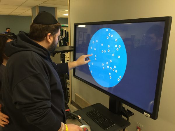
The machine can anticipate arm movement, and helps patients move their arm once the arm is in motion. The device is connected to a computer monitor that can simulate everyday activities such as picking an item off of a shelf at the supermarket and placing it in a shopping basket.

Balancing Acts

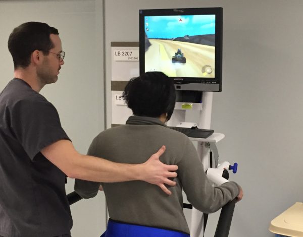
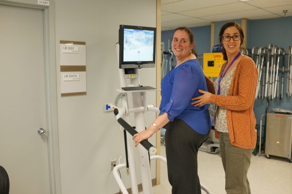
Balance, cognition, and memory are important functions that rehabilitation can help recover, and the team at NYU utilizes a device called the Bioness Integrated Therapy System that has several different programs. Patients stand in front of a large computer screen and point at images on different parts of the monitor, making them shift their weight in different directions, which ultimately helps them regain balance. Another program helps improve memory by requiring the patient to tap a series of objects on the screen, and then tap on the same series again to show they remember.
Once patients regain more balance, they move on to the Korebalance System. Patients stand on a flexible platform and shift their weight with their feet while playing a video game where they have to stay on a track. Instead of using their hands like they would if they were playing a typical video game, patients use their feet and allocate their weight on the platform to stay on the track. This system ultimately helps fine tune patients’ balance.

The Korebalance system
A patient (R) using the Korebalance System. (Courtesy of NYU Langone Hospital-Brooklyn)
The fourth interactive device patients use is called the Flint Rehab Glove, and it is designed to help patients regain mobility in their fingers. The glove is connected to a video game similar to Guitar Hero. Each finger corresponds to a colored button on the screen. Once the button appears, they touch the appropriate finger with their thumb and the game makes a sound, and the repetitive movement creates a song.

More Than a Game

According to PT Cavallaro, patients have reacted positively to these interactive games. The rehabilitation team works to ensure a patient has the ability to complete each game before moving onto the next in order to promote a positive, can-do attitude, and show that they can accomplish more than they previously believed.
“I think when you’re playing a game, I think there’s a natural tendency to want to score better each time you do it, and that gives you that visual input that comes off the screen that patients see, and want to, next time they’re on that machine, get a higher score or a longer distance time.”

Korebalance system
A patient (L) employing the Korebalance System. (Courtesy of NYU Langone Hospital-Brooklyn)
These interactive games give the physical therapist another means of treating their patients, and enhances the traditional rehabilitation methods. When patients succeed and families see them prevail in their treatment, they are less apprehensive when the patient returns home.
While the relationship between the patient and the physical therapist is the most important aspect of recovery, there is a lot to gain from technology.

A Needed Development

These new technologies come at a time when stroke recovery is a growing concern.
According to the Centers for Disease Control and Prevention, strokes are the fifth leading cause of death in the United States. Approximately 795,000 people suffer from a stroke annually. There are many factors that can lead to a stroke such as a history of strokes in the family. Medical conditions such as hypertension, diabetes, and Sickle Cell Anemia can also contribute to a stroke. However, there are behaviors that can make a stroke more likely such as alcohol abuse, tobacco use, lack of exercise, and an unhealthy diet. Fortunately, these behaviors can be moderated or avoided completely.
Following a stroke, patients may experience a variety of impairments including paralysis in some limbs or on one side of the body, difficulty understanding or constructing speech, trouble controlling or expressing emotion, numbness or sensation, trouble swallowing, and depression.
Successful rehabilitation can be the difference between years of suffering and dependency and a meaningful and active life.

Study: Memories of music cannot be lost to Alzheimer's and dementia

With your very likely chance of getting dementia poststroke maybe your doctor should prescribe music as a stroke therapy and possible preventative to dementia. But I'm not medically trained so don't listen to me. Listen to your doctors answer to; 'How do I get 100% recovered?"


Study: Memories of music cannot be lost to Alzheimer's and dementia 


The part of your brain responsible for ASMR catalogs music, and appears to be a stronghold against Alzheimer's and dementia.


Some music inspires you to move your feet, some inspires you to get out there and change the world. In any case, and to move hurriedly on to the point of this article, it's fair to say that music moves people in special ways. 
If you're especially into a piece of music, your brain does something called Autonomous Sensory Meridian Response (ASMR), which feels to you like a tingling in your brain or scalp. It's nature's own little "buzz", a natural reward, that is described by some as a "head orgasm". Some even think that it explains why people go to church, for example, "feeling the Lord move through you", but that's another article for another time. 
Turns out that ASMR is pretty special. According to a recently published study in The Journal of Prevention of Alzheimer's Disease (catchy name!), the part of your brain responsible for ASMR doesn't get lost to Alzheimer's. Alzheimer's tends to put people into layers of confusion, and the study confirms that music can sometimes actually lift people out of the Alzheimer's haze and bring them back to (at least a semblance of) normality... if only for a short while. ASMR is powerful stuff! 
This phenomenon has been observed several times but rarely studied properly. One of the most famous examples of this is the story of Henry, who comes out of dementia while listening to songs from his youth:  
Jeff Anderson, M.D., Ph.D., associate professor in Radiology at the Univerity of Utah Health and contributing author on the study, says  "In our society, the diagnoses of dementia are snowballing and are taxing resources to the max. No one says playing music will be a cure for Alzheimer's disease, but it might make the symptoms more manageable, decrease the cost of care and improve a patient's quality of life."

I am more than my stroke - UK Stroke Association

While the sentiment is good, this just allows them to not have to solve all the problems in stroke. Trying to make survivors feel good about not recovering 100%. I would suggest pushing back mightily.

I am more than my stroke

Power Spectral Density Analysis of Long-Term Motor Recovery in Patients With Subacute Stroke

My god, the fucking stupidity of stroke recovery prediction research.  Survivors don't care about prediction, they want RESULTS, 100% RECOVERY. Talk to a survivor sometime.

Power Spectral Density Analysis of Long-Term Motor Recovery in Patients With Subacute Stroke


First Published December 19, 2018 Research Article
Background. Prognostic measures of long-term motor recovery are important(NO,NO,NO they aren't)in patients with stroke presenting with severe hemiplegia.  
Objective. We aimed to investigate whether initial power spectral density (PSD) analysis of resting-state functional magnetic resonance (fMRI) data can provide a sensitive prognostic predictor in patients with subacute stroke with severe hand disability.  
Methods. Twelve patients with good recovery, 14 patients with poor recovery, and 12 healthy subjects were included. PSD analysis was performed using resting-state fMRI data. Contralesional and ipsilesional PSD in the motor cortex were measured. Pearson correlation analysis was performed to assess a possible association between the difference in ipsilesional versus contralesional PSD and motor outcomes. A receiver operating characteristic (ROC) curve was constructed to estimate the discriminative value of the difference between the ipsilesional PSD and the contralesional PSD for good versus poor recovery.  
Results. There were no differences in PSD between the contralesional and ipsilesional hemispheres in the good recovery group (P = .77). In contrast, there were significant differences in PSD between the 2 hemispheres in the poor recovery group (P = .07). The difference in PSD between the 2 hemispheres had a positive correlation with post Brunnstrom stage scores. ROC analysis showed that the difference in PSD between the 2 hemispheres was sensitive in discriminating good versus poor recovery.  
Conclusion. The present study suggests that PSD in the motor cortex may be a sensitive predictor of late-onset motor recovery following stroke.

Role of immune responses for extracellular matrix remodeling in the ischemic brain

What stroke leader are you going to propose this new direction? And where is the strategy you are updating? Just writing this in a journal does not relieve you of the responsibility of seeing that followup occurs.  Nothing will occur, stroke survivors will continue to be screwed by the lack of any stroke leadership.

Role of immune responses for extracellular matrix remodeling in the ischemic brain 

First Published December 17, 2018 Review Article
Neuroinflammation is one of the key components contributing to the devastating outcome of ischemic stroke. Starting with stroke onset, inflammatory processes contribute both to cell damage and tissue remodeling. The early release of alarmins triggers the upregulation of multiple proinflammatory cytokines, resulting in the compromised integrity of the blood–brain barrier. From this moment on, the infiltration of peripheral immune cells, reactive gliosis and extracellular matrix (ECM) alterations become intricately intertwined and act as one unit during the tissue remodeling. While the mechanisms of leukocyte and glia activation are amply reviewed, the field of ECM modification remains as yet under explored. In this review, we focus on the interplay between neuroinflammatory cascades and ECM in the ischemic brain. By summarizing the currently available evidence obtained by in vitro research, animal experimentation and human studies, we aim to propose a new direction for the future investigation of stroke recovery.

Sunday, December 23, 2018

Role of immune responses for extracellular matrix remodeling in the ischemic brain

Lots of big words but nothing I could see of use to recovery

Role of immune responses for extracellular matrix remodeling in the ischemic brain


Introduction Ischemic stroke remains a leading cause of death and disability in the adult population. Despite plenty of efforts devoted to understanding and treating the disease, most novel approaches have only a discouragingly limited impact on patients’ wellbeing. 1 We suggest that to improve the translation of scientific advances from bench to bedside, the pathophysiology of ischemic stroke should be investigated from complementary nb points of view. Today, most studies of stroke put the major focus on neuronal plasticity and repair, 2–4 blood–brain barrier (BBB) dysfunction, 5 and neuroinflammation. 6 In this review, we will address the relationship between the immune response and the reorganization of the extracellular matrix (ECM) during the acute and chronic phases of ischemic stroke. Although both aspects have been studied individually, their interaction is rarely considered in both experimental and clinical settings. We propose that the brain’s immune response and ECM regulation should be considered as a functional unit, as first proposed by Schönherr and Hausser, 7 opening new perspectives in stroke treatment. The ECM is a congregation of multiple adhesion molecules, polysaccharides, proteins and proteoglycans arranged three-dimensionally in the extracellular space. During development and adulthood, this complex fulfils various functions, such as regulating cell migration, proliferation, adhesion, differentiation, 8 synaptic plasticity, 9 maintenance of the BBB 10 and tissue architecture, integrity and homeostasis. 11 In the central nervous system (CNS), ECM can be divided into two compartments, the interstitial matrices and basement membranes (BMs). 12 The interstitial matrix is based on diffuse meshworks of hyaluronic acid (HA), which incorporate mainly collagens and proteoglycans. 13 The BMs are associated with the basal portion of cerebral endothelial cells and consist mainly of laminins, collagen IV, nidogens and heparan sulfate proteoglycans.

The role of neurogenesis in neurorepair after stroke

Where the fuck is the public writeup on this so the 10 million yearly stroke survivors  can use this to get recovered? Or do you not care about helping survivors?  Hide information in a research journal? 

Laziness? Incompetence? Or just don't care? No leadership? No strategy? Not my job?

The role of neurogenesis in neurorepair after stroke

Abstract

Stroke consists of an abrupt reduction of cerebral blood flow resulting in hypoxia that triggers an excitotoxicity, oxidative stress, and neuroinflammation. After the ischemic process, neural precursor cells present in the subventricular zone of the lateral ventricle and subgranular zone of the dentate gyrus proliferate and migrate towards the lesion, contributing to the brain repair. The neurogenesis is induced by signal transduction pathways, growth factors, attractive factors for neuroblasts, transcription factors, pro and anti-inflammatory mediators and specific neurotransmissions. However, this endogenous neurogenesis occurs slowly and does not allow a complete restoration of brain function. Despite that, understanding the mechanisms of neurogenesis could improve the therapeutic strategies for brain repair. This review presents the current knowledge about brain repair process after stroke and the perspectives regarding the development of promising therapies that aim to improve neurogenesis and its potential to form new neural networks.

KEYWORDS:

Stroke; brain ischemia; brain repair; neurogenesis
PMID:
30550812
DOI:
10.1016/j.semcdb.2018.12.003

Saturday, December 22, 2018

Blood donation today

17 so far in the last 5 years, missed a bunch because of high blood pressure, low iron. I'm O negative, universal donor so I'm quite popular there. I can only receive other O- blood, but luckily one of my friends has that, already told her I'm calling her if I get in an accident that needs blood.  Didn't realize all the requirements which make it impossible to use my affected left arm as the donation site.
  1. Arm must be able to lay completely flat.
  2. Arm must stay on table.
  3. Arm must not spastically contract for any reason.
  4. Must be able to squeeze rubber ball every 4 seconds. 
The major problem with using my good right arm for donation is the cotton swab and tape on the puncture site. The stretchy red elastic is usually wrapped at least 2-3 times around my arm. No tab is left free and I can't reach the tape with my teeth. My left fingers have zero ability to pick up anything and I can't even get the hand anywhere close. So I have to wedge a long knife in a kitchen drawer and saw the red elastic thru, hopefully without cutting myself.

Longitudinal Functional Brain Mapping in Supernormals

There is no one in the world that I can go to to give me protocols to assure I become a super ager. At age 90+ I still want to have all the social connections and drinking I do right now. Along with much international travel. For me, 8 days in Ecuador in Jan., 16 days in Italy in Feb.-Mar. 6 days in London in March. I'm sure there will be more for the rest of the year. 

Longitudinal Functional Brain Mapping in Supernormals


Cerebral Cortex, Volume 29, Issue 1, 1 January 2019, Pages 242–252, https://doi.org/10.1093/cercor/bhx322
Published:
23 November 2017
Article history

Abstract

Prevention of age-related cognitive decline is an increasingly important topic. Recently, increased attention is being directed at understanding biological models of successful cognitive aging. Here, we examined resting-state brain regional low-frequency oscillations using functional magnetic resonance imaging in 19 older adults with excellent cognitive abilities (Supernormals), 28 older adults with normative cognition, 57 older adults with amnestic mild cognitive impairment, and 26 with Alzheimer’s disease. We identified a “Supernormal map”, a set of regions whose oscillations were resistant to the aging-associated neurodegenerative process, including the right fusiform gyrus, right middle frontal gyrus, right anterior cingulate cortex, left middle temporal gyrus, left precentral gyrus, and left orbitofrontal cortex. The map was unique to the Supernormals, differentiated this group from cognitive average-ager comparisons, and predicted a 1-year change in global cognition (indexed by the Montreal Cognitive Assessment scores, adjusted R2 = 0.68). The map was also correlated to Alzheimer’s pathophysiological features (beta-amyloid/pTau ratio, adjusted R2 = 0.66) in participants with and without cognitive impairment. These findings in phenotypically successful cognitive agers suggest a divergent pattern of brain regions that may either reflect inherent neural integrity that contributes to Supernormals’ cognitive success, or alternatively indicate adaptive reorganization to the demands of aging.

Artificial intelligence system learns to diagnose, classify intracranial hemorrhage

Fascinating idea, remove the neurologist from the equation and speed up the diagnosis considerably, wouldn't even need to set up telehealth centers.  I'm all for it.

Artificial intelligence system learns to diagnose, classify intracranial hemorrhage


Mass.-General-developed system able to ‘explain’ reasons behind decisions based on CT scan images
IMAGE
Credit: Hyunkwang Lee, Harvard School of Engineering and Applied Sciences, and Sehyo Yune, MD, Massachusetts General Hospital Department of Radiology
A team of investigators from the Massachusetts General Hospital (MGH) Department of Radiology has developed a system using artificial intelligence to quickly diagnose and classify brain hemorrhages and to provide the basis of its decisions from relatively small image datasets. Such a system could become an indispensable tool for hospital emergency departments evaluating patients with symptoms of a potentially life-threatening stroke, allowing rapid application of the correct treatment. The team’s report has been published online in Nature Biomedical Engineering.
While ever-increasing computational power and the availability of big datasets have improved machine learning – the process by which computers analyze data, identify patterns and essentially teach themselves how to perform a task without the direct involvement of a human programmer – important obstacles can prevent such systems from being integrated into clinical decision making. These include the need for large and well annotated datasets – previously developed imaging analysis systems capable of duplicating the performance of a physician were trained with more than 100,000 images – and the “black box” problem, the inability of systems to explain how they arrived at a decision. The U.S. Food and Drug Administration requires any decision support system to provide data allowing users to review the reasons behind its findings.
“It is somewhat paradoxical to use the words ‘small data’ or ‘explainable’ to describe a study that used deep learning,” says Hyunkwang Lee, a graduate student at the Harvard School of Engineering and Applied Sciences, one of the two lead authors of the study. “However, in medicine, it is especially hard to collect high-quality big data. It is critical to have multiple experts label a dataset to ensure consistency of data. This process is very expensive and time-consuming.”
Co-lead author Sehyo Yune, MD, of MGH Radiology adds, “Some critics suggest that machine learning algorithms cannot be used in clinical practice, because the algorithms do not provide justification for their decisions. We realized that it is imperative to overcome these two challenges to facilitate the use in health care of machine learning, which has an immense potential to improve the quality of and access to care.
To train their system, the MGH team began with 904 head CT scans, each consisting of around 40 individual images, that were labeled by a team of five MGH neuroradiologists as to whether they depicted one of five hemorrhage subtypes, based on the location within the brain, or no hemorrhage. To improve the accuracy of this deep-learning system the team – led by senior author Synho Do, PhD, director of the MGH Radiology Laboratory of Medical Imaging and Computation and an assistant professor of Radiology at Harvard Medical School – built in steps mimicking the way radiologists analyze images. These include adjusting factors such as contrast and brightness to reveal subtle differences not immediately apparent and scrolling through adjacent CT scan slices to determine whether or not something that appears on a single image reflects a real problem or is a meaningless artifact.
Once the model system was created, the investigators tested it on two separate sets of CT scans – a retrospective set taken before the system was developed, consisting of 100 scans with and 100 without intracranial hemorrhage, and a prospective set of 79 scans with and 117 without hemorrhage, taken after the model was created. In its analysis of the retrospective set, the model system was as accurate in detecting and classifying intracranial hemorrhages as the radiologists that had reviewed the scans had been. In its analysis of the prospective set, it proved to be even better than non-expert human readers.
To solve the “black box” problem, the team had the system review and save the images from the training dataset that most clearly represented the classic features of each of the five hemorrhage subtypes. Using this atlas of distinguishing features, the system is able to display a group of images similar to those of the CT scan being analyzed in order to explain the basis of its decisions.
“Rapid recognition of intracranial hemorrhage, leading to prompt appropriate treatment of patients with acute stroke symptoms, can prevent or mitigate major disability or death,” says co-author Michael Lev, MD, MGH Radiology. “Many facilities do not have access to specially trained neuroradiologists – especially at night or over weekends – which can require non-expert providers to determine whether or not a hemorrhage is the cause of a patient’s symptoms. The availability of a reliable, ‘virtual second opinion’ – trained by neuroradiologists – could make those providers more efficient and confident and help ensure that patients get the right treatment.”
Co-author Shahein Tajmir, MD, MGH Radiology adds, “In addition to providing that much needed virtual second opinion, this system also could be deployed directly onto scanners, alerting the care team to the presence of a hemorrhage and triggering appropriate further testing before the patient is even off the scanner. The next step will be to deploy the system into clinical areas and further validate its performance with many more cases. We are currently building a platform to allow for the widespread application of such tools throughout the department. Once we have this running in the clinical setting, we can evaluate its impact on turnaround time, clinical accuracy and the time to diagnosis.”
###
The additional co-authors of the Nature Biomedical Engineering are Mohammad Mansouri, Myeongchan Kim, Claude E. Guerrier, MD, Sarah A. Ebert, MD, Stuart R. Pomerantz, MD, Javier M. Romero, MD, Shahmir Kamalian, MD, and Ramon G. Gonzalez, MD, PhD, all from MGH Radiology. Support for the study includes National Institutes of Health grant 5U01 EB025153.
Massachusetts General Hospital, founded in 1811, is the original and largest teaching hospital of Harvard Medical School. The MGH Research Institute conducts the largest hospital-based research program in the nation, with an annual research budget of more than $900 million and major research centers in HIV/AIDS, cardiovascular research, cancer, computational and integrative biology, cutaneous biology, genomic medicine, medical imaging, neurodegenerative disorders, regenerative medicine, reproductive biology, systems biology, photomedicine and transplantation biology. The MGH topped the 2015 Nature Index list of health care organizations publishing in leading scientific journals and earned the prestigious 2015 Foster G. McGaw Prize for Excellence in Community Service. In August 2018 the MGH was once again named to the Honor Roll in the U.S. News & World Report list of “America’s Best Hospitals.”
Media Contact
Terri Ogan Janos
tjanos@partners.org
617-726-0954

Friday, December 21, 2018

Why Coconut Oil Really is Healthy; Despite the American Heart Association's Views

I've got all these posts on coconut oil if you want to decide for yourself. Or you can trust your doctor to have read all these and more. I'm doing some, way too many pros vs. cons for me. But I'm not medically trained like that professor that called it pure poison with no research to back up her opinion.

  • coconut oil (54)


A Harvard professor just busted the myth that coconut oil is good for you, calling it 'pure poison'

Why Coconut Oil Really is Healthy; Despite the American Heart Association's Views

Thursday, December 20, 2018

Neuroprotective effect of Bacopa monniera on ischemia induced brain injury

Well nothing will ever be done for testing in humans. There is NO STROKE LEADERSHIP to talk to and NO STROKE STRATEGY.  Besides there have been 1000+ failures in trying neuroprotection therapies that worked in rodents that didn't work in humans. And no one has ever figured out why, so this is also destined to fail in humans. 


Neuroprotective effect of Bacopa monniera on ischemia induced brain injury


Abstract

Purpose

Brain stroke is a leading cause of death without effective treatment. B. monniera, an Indian herbal medicine, exerts antioxidant activity and antistress activity by modulating the antioxidative defence system. We wanted to test if B. monniera could alleviate the ischemia induced brain injury and cognitive dysfunction in Wistar rats.

Procedure

We studied the effect of B. monniera (120 mg kg− 1, 160 mg kg− 1 and 240 mg kg− 1 P.O.) on transient intracarotid artery (ICA) occlusion induced ischemia by testing the neurobehavioral and biochemical parameters on treated and control rats.

Findings

B. monniera attenuated the reduced transfer latency in ischemic rats in a step through test and showed a protective effect on ischemia induced memory impairment in the plus maze task. It also showed a marginal improvement in neurodeficit score and fore limb muscle grip strength. B. monniera reduced the infarct size in the ischemic brain. It also decreased nitrite, nitrate and lipid peroxidation and significantly improved catalase activity.

Conclusion

These observations suggest the neuroprotective and antioxidant activity of B. monniera on ischemia induced brain injury and pave the way for future investigations.

Research Highlights

►We studied the effect of B. monniera on transient intracarotid artery (ICA) occlusion induced ischemia. ►B. monniera attenuated ischemia induced brain injury. ►It also reversed impaired neurobehavioral and biochemical parameters in rats. ►This study observation suggests the neuroprotective and antioxidant activity of B. monniera.

Accuracy vs. Reproductibility in the National Institutes of Health Stroke Scale: When the NIHSS Does Not Assess All the Symptoms

You still don't know that the NIHSS subjective stroke scale is worthless? You can't do a goddamn thing with it; map recovery protocols to a objective starting point. No research is reproducible using this since it has nothing objective in it.


Accuracy vs. Reproductibility in the National Institutes of Health Stroke Scale: When the NIHSS Does Not Assess All the Symptoms


Elena Zapata-Arriaza, MD
@ElenaZaps

Eskioglou E, Huchmandzadeh Millotte M, Amiguet M, Michel P. National Institutes of Health Stroke Scale Zero Strokes: Immeasurable but Not Innocent. Stroke. 2018
The National Institutes of Health Stroke Scale (NIHSS) is the most widely employed deficit rating scale in modern neurology and has became the gold standard for stroke severity rating in recent published clinical trials. However, it is essential to know that the scale was not designed to serve as a bedside rating tool for widespread use outside of research trials. The goal of NIHSS is to assure reproducibility, not accuracy.
The authors performed a retrospective analysis of all acute ischemic stroke patients admitted to the stroke unit and intensive care unit of the Lausanne University Hospital from 2003 to 2013. The main goal of this paper was to highlight the characteristics of ischemic strokes with a NIHSS = 0, as well as to determine the long-term evolution of these patients. The originality of the paper is the aim itself, which makes us wonder whether the NIHSS is a sufficiently accurate scale to determine the severity of all ischemic strokes. The authors included all patients with a suspected stroke according to the classical World Health Organization definition. The sample was divided between NIHSS=0 and NIHSS > or equal to 1, and brain and vascular state was assessed mostly by computed tomography. During follow-up, mortality, stroke or transient ischemic attack recurrence and functional outcome (3 and 12 months) were collected.

Among 2997 included patients, 108 had a NIHSS=0. After multivariate analysis, NIHSS=0 subgroup patients showed statistically significant differences. The main findings are displayed in the Table below. The most outstanding clinical and radiological findings related to NIHSS=0 strokes were a lower prestroke disability, a delay in hospital arrival, lacunar and infratentorial presentation, higher ASPECTS at admission and less arterial stenosis or occlusion.
Table. Multivariate Analyses Showing Statistically Significant Differences Between Both Groups
Two NIHSS=0 patients died within 3 months from early stroke recurrence, and 5 others after 12 months (2 cancers, 1 pneumonia, 2 undetermined causes). Favorable outcome was clearly more frequent in NIHSS=0 patients. Recurrence rates were similar for strokes (6.6% in NIHSS=0 versus 8.8% in NIHSS ≥1) and for strokes and TIAs combined (11.4% versus 11.0%). About 28.5% NIHSS=0 patients had unfavorable 3 months outcome (mRS score of ≥2), and 46.7% had some remaining symptoms or handicap (mRS score of ≥1), mostly related in the multivariate analysis with infratentorial stroke localization and acute cerebellar symptoms.
Interesting assessment can be obtained from this paper. First, the NIHSS scale may be insufficient to detect certain neurological deficits with prognostic and functional implications. There may be patients with an NIHSS = 0 and disability in the post-stroke follow-up. This powerful message reveals the limitations in the clinical accuracy of a scale created to be reproducible and whose main application field are clinical trials. Likewise, the persistence of disability associated with cerebellar symptoms and stroke in the posterior territory highlights the underestimation of the NIHSS scale in this stroke localization. To determine a stroke patient’s severity, the complete and properly described neurological examination is closer to the real clinical patient state, and therefore will better guide us to the future evolution of the patient.
Secondly, the recurrence of stroke is similar in both subgroups, although patients with NIHSS = 0 have a CT without alterations. This should alert us to the following: Clinical examination is always the starting point that should lead the management and clinical follow-up. Patients with a non-quantifiable deficit are at risk of recurrence and deserve secondary prevention and adequate follow-up.
Finally, it is important to note that in this article, the image analysis is performed mainly by CT. Such assessment could misdiagnose mimics as strokes; MRI could identify situations where symptoms are not due to vascular causes. In any case, it is important to emphasize that the NIHSS is not infallible, and it must be taken into account the aim of its origin, which was reproducibility, not clinical accuracy.

One in four people will have a stroke

Used to be 1 in 6. Well, even with more people having strokes you are still completely screwed because there are NO protocols in stroke rehab to get you anywhere close to 100% recovery. The non-existent stroke leadership has done nothing for decades. They don't even acknowledge that there is a problem. This meme on World Stroke Day a couple of years ago proves how fucking incompetent they are. Yet all they talk about is prevention. Are they too fucking lazy or incompetent to tackle the

BHAGs(Big Hairy Audacious Goals) of 100% recovery for all survivors?

What a bald faced lie

 These minor problems still need solving. 

1. Only 10% of patients get to full recovery.
2. tPA only fully works to reverse the stroke 12% of the time. Known since 1996.
3. No protocols to prevent your 33% dementia chance post-stroke from an Australian study.
4. Nothing to alleviate your fatigue.
5. Nothing that will cure your spasticity.
6. Nothing on cognitive training unless you find this yourself.
7. No published stroke protocols.
8. No way to compare your stroke hospital results vs. other stroke hospitals.


One in four people will have a stroke

Alarming new research, published in the New England Journal of Medicine today, has revealed stroke is on the increase with one in four people globally to experience stroke in their lifetime.

Data released in 2006 had this estimate at one in six people. Research leading author Professor Valery Feigin said the sharp surge in stroke’s impact was due, in part, to an increasingly unhealthy lifestyle.

“The increase in stroke incidence globally comes down to more than an ageing and growing population. We know people are now experiencing strokes at younger ages,” Professor Feigin said.

“A total of 58 percent of all strokes now happen to people 70 years and under – and our modern lifestyle is to blame.

“We are spending our time scrolling throughout smart phones instead of walking outside. We are eating convenience foods instead of cooking for ourselves. We are stressed, we are not sleeping well. We are not looking after ourselves and we are suffering the consequences.”

Stroke attacks the brain, the human control centre, changing the lives of those impacted and their loved ones in an instant.

This year alone an estimated 56,000 strokes will be experienced by Australians – that is one stroke every nine minutes. Frighteningly, the country is on track for the number of strokes to more than double to 132,000 annually by 2050 – one stroke every four minutes.

Stroke Foundation Chief Executive Officer Sharon McGowan said this runaway train can and must be stopped immediately.

“We are at risk of leaving the next generation with a shorter life expectancy than our own unless decisive action is taken to reduce the burden of stroke,” Ms McGowan said.

“Stroke kills more men than prostate cancer, more women than breast cancer and leaves thousands with ongoing disabilities each year, yet 80 percent of strokes can be prevented. 

“Federal and State governments must act now, coming together to lead the way with targeted prevention programs to help people identify their stroke risk and do something about it.

“We must do more to support healthier communities.

“An investment in prevention is just that, an investment, the return is lives saved and a reduction in stroke’s burden on our community and health system.”

Ms McGowan also encouraged people to visit their General Practitioner (GP), ask for a health check and take steps to control their stroke risk. GPs can help people identify and manage key risk factors like blood pressure, cholesterol and atrial fibrillation (irregular heart beat). They can also offer advice on a healthy diet, exercise routine and on how to quit smoking and reduce alcohol intake.


Variation of Finger Activation Patterns Post-stroke Through Non-invasive Nerve Stimulation

Way too many big words used to have any clue on how to explain this to therapists and doctors for our use.  I see no objective damage diagnosis that would point to which patients this would work on. Without that, this research is not repeatable and thus useless. 

Variation of Finger Activation Patterns Post-stroke Through Non-invasive Nerve Stimulationh

  • Joint Department of Biomedical Engineering, University of North Carolina at Chapel Hill and North Carolina State University, Raleigh, NC, United States
Purpose: A transcutaneous proximal nerve stimulation technique utilizing an electrode grid along the nerve bundles has previously shown flexible activation of multiple fingers. This case study aimed to further demonstrate the ability of this novel stimulation technique to induce various finger grasp patterns in a stroke survivor.
Methods: An individual with chronic hemiplegia and severe hand impairment was recruited. Electrical stimulation was delivered to different pairs of an electrode grid along the ulnar and median nerves to selectively activate different finger flexor muscles, with an automated electrode switching method. The resultant individual isometric flexion forces and forearm flexor high-density electromyography (HDEMG) were acquired to evaluate the finger activation patterns. A medium and low level of overall activation were chosen to gauge the available finger patterns for both the contralateral and paretic hands. All the flexion forces were then clustered to categorize the different types of grasp patterns.
Results: Both the contralateral and paretic sides demonstrated various force clusters including single and multi-finger activation patterns. The contralateral hand showed finger activation patterns mainly centered on median nerve activation of the index, middle, and ring fingers. The paretic hand exhibited fewer total activation patterns, but still showed activation of all four fingers in some combination.
Conclusion: Our results show that electrical stimulation at multiple positions along the proximal nerve bundles can elicit a select variety of finger activation patterns even in a stroke survivor with minimal hand function. This system could be further implemented for better rehabilitative training to help induce functional grasp patterns or to help regain muscle mass.

Introduction

Following a stroke, a majority of individuals have paresis due to a loss of excitatory input and subsequent complications, such as disuse atrophy (1) and altered spinal organization (24). This loss of voluntary control of muscle activation often limits activities of daily living. Neuromuscular electrical stimulation (NMES) has been widely utilized both in the clinic and in research settings to help restore atrophied muscle and lost functions (57). Electrical stimulation has been particularly successful with post-stroke survivors for functional recovery (810). Research in NMES also aims to restore functional activation of muscles, such as the restoration of hand grasps (11).
Traditionally, NMES uses large electrode pads, targeting the distal branches of the nerve, known as the motor point stimulation (12). Although stimulation of the motor point is straightforward methodologically, NMES is limited to localized muscle activation, which limits its functional efficacy and also leads to rapid muscle fatigue (13). Advances in NMES techniques to alleviate these issues involve various multi-electrode techniques, which can stimulate multiple small regions of the muscle to help distribute the current and potentially activate more muscle fibers (14, 15). Crema et al. has also demonstrated flexible activation of multiple fingers using a multi-electrode array across the forearm and hand (16). Other approaches to NMES involve stimulation of the nerve bundle prior to branching and innervating a muscle, which has shown to allow for a larger area of muscle activation and potentially reduce long-term fatigue effects (1719).
Recent developments have demonstrated the capabilities of an alternative non-invasive transcutaneous electrical nerve stimulation method targeting the ulnar and median nerves proximal to the elbow to flexibly activate individual and multiple fingers (20, 21). In addition, this technique shows the ability to delay the force decline (22, 23). A stimulation electrode grid placed along the two nerves allows us to activate different muscles or muscle portions to elicit varied desired movements, but manually switching between different electrode pairs is time-consuming. To shorten this process, an automated electrode pair searching method has been developed and tested on intact control subjects (24). This new method can further categorize the total available sets of finger activation patterns across the entire electrode grid, providing valuable information on electrode selection and the force generation capacity of stroke muscles. However, the efficiency of this method has not been tested on stroke survivors. Therefore, this case study recruited a control subject and a stroke survivor with severe weakness of the right arm, and evaluated the available finger activation patterns of the subjects. Our results showed varied activation of multiple fingers from both subjects. Further development of this stimulation technique can provide valuable alternatives to current rehabilitation for the restoration of hand movements.

Arm Ability Training (AAT) Promotes Dexterity Recovery After a Stroke—a Review of Its Design, Clinical Effectiveness, and the Neurobiology of the Actions

Where the fuck is the protocol for this located? You left out treatment options for severe arm paresis. Damn it all, quit cherry picking easier patients to treat. 

This is the closest I could get to finding a protocol; you'll have to extrapolate a lot.


Arm Ability Training (AAT) Promotes Dexterity Recovery After a Stroke—a Review of Its Design, Clinical Effectiveness, and the Neurobiology of the Actions


  • 1BDH-Klinik Greifswald, Centre for Neurorehabilitation, Intensive and Ventilation Care, Spinal Cord Injury Unit, University of Greifswald, Greifswald, Germany
  • 2Functional Imaging Unit, Center for Diagnostic Radiology, University of Greifswald, Greifswald, Germany
Arm Ability Training (AAT) has been specifically designed to promote manual dexterity recovery for stroke patients who have mild to moderate arm paresis. The motor control problems that these patients suffer from relate to a lack of efficiency in terms of the sensorimotor integration needed for dexterity. Various sensorimotor arm and hand abilities such as speed of selective movements, the capacity to make precise goal-directed arm movements, coordinated visually guided movements, steadiness, and finger dexterity all contribute to our “dexterity” in daily life. All these abilities are deficient in stroke patients who have mild to moderate paresis causing focal disability. The AAT explicitly and repetitively trains all these sensorimotor abilities at the individual's performance limit with eight different tasks; it further implements various task difficulty levels and integrates augmented feedback in the form of intermittent knowledge of results. The evidence from two randomized controlled trials indicates the clinical effectiveness of the AAT with regard to the promotion of “dexterity” recovery and the reduction of focal disability in stroke patients with mild to moderate arm paresis. In addition, the effects have been shown to be superior to time-equivalent “best conventional therapy.” Further, studies in healthy subjects showed that the AAT induced substantial sensorimotor learning. The observed learning dynamics indicate that different underlying sensorimotor arm and hand abilities are trained. Capacities strengthened by the training can, in part, be used by both arms. Non-invasive brain stimulation experiments and functional magnetic resonance imaging data documented that at an early stage in the training cortical sensorimotor network areas are involved in learning induced by the AAT, yet differentially for the tasks trained. With prolonged training over 2 to 3 weeks, subcortical structures seem to take over. While behavioral similarities in training responses have been observed in healthy volunteers and patients, training-induced functional re-organization in survivors of a subcortical stroke uniquely involved the ipsilesional premotor cortex as an adaptive recruitment of this secondary motor area. Thus, training-induced plasticity in healthy and brain-damaged subjects are not necessarily the same.

Motor Deficits of Stroke Survivors With Mild to Moderate Arm Paresis

I think I have severe since both my pre-motor and motor cortex for arm use are mostly dead.
Arm paresis post stroke shows a bi-modal distribution. Many stroke survivors have either severe arm paresis and are only able to use their arms functionally in everyday life to a very limited extent, if at all, or mild to moderate arm paresis with the ability to use their paretic arm for functional tasks, yet with a lack of dexterity (1, 2). Thus, the motor control deficits of these subgroups are quite different and hence so too are their therapeutic needs.
Clinically, stroke survivors with mild to moderate arm paresis have reduced strength and endurance of their paretic arm and are functionally limited by a lack of speed, accuracy and co-ordination of arm, hand, and finger movements and a lack of dexterity when handling objects. Key to understanding any functional deficits and the need and opportunities to improve function by training is a focused analysis of the specific motor control deficits involved in this clinical syndrome. A way to do this is to test various domains of sensorimotor control that have been shown to be independent by factorial analysis (3, 4).
When motor performance of healthy people across various tasks has been analyzed by factorial analysis certain independent arm motor abilities have been documented. These are different independent sensorimotor capacities that together contribute to our skilfulness in everyday life. What are these abilities? They are our ability to make fast selective wrist and finger movements (wrist-finger speed), to manipulate small objects (finger dexterity) or larger objects (manual dexterity) efficiently, our ability to keep our arm steady (steadiness), to move our arm quickly and precisely to an intended target (aiming), or to move it under constant visual control along a line (tracking) (5).
When tested among stroke survivors with mild to moderate arm paresis all these abilities are deficient, indicating the complex nature of sensorimotor control deficits in this clinical condition (6, 7).

Young Stroke Survivors With No Early Recurrence at High Long‐Term Risk of Adverse Outcomes

Well, I guess I don't have to worry I was 50 at my stroke. Ok they have identified a possible problem(quantify risks) but it is pretty much useless with no solution provided.  And their mentors and senior researchers allowed such useless research to go ahead.

Young Stroke Survivors With No Early Recurrence at High Long‐Term Risk of Adverse Outcomes


Originally publishedhttps://doi.org/10.1161/JAHA.118.010370Journal of the American Heart Association. 2018;8:e010370

Abstract

Background

Approximately 8% to 21% of strokes affect adults aged <45 years. Although early stroke recurrence conveys the largest risk, long‐term risks for young survivors with no early complications are unclear.

Methods and Results

Longitudinal matched case‐control study (2003–2013). Consecutive patients with ischemic stroke or transient ischemic attack (young, ≤44 years) discharged from emergency or regional stroke centers in Ontario, Canada, with no death, recurrent stroke/transient ischemic attack, myocardial infarction, all‐cause hospitalization, or admission to a long‐term or continuing care facility (≤90 days) were matched 10:1 to general population controls on age (±1 year), sex, income, geography, and case date (±50 days). The primary outcome was a composite of death, stroke, myocardial infarction, and long‐term or continuing care facility admission at 1, 3, and 5 years. Absolute event rates for young stroke/transient ischemic attack patients were lower than for older patients at 1 (2.2% versus 9.9%), 3 (4.7% versus 24.6%), and 5 (7.1% versus 37.2%) years. However, piecewise constant hazard modeling revealed that, even after adjustment for vascular comorbidities, young patients showed a 7‐fold increased hazard of the composite outcome compared with young controls at 1 year (hazard ratio, 7.3; 95% CI, 4.0–13.6). Adjusted 5‐year piecewise hazard also remained >5× that of young controls (hazard ratio, 5.2; 95% CI, 2.8–9.4), compared with a 30% increase at 5 years for older patients (hazard ratio, 1.3; 95% CI, 1.3–1.4).

Conclusions

Young stable stroke/transient ischemic attack survivors show a higher long‐term hazard of adverse outcomes compared with matched controls than older patients. Findings support the need for long‐term follow‐up and aggressive risk reduction in young survivors and suggest secondary prevention guidelines for these patients are required.

Clinical Perspective

What Is New?
  • This study provides new evidence that long‐term risks of major vascular events and adverse complications are elevated for young stroke survivors, even if they are clinically stable in the early high‐risk period after stroke/transient ischemic attack.
What Are the Clinical Implications?
  • This work has implications for the management of young stroke/transient ischemic attack patients, supporting aggressive long‐term risk reduction, and suggests that guidelines for secondary prevention in these young high‐risk patients are required.

Introduction

Unlike the decline in ischemic stroke rates observed for the general population,1 the incidence of stroke among young adults is increasing,2 with ≈8% to 21% affecting adults aged <45 years.3 These strokes are associated with high early mortality4 and disability5 and, for working‐age adults, have a particularly significant economic impact.6 Although, for young patients, early recurrence after stroke or transient ischemic attack (TIA) conveys the largest risk,7 less is known about long‐term morbidity and mortality among stable young stroke/TIA patients who show no complications during the early high‐risk period after stroke or TIA and, specifically, whether long‐term risk returns to baseline in these patients. The purpose of this study was to quantify long‐term risks of death, major cardiovascular outcomes, and institutionalization among young stroke/TIA patients with no early complications versus young matched control adults, compared with risks for older stroke/TIA patients with no early complications compared with older matched control adults.