Changing stroke rehab and research worldwide now.Time is Brain! trillions and trillions of neurons that DIE each day because there are NO effective hyperacute therapies besides tPA(only 12% effective). I have 523 posts on hyperacute therapy, enough for researchers to spend decades proving them out. These are my personal ideas and blog on stroke rehabilitation and stroke research. Do not attempt any of these without checking with your medical provider. Unless you join me in agitating, when you need these therapies they won't be there.

What this blog is for:

My blog is not to help survivors recover, it is to have the 10 million yearly stroke survivors light fires underneath their doctors, stroke hospitals and stroke researchers to get stroke solved. 100% recovery. The stroke medical world is completely failing at that goal, they don't even have it as a goal. Shortly after getting out of the hospital and getting NO information on the process or protocols of stroke rehabilitation and recovery I started searching on the internet and found that no other survivor received useful information. This is an attempt to cover all stroke rehabilitation information that should be readily available to survivors so they can talk with informed knowledge to their medical staff. It lays out what needs to be done to get stroke survivors closer to 100% recovery. It's quite disgusting that this information is not available from every stroke association and doctors group.

Wednesday, May 29, 2019

These Magnetic Microbots Will Scrub Your Teeth Clean

Your doctor will need to be following this because of this:

Direct Link Between Oral Bacteria and Stroke 

But that does make the assumption that your doctor is competently following and implementing research that is related to stroke. 


These Magnetic Microbots Will Scrub Your Teeth Clean

‘They can be swarmed to work like a Roomba, you can program them to move in specific movements and locomotion to create scrubbing.’



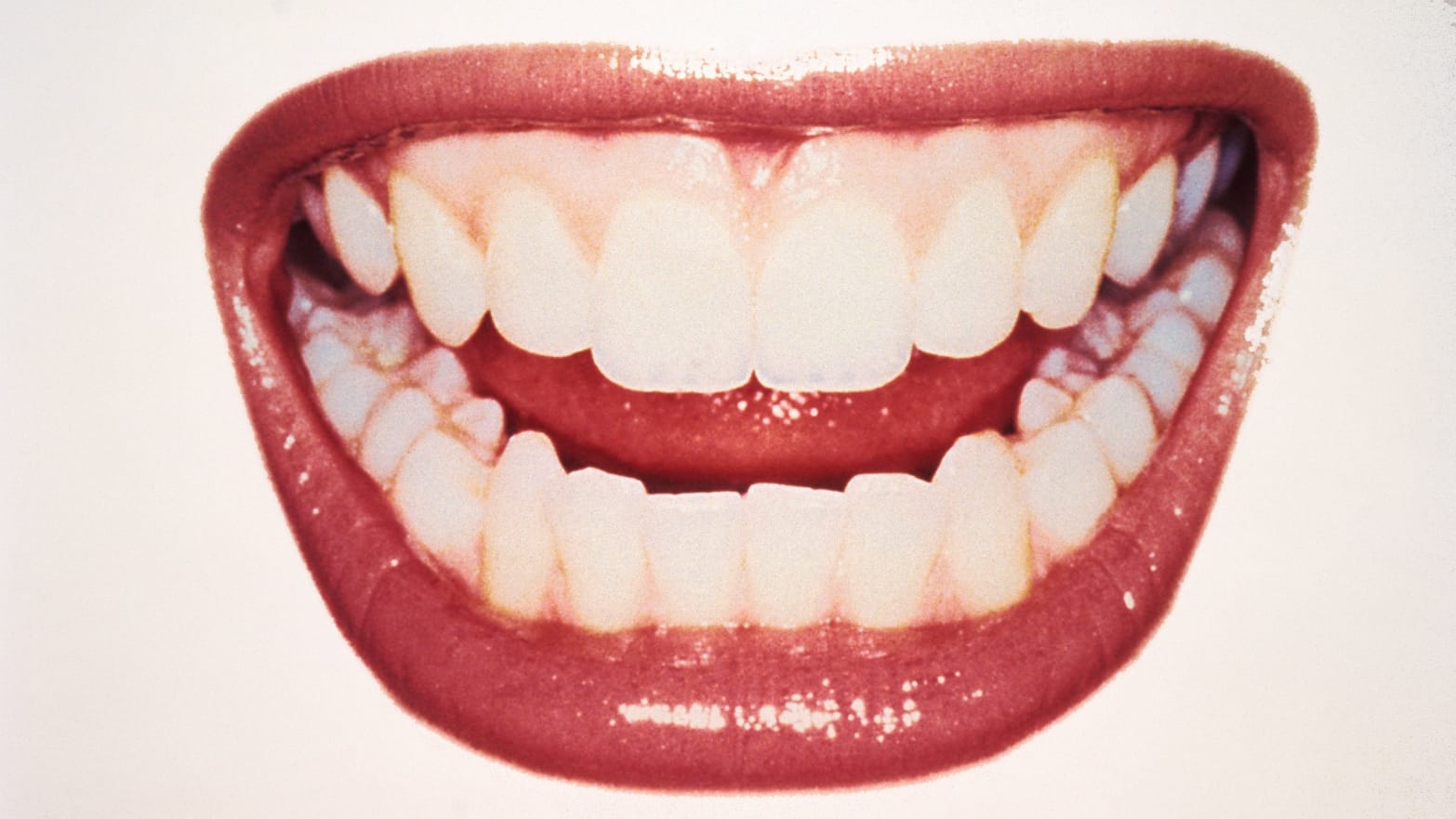
close up of grinning mouth teeth millennial pink gums micro robots microorganism gum dentist dental
Getty
Dentist offices are often terrifying, mostly because fighting microscopic bacteria is a serious task. They join together in groups using a sticky matrix and form colonies (like bricks stuck together with mortar) and attach themselves to your chompers.
They’re so stubbornly clingy, in fact, that dentists have to use scrapers and water jets to clean your teeth. Getting bacterial nets, known as biofilms, out of there is not an easy task.
“Certain bacteria, when they arrive on a surface, they know in order to survive there they need to cling on it,” Hyun (Michel) Koo, a dental researcher, told The Daily Beast. “So they secrete a matrix and stick with each other and multiply. As they firm up on the surface they cover each other up with this glue-like polymer.”


It’s a process that’s unpleasant, inefficient, and causes fear and panic—ripe for disruption. So researchers at the University of Pennsylvania are using nanotechnology to turn microscopic particles of iron oxide into robots that can do the work theoretically without any of the pain.
Once the bacteria are covered in their protective shielding, Koo said antimicrobials can’t penetrate to kill them so they need to be physically removed. “It’s very cumbersome. You need to disrupt them, kill them, and remove them. That’s true not just of the tooth but of biofilms on other parts of the body.”
Koo and his colleagues created nanoparticles of iron-oxide, which are safe to ingest and are easily digested in the stomach (they are regularly used in iron supplements). That means these particles have some very useful properties. They are magnetic, so doctors can use them in swarms and move them around to specific locations using magnets. The particles can also be a catalyst to produce hydrogen peroxide, breaking down the biofilm’s matrix to kill the bacteria inside. And, perhaps most importantly, they can scrape the dead bacteria off of teeth when they’re done.



Essentially, your teeth could be cleaned with a magnetic microcleaning machine.
“They can be swarmed to work like a Roomba, you can program them to move in specific movements and locomotion to create scrubbing,” Koo said.
Koo noted this control mechanism is one of the many technical challenges the team is still working to solve. But the team is moving forward on a solution to that, according to Koo: “They can be very creative and they are making ways so the magnetic field can be all around the surface of the tooth.”
The team also devised a way to 3D print the nanoparticles together into certain shapes to achieve goals other than cleaning the outside of the teeth. For example, Koo said, they can build them in the shape of a drill bit or a vein with fins on it. Theoretically, if a hole is drilled by the dentist through the outside of the tooth, these pre-shaped microbots could be inserted into a tooth and perform the cleaning necessary for a root canal. “The idea is we can use the robots to go inside and do the cleaning for the dentist,” he says.
Much of this work is still in its early stages. Koo says it will be at least two to three years before they have built a proof-of-concept version of a teeth-cleaning device (and it will take several years after that before it can become a prototype and undergo clinical testing). But when it’s ready he says he hopes that it will operate like playing a video game.
Koo says he doesn’t believe this innovation will put dentists out of business but rather, it will be another tool they can add to their kit.


“Technology will always provide new solutions,” he said. “Dentists are just advancing this so it can provide better or new ways to address the issues we have in dentistry.”


How are we improving outcomes for those affected by stroke? UK

You're not. You are completely failing stroke survivors by not creating rehab protocols. Guidelines don't count. Your initial management doesn't say a damn thing about trying to save all the neurons dying during the neuronal cascade of death. You can have your incompetence two ways; not knowing about the neuronal cascade of death, OR not doing anything about it. I think they hit both.

How are we improving outcomes for those affected by stroke?

Professor Gillian Leng, deputy chief executive at the National Institute for Health and Care Excellence (NICE), provides her view on the newly published NICE impact report on stroke.
Stroke is one of the greatest health challenges of our time. There are a reported 38,000 deaths occurring every year in the UK from more than 100,000 strokes. Without better access to treatment and rehabilitation, more people will lose their lives or be left with very serious life-impacting disabilities. The cost of stroke to health and social care is also rising and is predicted to reach as much as £91bn by 2035.
NICE has an important role in producing high quality, evidence-based recommendations for those who are at risk of or who have had a stroke. We published our first guideline on the diagnosis and initial management of stroke in 2008 and since then we have developed 2 guidelines covering stroke rehabilitation and stroke and transient ischaemic attack in over 16s, a quality standard on stroke in adults to drive improvements, and we have recommended two new drugs; alteplase and clopidogrel.
To bring together NICE’s guidance and to show how our recommendations are making a difference in priority areas of stroke care, we have published a new impact report on stroke. The report looks at where improvements have been made in health and social care but also identifies areas where more progress is needed.
One area the impact report considers is how we are working together to prevent stroke. In 2014, NICE updated its guideline on the prevention of stroke in people with atrial fibrillation (AF) to include a new risk assessment tool. The tool identifies those with AF who are at a high risk of stroke and need anticoagulation, and those with a lower risk who do not. The updated recommendations subsequently became part of the Quality and Outcomes Framework (QOF) and by 2017-18, 94% of people with atrial fibrillation were risk assessed using this tool.
Another area the report highlights is the impact of NICE’s recommendations on improving outcomes for those receiving acute care. In NICE’s guideline on the diagnosis and initial management of stroke, we focus on the importance of performing brain imaging as soon as possible. The evidence shows that by performing brain imaging within one hour, we can determine the most effective treatment for those with acute stroke. Since 2013/14, there have been improvements in the number of people who are scanned within 1 hour from 42% to 53% in 2017/18.
This is further evidence of how important implementation of NICE recommendations is in improving care for those who need it the most. However, there is more that needs to be done.
For example, an area that requires specific attention is waiting times for admissions to acute stroke units. NICE’s recommendations state that admissions must be within four hours to speed up treatment and to help prevent complications. However data from SSNAP shows that only 58% of patients overall across the UK were admitted to a stroke unit within four hours. Worryingly, this figure has not changed over the last five years, which could be due to long waiting times in Accident and Emergency and the availability of beds.
A new development in stroke treatment is another important focus of our impact report. Thrombectomy involves using a device to remove the blood clot directly from the blocked artery and restore blood flow, thus preventing a stroke. NICE guidance produced in 2016 on mechanical clot retrieval for treating acute ischaemic stroke provides evidence of its safety and efficacy.
SSNAP analysis indicates that in trials the procedure has shown real improvement in eligible patient outcomes, if performed within a few hours of a stroke. In March 2018, NHS England commissioned mechanical thrombectomy for acute ischaemic stroke and the NHS Long Term Plan aims to increase availability of mechanical thrombectomy from 1% to 10% of stroke patients. The updated NICE stroke guideline, which published this month, includes recommendations on the use of thrombectomy.
Our impact report clearly demonstrates that we are making significant strides in improving the lives of those affected by, or at risk of, stroke. However, there is still room for improvement. We need to work together to implement our evidence-based recommendations, to reduce the ‘postcode lottery’ of access to rehabilitation services, and to provide better support for people once they leave hospital. We will then be able to see a brighter, healthier future for all of those who have been affected by stroke, alongside significant advances in the health and social care services that support them.

"Best Stroke Blogs on the Planet" list

I'm only #4, but they are wrong about frequency, it is more like 70 posts a week

"Best Stroke Blogs on the Planet" list

SPG Stimulation 'Likely' of Benefit for Some Acute Strokes

So WHOM is going to followup to see if this treatment as a standalone immediately post tPA or thrombectomy would benefit stroke survivors? Or is nobody thinking that far ahead and prefer to stay with the failed status quo of stroke? I'm guessing that absolutely no followup will be done because we don't have two functioning neurons in stroke leadership.

SPG Stimulation 'Likely' of Benefit for Some Acute Strokes


Trial narrowly misses endpoint in confirmed cortical involvement group

  • by Staff Writer, MedPage Today
Acute ischaemic stroke patients ineligible for thrombolytic therapy and with confirmed cortical involvement (CCI) may derive benefit from an injectable neurostimulator implant that works on the sphenopalatine ganglion (SPG), a sham-controlled randomized trial found.
In the modified intent-to-treat population, the proportion of patients whose disability level was better than anticipated at 90 days was 49% in the SPG stimulation group versus 45% in the sham group (OR 1.14, P=0.31), reported Jeffrey Saver, MD, of the University of California Comprehensive Stroke Center in Los Angeles, and colleagues.
In the CCI population, 50% of the intervention group had better than expected outcomes compared with 40% of the control group (OR 1.48, P=0.0258), which narrowly missed statistical significance (P<0.025 required), as described in The Lancet.
The research was also presented at the European Stroke Organisation Conference in Milan.
"Although the improvement in 90-day functional outcome among patients with imaging evidence of cortical involvement at presentation was not significant in this trial alone, study findings indicate the use of sphenopalatine ganglion stimulation is likely to be beneficial, on the basis of consistent effects across the primary and all secondary endpoints, and of the presence of an inverted U-shaped dose-response curve," Saver's group wrote.
When assessing the U-shaped dose-response relationship between the primary outcome and SPG stimulation intensity in the CCI group, the proportion with favorable outcomes went up from 40% to 70% at low-midrange intensity and declined back to 40% during high-intensity stimulation (P=0.0034).
"Pooled analysis of all completed trials indicated that, among patients with imaging evidence of cortical involvement at presentation, sphenopalatine ganglion stimulation improves functional outcome," the researchers concluded.
IMPACT-24B (Implant Augmenting Cerebral Blood Flow Trial-24B) was a pivotal, international, double-blind study that randomized 1,078 patients with anterior-circulation acute ischaemic stroke, who were not receiving reperfusion treatment, to SPG stimulation (n=481) or sham treatment (n=519). Median patient age was about 70 years and 51% were women. Among these, 52% of patients had CCI -- 244 in the intervention group and 276 in the sham group. No differences in serious adverse events or mortality were seen between the two groups.
Patients were eligible if they had radiological and clinical evidence of acute ischemic stroke in the anterior cerebral circulation, had National Institutes of Health Stroke Scale scores between 7 and 18, and were able to start treatment 8 to 24 hours from stroke onset. Age requirements were 40-85 for women and 40-80 years for men.
Exclusion criteria included pregnancy, oral cavity conditions that might hinder device implantation, uncontrolled high blood pressure, bilateral stroke, bleeding propensity, radiological evidence of intracranial haemorrhage, and others.
Results of the present investigation fall into line with previous pilot trial (IMPACT-24A), which also looked at SPG stimulation within 1 day of stroke onset.
"Additional studies are planned or underway in patients receiving intravenous thrombolytic therapy and endovascular mechanical thrombectomy, to assess whether adding sphenopalatine ganglion stimulation improves outcomes in these patients as well," the authors wrote.
The study was funded by BrainsGate.
Saver disclosed relationships with BrainsGate.

Tuesday, May 28, 2019

Diurnal Variations in the First 24/7 Mobile Stroke Unit

My God, the stupidity here is incredible. You are not even measuring how many got to 100% recovery. Are you that goddamn fucking stupid? Your mentors and senior researchers need to be fired. 

Diurnal Variations in the First 24/7 Mobile Stroke Unit


Originally publishedhttps://doi.org/10.1161/STROKEAHA.119.024950Stroke. ;0

Background and Purpose—

Mobile Stroke Units (MSUs) provide innovative prehospital stroke care but their 24/7 operation has not been studied. Our study investigates 24/7 MSU diurnal variations related to transport frequency, patient characteristics, and stroke treatments. (The whole purpose of these mobile units is to get patients recovered, not just deliver tPA. You do have to measure that recovery. Patients care about recovery. Maybe you should talk to a couple.)

Methods—

We compared transportation frequency, demographics, thrombolytic and mechanical thrombectomy administration, and treatment metrics across 8-hour shifts (morning, evening, and nocturnal) from our 24/7 MSU in Northwest Ohio prospective database.

Results—

One hundred ninety-five patients were transported by the MSU. Most transports occurred during the morning shift (52.3%) followed by evening shift (35.8%) and nocturnal shift (11.9%; Ptrend<0.001). Twenty-three patients (11.9%) received intravenous thrombolytic in the MSU, most frequently in the morning shift (56.5%). No cases of mechanical thrombectomy were performed on MSU patients in the nocturnal shift.

Conclusions—

Morning and evening shifts account for the majority of our MSU transports (88.1%) and therapeutic interventions. Understanding temporal variations in a resource-intensive MSU is critical to its worldwide implementation.

Footnotes

*Drs Zaidat and Changal are co-first authors.
Correspondence to Osama O. Zaidat, MD, MS, FAHA, Northeast Ohio Medical University (NeoMed), St. Vincent Hospital, M200, Toledo, OH 43608. Email

Analysis of Brain Lesion Impact on Balance and Gait Following Stroke

I was tested in one of these called a Balance Master 4 weeks post-stroke. The force plates I was standing on could move side to side, front to back and tilt in various directions, all the while the three sides; front of you and each side were moving around. You were strapped into a harness to catch you when you fall. After it was all done my PT ran my scores thru the universe of  results and at that time my age of 50 results were better than the average 50 year old male. Luckily when I've fallen my left hip was strong enough not to break, but then I take insane chances in life. 

Analysis of Brain Lesion Impact on Balance and Gait Following Stroke

  • 1Department of Physical Therapy, Faculty of Health Sciences, Ben-Gurion University of the Negev, Beer-Sheva, Israel
  • 2Loewenstein Rehabilitation Hospital, Ra’anana, Israel
  • 3Sackler Faculty of Medicine, Tel-Aviv University, Tel-Aviv, Israel
Falls are a leading cause of serious injury and restricted participation among persons with stroke (PwS). Reactive balance control is essential for fall prevention, however, only a few studies have explored the effects of lesion characteristics (location and extent) on balance control in PwS. We aimed to assess the impact of lesion characteristics on reactive and anticipatory balance capacity, gait, and hemiparetic lower limb function, in PwS. Forty-six subacute PwS were exposed to forward, backward, right and left unannounced horizontal surface translations in six increasing intensities while standing. Fall threshold (i.e., perturbation intensity that results in a fall into the harness system) was measured. In addition, the Berg Balance Scale (BBS), 6 Minute Walk Test (6MWT) and Lower Extremity Fugl-Meyer (LEFM) were measured. Lesion effects were analyzed separately for left and right hemisphere damaged (LHD, RHD) patients, using voxel-based lesion-symptom mapping (VLSM). Our results show that voxel clusters where damage exerted a significant impact on balance, gait and lower-limb function were found in the corticospinal tract (CST), in its passage in the corona radiata and in the posterior limb of the internal capsule. An additional significant impact was found to lesions affecting the putamen and the external capsule (EC). Balance, gait, and hemiparetic lower limb function showed much overlap of the corresponding “significant” voxel clusters. Test scores of RHD and LHD patients were affected largely by damage to homologous regions, with the LHD group showing a wider distribution of “significant” voxels. The study corroborates and extends previous findings by demonstrating that balance control, gait, and lower limb function are all affected mainly by damage to essentially the same brain structures, namely—the CST and adjacent structures in the capsular-putaminal region.

Introduction

Falls occur in up to 70% of stroke victims during the first 6 months after discharge from hospital or rehabilitation setting (Forster and Young, 1995; Davenport et al., 1996; Weerdesteyn et al., 2008; Batchelor et al., 2012). Compared with a general population of older adults who fall, persons with stroke (PwS) who fall are twice as likely to sustain a hip fracture (Forster and Young, 1995; Langhorne et al., 2000; Pouwels et al., 2009; Winstein et al., 2016). In addition to physical consequences associated with fractures and related injuries, falls may have serious psychological and social consequences such as functional decline, poor quality of life, dependency, social isolation and depression (Winstein et al., 2016).
Balance control is a strong predictor of functional recovery, walking capacity and fall risk after stroke (Michael et al., 2005; Belgen et al., 2006; Simpson et al., 2011; van Duijnhoven et al., 2016; Xu et al., 2018). Commonly used clinical measures [e.g., the Berg Balance Scale (BBS), Dynamic Gait Index (DGI) and Timed Up and Go (TUG)] focus on anticipatory balance control, that is essential for the maintenance of postural stability prior to voluntary movement by compensating for destabilizing forces associated with the movement. However, in situations of unexpected loss of balance, the ability to respond effectively (i.e., reactive balance control), is crucial for fall prevention (Maki and McIlroy, 1997). After small external disturbances, we can usually regain balance while keeping the feet in place. However, falls often occur from large external disturbances (Maki and McIlroy, 2006) which require a rapid step response to alter the base of support. Recent studies assessed reactive balance control abilities by exposure to external perturbations delivered from a movable platform (Salot et al., 2015; Honeycutt et al., 2016; de Kam et al., 2017). In this paradigm, the time, direction and intensity of perturbation is unpredicted, thus simulating situations in real life where loss of balance is unexpected. PwS have shown substantially impaired reactive balance responses compared to healthy individuals, characterized by increased need for external assistance, difficulty initiating protective stepping with either lower limb, increased usage of multiple step strategy, and more falls into the harness system (Marigold and Eng, 2006; Mansfield et al., 2013; Martinez et al., 2013; Inness et al., 2014; Salot et al., 2015; Honeycutt et al., 2016; de Kam et al., 2017).
Although impairments in balance control following stroke have been studied extensively and their impact on the risk of falls and fractures has been established, relatively few studies have explored the associations between these impairments and damage to specific brain structures. Voxel-based lesion symptom mapping (VLSM) is a commonly used method for analyses of the neural basis underlying different types of impairment described by Bates et al. (2003). Use of VLSM for analysis of lesion characteristics in lower-limb paresis, gait instability and impaired balance, is much less prevalent compared with its use in analyses focusing on the hemiparetic upper limb. Using VLSM, Reynolds et al. (2014) found that lower BBS scores were associated with damage in the precentral gyrus, putamen, caudate and pallidum, cuneus, frontal operculum, and also damage to some thalamic structures. They also found that TUG scores were associated with lesions in the postcentral gyrus, insular cortex, superior temporal cortex, and the inferior parietal lobule. Lee et al. (2017) found that lesions involving the corona radiata, internal capsule, globus pallidus, putamen, primary motor cortex and caudate nucleus are associated with poor recovery of gait, as measured with the functional ambulation category (FAC) 6 months after stroke onset. In contradiction to the above findings, Moon et al. (2016) found no specific lesion locations in association with poor BBS and FAC scores. Poor gait speed was found to associate with damage to the putamen, insula, caudate, corona radiata and external capsule (EC; Reynolds et al., 2014; Jones et al., 2016). Alexander et al. (2009) found that damage to the putamen, insula and EC was related to gait asymmetry in chronic PwS. Lower Extremity Fugl-Meyer (LEFM) scores were found to be associated with damage in the corona radiata, putamen, globus pallidus, caudate, insula and internal capsule (Reynolds et al., 2014; Moon et al., 2016).
Lesion studies investigating the effects of stroke location on motor ability often address the right and left hemispheres as two parallel and analogous systems, and flip lesions onto a single hemisphere template (Lo et al., 2010; Zhu et al., 2010; Cheng et al., 2014; Meyer et al., 2015). This practice may obscure important differences between the hemispheres. A recent VLSM study showed that LEFM scores are affected by a wider lesion distribution in the left hemisphere compared to the right hemisphere (Moon et al., 2016). In contrast, a post hoc VLSM analysis aimed to assess hemispheric effects (Jones et al., 2016), revealed no “significant” voxel clusters in either hemisphere. Considering the relative paucity of studies addressing lesion effects on balance control and gait and the fact that most of the existing studies did not analyze right and left hemisphere damage separately, our objective in the current study was to explore the impact of lesion location, in each hemisphere, on reactive and anticipatory balance capacity, gait, and hemiparetic lower limb function, in PwS.

More at link. 

Putting the “Sensory” Into Sensorimotor Control: The Role of Sensorimotor Integration in Goal-Directed Hand Movements After Stroke

But is this better than the Margaret Yekutiel  book about this from 2001, 'Sensory Re-Education of the Hand After Stroke'? The objective should have been to update the sersorimotor recovery protocol with the newest research results.  Not whatever the fuck this review was for.  Lots of big words in here but I see nothing that even remotely looks like useful rehab interventions. And the conclusion is more research needed.


Putting the “Sensory” Into Sensorimotor Control: The Role of Sensorimotor Integration in Goal-Directed Hand Movements After Stroke

  • 1Neuroscience Graduate Program, Graduate Division of Biological and Biomedical Sciences, Emory University, Atlanta, GA, United States
  • 2Department of Rehabilitation Medicine, Laney Graduate School, Emory University, Atlanta, GA, United States
  • 3Department of Neurology, Emory University, Atlanta, GA, United States
  • 4Department of Radiology and Imaging Sciences, School of Medicine, Emory University, Atlanta, GA, United States
Integration of sensory and motor information is one-step, among others, that underlies the successful production of goal-directed hand movements necessary for interacting with our environment. Disruption of sensorimotor integration is prevalent in many neurologic disorders, including stroke. In most stroke survivors, persistent paresis of the hand reduces function and overall quality of life. Current rehabilitative methods are based on neuroplastic principles to promote motor learning that focuses on regaining motor function lost due to paresis, but the sensory contributions to motor control and learning are often overlooked and currently understudied. There is a need to evaluate and understand the contribution of both sensory and motor function in the rehabilitation of skilled hand movements after stroke. Here, we will highlight the importance of integration of sensory and motor information to produce skilled hand movements in healthy individuals and individuals after stroke. We will then discuss how compromised sensorimotor integration influences relearning of skilled hand movements after stroke. Finally, we will propose an approach to target sensorimotor integration through manipulation of sensory input and motor output that may have therapeutic implications.

Introduction

Goal-directed movements of the hand are required to perform most tasks of daily living, such as tying a shoe, buttoning a shirt, and typing, among others. These highly coordinated voluntary movements involve interacting with and manipulating objects in the environment and rely on sensorimotor integration. Sensorimotor integration is the ability to incorporate sensory inputs that provide information about one’s body and the external environment to inform and shape motor output (Wolpert et al., 1998). More specifically, sensory inputs for goal-directed hand movements provide information in an egocentric reference frame detailing location, size, weight, and shape of an object. In addition, kinematic information about the hand and upper extremity, including the trajectory needed to interact with the object, is provided. Successful integration of information contributes to generating the most efficient motor plan to execute a given task. Additionally, ongoing sensory feedback during motor performance refines the motor plan to optimize current and future performance. This process of sensorimotor integration is often disrupted in neurological disorders, such as stroke.
Stroke is defined as infarction of central nervous system tissue attributable to ischemia, based on neuropathological, neuroimaging, and/or clinical evidence of permanent injury (Sacco et al., 2013). Stroke is the fourth leading cause of death and remains the number one leading cause of long-term adult disability (Benjamin et al., 2017). Furthermore, the loss of productivity after stroke currently costs the United States an average of $33.9 billion per year and is expected to reach $56 billion by 2030 (Ovbiagele et al., 2013), making stroke a public health crisis. A primary contributor to persistent disability after stroke is incomplete motor recovery (Lai et al., 2002). Spontaneous biological recovery of motor function occurs during the first months after stroke (Cramer, 2008), underlying a current emphasis on intensive early intervention, although results are often mixed and complex (Bernhardt et al., 2017a). Despite intensive therapy, upper extremity impairment resolves up to 70% of baseline function for a given patient with some patients showing even less recovery than predicted (Winters et al., 2015). Most stroke survivors are left with a limited ability to perform skilled hand movements necessary for daily functioning (Lang et al., 2013). To reduce disability after stroke, there is a need to improve our understanding of the neuronal network physiology necessary to regain skilled functional hand use.
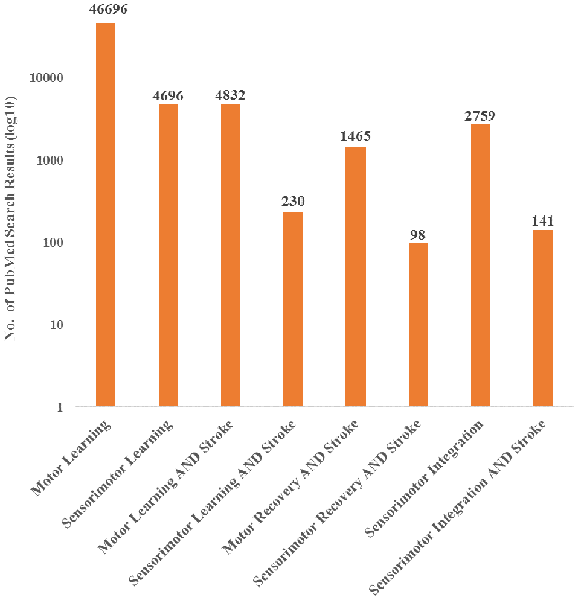
Currently, the field has primarily investigated motor deficits and motor learning with limited consideration of the role of sensory information, even though it is recognized that integration of sensory information is a critical component of motor control (Borich et al., 2015; Bolognini et al., 2016). Furthermore, evidence has shown that sensory input is important for recovery after stroke. In a systematic review, Meyer et al. found that across six studies, the extent of deficits in proprioception and light touch of the arm and hand were significantly related to recovery after stroke (Meyer et al., 2014). Despite evidence that sensory input is a critical component to motor execution, research nomenclature has been primarily focused on motor characteristics post-stroke and has therefore not capitalized fully on the information a sensorimotor perspective could provide. This observation is supported by a literature search showing an emphasis towards motor recovery and learning after stroke, over sensorimotor recovery and learning, with limited focus on sensorimotor integration (Figure 1). While it is possible that authors may use these terms interchangeably, the literature search terminology suggests that there is potential bias towards motor contributions. Therefore, there is an important gap in our understanding of the contributions of sensorimotor integration to recovery.
FIGURE 1
www.frontiersin.org Figure 1. PubMed search results for both motor and sensorimotor aspects of learning and stroke recovery. More publications focused on motor learning and recovery than on both motor and sensory components of learning and recovery. Furthermore, there were a relatively small number of publications involving sensorimotor integration and stroke compared to sensorimotor integration overall. “Sensorimotor,” “Sensori-motor,” and “Sensory motor” were all used to ensure differences in terminology did not affect the search results. Additionally, “Sensory motor” and “Sensory-motor” produced the same search results.
In the following brief review, we will highlight the importance of processing and integrating sensory and motor information that underlies skill performance and learning with an emphasis on skilled hand movements in stroke. We will focus primarily on three cortical regions: primary motor cortex (M1), posterior parietal cortex (PPC) and primary somatosensory cortex (S1) while briefly mentioning other cortical and subcortical brain areas also involved in sensorimotor integration. These brain regions are highlighted due to our focus on the integration of sensory and motor information at the level of the cortex, but also because these cortical areas receive blood supply from the middle cerebral artery (MCA), which is the most common type of stroke (Walcott et al., 2014). Furthermore, all three brain regions contribute to the corticospinal tract (CST) that provide necessary contributions to executing and controlling skilled hand movements routinely used in daily life. It should be noted that strokes occur in other brain regions but usually have less of an impact on sensorimotor integration underlying goal-directed, skilled hand movements and are outside the primary scope of this review article.
In the first section of this review article, we will discuss the role of sensorimotor integration via M1, PPC, and S1 in normal, skilled hand movements. We will then discuss how sensorimotor integration is affected by stroke and how impaired sensorimotor integration can impact relearning of skilled hand movements. Last, we propose an approach to target sensorimotor integration by manipulating sensory input and restricting motor output that may have therapeutic implications for stroke recovery.

More at link. 

Redundancy Among Parameters Describing the Input-Output Relation of Motor Evoked Potentials in Healthy Subjects and Stroke Patients

I got absolutely nothing out of this that would help survivors recover. 

Redundancy Among Parameters Describing the Input-Output Relation of Motor Evoked Potentials in Healthy Subjects and Stroke Patients

Claire Kemlin1, Eric Moulton1, Sara Leder2, Marion Houot3, Sabine Meunier1, Charlotte Rosso1,2 and Jean-Charles Lamy1*
  • 1Institut du Cerveau et de la Moelle épinière, ICM, Inserm U 1127, CNRS UMR 7225, Sorbonne Université, Paris, France
  • 2APHP, Urgences Cérébro-Vasculaires, Hôpital de la Pitié Salpêtrière, Paris, France
  • 3AP-HP, Department of Neurology, Hôpital de la Pitié-Salpêtrière, Centre of excellence of neurodegenerative disease (CoEN), Institute of Memory and Alzheimer's Disease (IM2A), ICM, CIC Neurosciences, Paris, France
Background: Transcranial magnetic stimulation (TMS) is widely used to probe corticospinal excitability through Motor Evoked Potential (MEP) amplitude measurements. The input-output (I/O) curve is a sigmoid-shaped relation between the MEP amplitude at incremented TMS intensities. The aim of this study was to examine the relationships between seven parameters derived from the sigmoid function.
Methods: Principal Component Analysis and Spearman's rank correlation matrices were used to determine if the seven I/O curve parameters capture similar or, conversely, different aspects of the corticospinal excitability in 24 healthy subjects and 40 stroke survivors with a hand motor impairment.
Results: Maximum amplitude (MEPmax), peak slope, area under the I/O curve (AUC), and MEP amplitude recorded at 140% of the resting motor threshold showed strong linear relationships with each other (ρ > 0.72, p < 0.001). Results were found to be similar in healthy subjects and in both hemispheres of stroke patients. Our results did not support an added benefit of sampling entire I/O curves in both healthy subjects and stroke patients, with the exception of S50, the stimulus intensity needed to obtain half of MEPmax amplitude.
Conclusions: This demonstrates that MEP elicited at a single stimulus intensity allows to capture the same characteristics of the corticospinal excitability as measured by the AUC, MEPmax and the peak slope, which may be of interest in both clinical and research settings. However, it is still necessary to plot I/O curves if an effect or a difference is expected at S50.

Introduction

Transcranial magnetic stimulation (TMS) is widely used to probe corticospinal excitability in both healthy subjects and in a broad range of neuropsychiatric conditions. A common approach from basic research to pivotal clinical trials is to compare recruitment curves of TMS-induced motor evoked potentials (MEPs) between groups of subjects or before and after different types of interventions aimed at promoting brain plasticity (i.e., pharmacotherapy or non-invasive brain stimulation).
The input-output (I/O) relation in the corticospinal pathway is assessed by plotting MEP amplitude vs. stimulus intensity and fitting the data with the following sigmoid function equation (14):
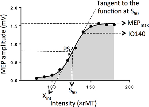
, where MEP(s) is the MEP amplitude at the stimulation intensity s, MEPmax is the maximum MEP amplitude, S50 is the stimulus intensity needed to obtain 50% of MEPmax amplitude, and m is the slope parameter of the sigmoid function, i.e., the global slope of the function (Figure 1). Three additional parameters can be derived from the I/O curve: (1) the peak slope (PS), i.e., the instantaneous slope of the ascending limb of the curve at S50, which reflects the recruitment gain of motoneurons and is given by the formula: PS = m x MEPmax/4, (2) the x-intercept (Xint) of the tangent at S50, and (3) the area under the I/O curve (AUC) usually calculated using the trapezoidal area method (5).
FIGURE 1
www.frontiersin.orgFigure 1. Electrophysiological parameters extracted from an example of an input output curve (I/O curve) fitted by a sigmoid function. Are shown the following variables: Xint: X intercept, IO140: Motor Evoked Potential amplitude recorded at 140% rMT, PS, peak slope; AUC, area under the I/O curve in gray; S50, stimulus intensity needed to obtain 50% of the maximum response; MEPmax, maximum value of the sigmoid function.
To date, the inter-dependency between all these parameters are not fully understood. Indeed, although the PS depends on both m parameter and MEPmax, it does not mean these three parameters are correlated together. Same for Xint, which depends on m parameter and S50. The question arises whether these variables capture similar or, conversely, different aspects of the corticospinal excitability and if so, how each of them relates to one other. To clarify the interdependency between these parameters, we estimated I/O curves from the dominant hemisphere of healthy volunteers and performed Principal Component Analyses (PCA) in addition to correlation matrices to summarize the most important linear relationships between variables. PCA is a tool capable of summarizing the most important linear relationships between variables and computing synthetic variables from the original variables named principal components (PCs). PCA provides a visual and geometric representation of the correlation matrix (6, 7). In a second step, to test whether our results could be extrapolated to patients suffering from neurological conditions, we performed the same analyses on data collected in both the affected and unaffected hemispheres of stroke patients given that this population represents the most frequent brain damaged disease worldwide. Indeed, the sigmoid function has been previously shown to be a reliable method to plot IO curve in stroke patients (4).

More at link. 

Cross-Reactivity as a Mechanism Linking Infections to Stroke

This is way out of my league.

Cross-Reactivity as a Mechanism Linking Infections to Stroke

Guglielmo Lucchese1,2*, Agnes Flöel1 and Benjamin Stahl1,3,4,5
  • 1Department of Neurology, University of Greifswald, Greifswald, Germany
  • 2Department of Computing, Goldsmiths, University of London, London, United Kingdom
  • 3Department of Neurology, Charité Universitätsmedizin Berlin, Berlin, Germany
  • 4Department of Neurophysics, Max Planck Institute for Human Cognitive and Brain Sciences, Leipzig, Germany
  • 5Psychologische Hochschule Berlin, Berlin, Germany
The relevance of infections as risk factor for cerebrovascular disease is being increasingly recognized. Nonetheless, the pathogenic link between the two entities remains poorly understood. Consistent with recent advances in medicine, the present work addresses the hypothesis that infection-induced immune responses may affect human proteins associated with stroke. Applying established procedures in bioinformatics, the pathogen antigens and the human proteins were searched for common sequences using pentapeptides as probes. The resulting data demonstrate massive peptide sharing between infectious pathogens—such as Chlamydia pneumoniae, Streptococcus pneumoniae, Tannerella forsythia, Haemophilus influenzae, Influenza A virus, and Cytomegalovirus—and human proteins related to risk of ischemic and hemorrhagic stroke. Moreover, the shared peptides are also evident in a number of epitopes experimentally proven immunopositive in the human host. The present findings suggest cross-reactivity as a potential mechanistic link between infections and stroke.

Introduction

When considered separately from other cardiovascular diseases, stroke ranks fifth among all causes of death (1) and, critically, its incidence is on the rise (2).
The etiology of stroke is multifactorial with various environmental and genetic risk factors. Hypertension, diabetes and insulin resistance, smoking, dyslipidemia, obesity, heavy alcohol consumption, atrial fibrillation, and carotid stenosis are all established and well-investigated modifiable risk factors of stroke (35).
Additionally, there is evidence that environmental factors may also increase risk of stroke, including viral and bacterial infections, such as periodontitis (6) and respiratory infections (7), and infection with Chlamydia pneumoniae (8) or Cytomegalovirus (9). However, relatively little is known so far about the role of different pathogens as well as the molecular basis and the mechanisms that potentially link infections to stroke.
Here we set out to investigate whether or not infections can induce immune responses capable of cross-reacting with human proteins that, when altered, have been associated with stroke. Our hypothesis was that immune responses induced by infectious agents might cross-react with crucial stroke-related proteins, thus contributing to the multifactorial pathogenesis of cerebrovascular disease.
To address this hypothesis, we analyzed pathogens, as well as proteins that are known to be associated with increased risk of ischemic and hemorrhagic stroke by searching for common peptides that might underlie cross-reactions.
Specifically, we analyzed antigens from the following pathogens that have been reported to have a possible influence on stroke: the periodontal bacterium Tannerella forsythia (10), Haemophilus influenza (11), Streptococcus pneumoniae (7), Chlamydia pneumoniae (8), Influenza A viruses (12, 13), and Human Cytomegalovirus (9).

Methods

We analyzed the amino acid (aa) primary sequence of pathogen antigens (with short name and UniProt ID in parentheses):
• Surface antigen repeat/outer membrane protein (OMP; UniProtKB: A0A0F7WYE8_CHLPN) from Chlamydia pneumoniae;
• Pneumococcal vaccine antigen A (PVAA;UniProtKB: PVAA_STRR6) from Streptococcus pneumoniae;
• Surface antigen BspA (BspA; UniProtKB: O68831_TANFO) from Tannerella forsythia;
• Outer membrane antigenic lipoprotein B (LPPB; UniProtKB: LPPB_HAEIN) from Haemophilus influenzae (strain ATCC 51907);
• Hemagglutinin (HA H1N1; UniProtKB: HEMA_I34A1) from Influenza A virus (strain A/Puerto Rico/8/1934 H1N1);
• Hemagglutinin (HA H5N1; UniProtKB: HEMA_I96A0) from Influenza A virus (strain A/Goose/Guangdong/1/1996 H5N1);
• Hemagglutinin (HA H3N2; UniProtKB: HEMA_I68A6) from Influenza A virus (strain A/Northern Territory/60/1968 H3N2); and
• 65 kDa phosphoprotein (pp65; UniProtKB: PP65_HCMVM) from Human Cytomegalovirus (HCMV; strain Merlin).
The primary sequence of pathogen antigens was dissected into partially overlapping pentapeptides with a one-residue-offset: i.e., MFKRI, FKRIR, KRIRR, and so on. Then, each pentapeptide was analyzed for occurrences within a library consisting of primary sequences of human proteins involved in stroke. The human protein library was a priori chosen from the UniProtKB Database (https://www.uniprot.org) (14) using the keyword “stroke.” We obtained an unbiased list of 74 human proteins (in)directly associated with stroke (Table S1). Stroke-related proteins are indicated as UniProtKB entry names throughout the present article, except when discussed in detail. The pathogen antigens and the human proteins were searched for common sequences using the pentapeptide as a probe unit because a pentapeptide is an immunobiological determinant sufficient for epitope-paratope interaction and for inducing specific immune responses (1518).
The immunologic potential of the shared peptides was analyzed using the Immune Epitope Database (IEDB; www.iedb.org) (19). All evaluations were based only on epitopic sequences that had been experimentally validated as immunopositive in the human host.
This linear peptide similarity analysis procedure has been used and described before (20, 21).

Results

In a detailed overview, Table 1 shows that 49 out of the 74 human stroke-related proteins share peptide sequences with antigens from pathogens that proved to be (in)directly involved in stroke (610). It can be seen that
• The pathogen vs. human peptide overlap is unexpectedly high when considering that the probability for two proteins to share a pentapeptide is 1 out of 20−5, that is, 0.0000003125 or close to zero.
• The peptide overlap varies widely, with T. forsythia BspA and Influenza A HA H3N2 being the pathogen more and less involved in the peptide sharing, respectively.
• The high number of stroke-related proteins involved in the viral peptide overlap precludes a detailed protein-by-protein analysis. However, an example worth noting is the human ATP-binding cassette sub-family C member nine (ABCC9 or SUR2) that shares peptide sequences with all of the pathogen antigens analyzed, with the exception of the Influenza A HA H3N2 virus. ABCC9 is a subunit of ATP-sensitive potassium channels (KATP) that can form cardiac and smooth muscle-type KATP channels with KCNJ11 and mediates neuroprotection (22).

Antibiotic Class and Outcome in Post-stroke Infections: An Individual Participant Data Pooled Analysis of VISTA-Acute

With a 30% occurrence rate post stroke, you can tell your stroke hospital's competence if they have a infection prevention protocol and an infection protocol. Or are you OK with them just 'winging it'?

Antibiotic Class and Outcome in Post-stroke Infections: An Individual Participant Data Pooled Analysis of VISTA-Acute

Craig J. Smith1,2, Calvin Heal3, Andy Vail3, Adam R. Jeans4, Willeke F. Westendorp5, Paul J. Nederkoorn5, Diederik van de Beek5, Lalit Kalra6, Joan Montaner7,8, Mark Woodhead9, and Andreas Meisel10* on behalf of the VISTA Collaboration and PISCES Group
  • 1Greater Manchester Comprehensive Stroke Centre, Manchester Academic Health Science Centre, Salford Royal NHS Foundation Trust, Salford, United Kingdom
  • 2Division of Cardiovascular Sciences, School of Medical Sciences, University of Manchester, Manchester, United Kingdom
  • 3Centre for Biostatistics, Manchester Academic Health Science Centre, University of Manchester, Manchester, United Kingdom
  • 4Division of Clinical Support Services and Tertiary Medicine, Department of Microbiology, Salford Royal NHS Foundation Trust, Salford, United Kingdom
  • 5Department of Neurology, Amsterdam Neuroscience, Academic Medical Center, University of Amsterdam, Amsterdam, Netherlands
  • 6Clinical Neurosciences, King's College Hospital NHS Foundation Trust London, London, United Kingdom
  • 7Neurovascular Research Laboratory, Vall d' Hebron Institute of Research, Barcelona, Spain
  • 8Stroke Research Program, Department of Neurology, Institute de Biomedicine of Seville, Hospital Universitario Virgen Macarena, IBiS/Hospital Universitario Virgen del Rocío/CSIC/University of Seville, Seville, Spain
  • 9Faculty of Biology, Medicine and Health, Manchester Academic Health Science Centre, University of Manchester, Manchester, United Kingdom
  • 10Department of Neurology, NeuroCure Clinical Research Center, Center for Stroke Research Berlin, Charité Universitaetsmedizin Berlin, Berlin, Germany
Introduction: Antibiotics used to treat post-stroke infections have differing antimicrobial and anti-inflammatory effects. Our aim was to investigate whether antibiotic class was associated with outcome after post-stroke infection.
Methods: We analyzed pooled individual participant data from the Virtual International Stroke Trials Archive (VISTA)-Acute. Patients with ischemic stroke and with an infection treated with systemic antibiotic therapy during the first 2 weeks after stroke onset were eligible. Antibiotics were grouped into eight classes, according to antimicrobial mechanism and prevalence. The primary analysis investigated whether antibiotic class for any infection, or for pneumonia, was independently associated with a shift in 90 day modified Rankin Scale (mRS) using ordinal logistic regression.
Results: 2,708 patients were eligible (median age [IQR] = 74 [65 to 80] y; 51% female; median [IQR] NIHSS score = 15 [11 to 19]). Pneumonia occurred in 35%. Treatment with macrolides (5% of any infections; 9% of pneumonias) was independently associated with more favorable mRS distribution for any infection [OR (95% CI) = 0.59 (0.42 to 0.83), p = 0.004] and for pneumonia [OR (95% CI) = 0.46 (0.29 to 0.73), p = 0.001]. Unfavorable mRS distribution was independently associated with treatment of any infection either with carbapenems, cephalosporins or monobactams [OR (95% CI) = 1.62 (1.33 to 1.97), p < 0.001], penicillin plus β-lactamase inhibitors [OR (95% CI) = 1.26 (1.03 to 1.54), p = 0.025] or with aminoglycosides [OR (95% CI) = 1.73 (1.22 to 2.46), p = 0.002].
Conclusion: This retrospective study has several limitations including effect modification and confounding by indication. Macrolides may have favorable immune-modulatory effects in stroke-associated infections. Prospective evaluation of the impact of antibiotic class on treatment of post-stroke infections is warranted.

Introduction

Infections frequently complicate stroke, occurring in up to 30% of patients, and increase the likelihood of death and unfavorable outcomes in survivors (13). Whilst antibiotics are the mainstay of treatment, the microbiological etiology of common infections complicating stroke, such as pneumonia or urinary tract infection, are poorly characterized. Further, there are no antibiotic treatment trials of infections complicating stroke. The effectiveness of different antibiotic classes is therefore uncertain, there is a lack of evidence to inform antibiotic guidelines (e.g., for pneumonia complicating stroke) and empirical antibiotic treatment is variable (4, 5).
Inflammatory and immune responses play a central role in the pathophysiology of stroke and associated clinical outcomes (6). Post-stroke infections exacerbate deleterious inflammatory and immune responses, which may impact further on adverse outcomes (7). Antibiotics used to treat post-stroke infections can modulate the pathophysiology of experimental stroke independent of their anti-microbial effects, by modulating inflammatory or excitotoxic pathways (814). Randomized trials of prophylactic antibiotics in acute stroke have failed to improve clinical outcomes or prevent pneumonia (3, 5, 1517), and had varying effects in preventing urinary tract infections. This has raised questions about the potential effectiveness of some antibiotic classes commonly used for post-stroke infections, particularly pneumonia (18).
Taken together, these data suggest that choice of antibiotic class for post-stroke infections could have important implications for clinical outcomes. We therefore hypothesized that antibiotic class influences outcome after stroke relating to spectrum of antimicrobial coverage and to other (e.g., inflammatory) mechanisms independent of antimicrobial effects. The aim of this study was to investigate whether class of antibiotic used to treat clinically diagnosed pneumonia or any infection in the first 2 weeks after stroke was associated with clinical outcomes.

More at link.