Changing stroke rehab and research worldwide now.Time is Brain! trillions and trillions of neurons that DIE each day because there are NO effective hyperacute therapies besides tPA(only 12% effective). I have 523 posts on hyperacute therapy, enough for researchers to spend decades proving them out. These are my personal ideas and blog on stroke rehabilitation and stroke research. Do not attempt any of these without checking with your medical provider. Unless you join me in agitating, when you need these therapies they won't be there.

What this blog is for:

My blog is not to help survivors recover, it is to have the 10 million yearly stroke survivors light fires underneath their doctors, stroke hospitals and stroke researchers to get stroke solved. 100% recovery. The stroke medical world is completely failing at that goal, they don't even have it as a goal. Shortly after getting out of the hospital and getting NO information on the process or protocols of stroke rehabilitation and recovery I started searching on the internet and found that no other survivor received useful information. This is an attempt to cover all stroke rehabilitation information that should be readily available to survivors so they can talk with informed knowledge to their medical staff. It lays out what needs to be done to get stroke survivors closer to 100% recovery. It's quite disgusting that this information is not available from every stroke association and doctors group.

Friday, October 2, 2026

Brain Age Offers Clues to Aphasia Outcomes After Stroke

 Don't let your incompetent? doctor use this as an excuse for failing to get you recovered! Screaming at them might be required, referencing their incompetence!

Predicting failure to recover is the sign of PURE INCOMPETENCE!

Brain Age Offers Clues to Aphasia Outcomes After Stroke

Following stroke, patterns of accelerated brain aging in the intact hemisphere may help explain differences in chronic aphasia severity and predict response to language rehabilitation.

Using an online tool that compares brain scans with age-based reference data, researchers found that brain-aging patterns in the intact right hemisphere explained an additional 7% of the variation in aphasia severity beyond age, lesion volume, and lesion location.

In a smaller group of patients who received speech therapy paired with noninvasive brain stimulation, brain-age measures taken before treatment were also associated with language improvement 6 months later.

photo of  Nicholas Riccardi, PhD
Nicholas Riccardi, PhD

“The most important takeaway for clinicians and people with aphasia, is that recent breakthroughs in neuroimaging will allow groundbreaking advances in understanding the mechanisms of recovery after stroke in the coming years, which will ultimately help people with aphasia and their families,” lead investigator Nicholas Riccardi, PhD, research assistant professor, Department of Pharmacology, Physiology, and Neuroscience, University of South Carolina Floyd School of Medicine in Columbia, South Carolina, told Medscape Medical News.

Article Key Points
  • Right-hemisphere brain age explained +7% aphasia severity variance beyond lesion factors.
  • Older age, larger lesions, temporal lesions → lower WAB-R AQ scores.
  • Medial prefrontal + cerebellar/ventral brain age linked to worse language function.
  • Brain age added +7% variance in naming; medial prefrontal pattern predicted naming.
  • Pre-treatment brain age predicted 6-month naming gain after speech therapy + tDCS.
Dive Deeper
Which brain-age patterns predict aphasia recovery after stroke?
How modifiable are contralesional brain-age signatures after stroke?
Does brain age outperform lesion metrics for aphasia prognosis?

The study was published online on September 28 in The Journal of Neuroscience.

Assessing the Intact Brain 

Stroke size and location do not fully explain why some patients have more severe or persistent aphasia. 

Previous studies suggest that the health of brain tissue spared by the stroke may also affect language recovery. Brain-age estimates offer one way to assess this. 

Using structural MRI, researchers can estimate how old a person’s brain tissue appears compared with their actual age. Riccardi and colleagues focused on age-related patterns in the intact right hemisphere to examine whether these patterns were linked to aphasia severity and treatment response.

They analyzed 188 adults (mean age, 61 years; 67 women, 121 men) with chronic left hemisphere stroke. Participants were at least 6 months poststroke, with an average time since stroke of approximately 50 months.

Aphasia severity was assessed using the Western Aphasia Battery-Revised Aphasia Quotient (WAB-R AQ), which ranges from 0 to 100, with higher scores indicating better language function. Naming ability was assessed with the Philadelphia Naming Test (PNT) in 165 participants.

Because the left hemisphere contained stroke damage, the researchers focused their brain-age analyses on the right, contralesional hemisphere. They digitally filled in damaged areas by mirroring corresponding tissue from the intact right hemisphere, allowing the scans to be processed by the brain-age tool.

They used the volBrain BrainStructureAges pipeline to estimate brain age from routine MRI scans, then grouped the regional estimates into four broader brain-aging patterns in the right hemisphere: medial prefrontal, frontotemporoparietal, cerebellar/ventral, and dorsal.

Why Brain Age Matters 

Brain aging measures in the right hemisphere accounted for an additional 7% of the variance in language scores after adjusting for patients’ age, sex, and stroke size and location. When all of these factors were considered, they accounted for 46% of the variance in participants’ WAB-R AQ scores.

Older age, larger lesions, and a temporal lesion pattern were associated with lower WAB-R AQ scores. Higher brain-age estimates in the right hemisphere’s medial prefrontal and cerebellar/ventral areas were also tied to lower scores (beta = -.31; P < .001 and beta = -.18; P = .009, respectively).

In an analysis of 131 participants, brain aging measures accounted for an additional 7% of the variance in naming performance after adjustment for age and stroke characteristics. The medial prefrontal pattern was independently associated with naming ability (beta = -.30; P = .008).

The study could not determine whether the differences in brain age were related to the stroke or reflected preexisting differences in brain health.

The researchers also looked at whether regional brain-age estimates were associated with longer-term language improvement. The analysis included 34 participants who had received 3 weeks of speech therapy paired with anodal transcranial direct-current stimulation.

Mean PNT scores increased from 60.2 at baseline to 68.2 at 6 months. Age, lesion volume, and the medial prefrontal brain-age measure together explained 23% of the differences in naming-score change at 6 months. The medial prefrontal measure was associated with naming-score change (beta = -.47; P = .03).

Overall right-hemisphere gray-matter volume was not significantly associated with naming-score changes.

Brain Health and Recovery 

Recovery after stroke may depend not only on the area that had been directly damaged but also on the health of brain tissue that remains intact, Riccardi said.

“More and more, we are learning that overall brain health is extremely important for resilience and recovery after brain injury.”

This includes factors such as high blood pressure, diabetes, and other cardiometabolic risks that can affect brain health, he added. The current study did not examine these factors, but determining whether they can be modified to support brain health is a next step.

The researchers also want to better understand what underlies the brain aging signature associated with treatment response.

“We found a brain signature of people who will respond to this therapy, so now we need to better understand what causes this brain signature,” Riccardi said. If the signature is influenced by modifiable health or lifestyle factors, it could eventually have implications for treatment planning.

The findings will need to be replicated in other groups of people with aphasia and tested with different rehabilitation approaches, as the treatment subgroup received a specific combination of speech therapy and brain stimulation. Because the approach relies on routine MRI and a freely available online tool, it could be practical to study in other settings.

“This type of ‘ease of use’ is very important for approaches that could eventually make it into the clinic,” Riccardi said.

Disclosure information for study authors is available in the original study publication.

Scientists had people eat tomato paste every day. This is what their brain scans looked like after 3 months

 Didn't your competent? doctor already figure out your daily protocol needs of lycopene via tomatoes? And BDNF needs?

  • lycopene (28 posts to October 2012)
  • tomatoes (21 posts to October 2012)
  • BDNF (216 posts to April 2011)

NO? Nothing was done! THEN PURE INCOMPETENCE!

Scientists had people eat tomato paste every day. This is what their brain scans looked like after 3 months

Researchers looked at everything from blood markers to attention for clues about how the brain might respond to consuming tomato paste regularly.


A small study found that healthy adults ages 40 to 55 performed better on measures of concentration and processing speed after consuming about 2 tablespoons of tomato paste daily for three months.

  • Daily tomato paste consumption was associated with higher blood levels of lycopene and modestly higher levels of BDNF, a protein tied to how brain cells adapt.
  • Researchers called the findings preliminary, noting that participants were healthy and mostly well-educated and that compounds in tomato paste besides lycopene may have contributed to the observed effects.

Science is giving you one more reason to enjoy an extra scoop of tomato sauce on your spaghetti tonight.

In May 2026, researchers from Barcelona published their study in Antioxidants, examining the effects of daily tomato paste consumption on the human brain.

To understand what tomato paste might do for us, the team enrolled 47 healthy adults between the ages of 40 and 55 (42 completed the study) and had each of them follow two diets for three months each, in random order, with a one-month break in between. On the tomato paste diet, volunteers kept their regular eating habits and added tomato paste, dosed by body weight, averaging about 35 grams a day, or roughly 2 tablespoons. On the control diet, they ate their usual foods but cut back on tomatoes and other lycopene-rich foods (lycopene is the compound that gives tomatoes their gorgeous red hue), such as watermelon, papaya, grapefruit and guava.

Related: I Tested 8 Ways to Store Tomatoes — This Simple Trick Kept Them Juicy and Mold-Free for Over a Week

 Related video: How eating an avocado every day can affect your heart, gut and cholesterol (Welltica+) 

Before and after each phase, the researchers collected blood samples from the entire group, performed brain scans on some participants, and measured attention with a pencil-and-paper test. Participants scanned rows of the letters “d” and “p,” each marked with tiny dashes, and marked only the d’s with exactly two dashes, at 20 seconds per line. After the tomato intervention, the team found that participants scored about seven points higher on concentration and eight points higher on processing speed than during the control months on a low-lycopene diet.

The team also looked for clues as to why this change may occur and found that blood levels of lycopene climbed during the tomato months, and so did a protein tied to how brain cells adapt, called BDNF, albeit modestly. Brain scans of 14 of the volunteers also suggested that after the tomato paste intervention, two networks (the frontoparietal and auditory networks) in the brain were less tightly linked to other regions while at rest, which the researchers suggested could mean a more efficient pattern of brain connectivity at rest, which could mean a more streamlined way of communicating.

The results are promising, but the team is quick to note that they are “preliminary.” Additionally, the study was conducted on healthy, mostly well-educated people, which may influence their memory and attention.

What is lycopene?

Lycopene belongs to a family of plant compounds called carotenoids and acts as an antioxidant that helps protect cells from oxidative damage. It is the natural pigment that gives tomatoes, watermelon, and several other fruits their red or pink color. Common food sources include:

  • Tomatoes and tomato products, including tomato paste
  • Watermelon
  • Pink and red grapefruit
  • Guava
  • Papaya

Cooking and processing tomatoes can make their lycopene easier for the body to absorb. Lycopene is also fat-soluble, so eating tomato products with olive oil, cheese, avocado, or another fat source can help the body absorb it.

While lycopene seems like a likely suspect for these changes, the team also noted that the study used tomato paste, so they can’t rule out the possibility that other compounds may play a role. Still, it’s a start. As the researchers wrote in the paper, “These findings provide evidence that tomato consumption may support cognitive function and modulate brain connectivity in healthy middle-aged adults.”

Related: Check the Label Before Buying Canned Tomatoes — This Ingredient Can Ruin Your Sauce

While this study has caveats, ScienceAlert noted it’s not the only study to reach similar conclusions. A previous pilot study on older adults also found that eating a congee made with tomato powder for eight weeks improved cognitive function. So go ahead and try one of these tomato paste-rich recipes tonight, just in case.

Reviewed by

Lauren Manaker MS, RDN, LD, CLEC: Lauren is an award-winning registered dietitian and three-time book author, with more than 22 years in the field.

​

Read the original article on Food & Wine

This Is What Science Says About Optimism & Your Health


 I'd rather be an optimist and die joyfully than be miserable and live longer. I make sure I don't engage with pessimists and whiners.  But I'm also helping prevent dementia by my consumption of coffee and social connections. Social connections are lubricated by happy persons, nobody wants to be around a whiner.

Pessimists May Live Longer  

The latest here, which I think completely nullifies the above link to pessimists:

This Is What Science Says About Optimism & Your Health

Optimism is more than a mindset — new studies suggest it may tangibly improve healthspan.

The glass-half-full people might be onto something after all. Optimism is more than a worldview. There’s mounting momentum behind the idea that an optimistic outlook may be a legitimate health factor. Most recently, a study published this year in the Journal of the American Geriatrics Society contained a startling finding:

The more optimistic you are, the lower your likelihood of developing dementia in old age. Can you truly smile your way to sidestepping dementia? Maybe. But this paper is more than one point of data. The clinical case for optimism has been building for more than two decades.

What You Should Know About Optimism

Optimism is the belief that favorable outcomes are possible, that’s nothing new. It’s also not revelatory to suggest that your worldview can influence your behavior — what’s different here is the clinical backing. In fact, researchers have conducted long-term studies for decades to test the impact of optimism on real-world health outcomes.

The result? Consistent and clear: People who report higher scores on optimism scales tend to have better health outcomes across chronic disease, longevity, and mortality risk. 

Going Deeper on Optimism

Stenlund & colleagues looked at how optimism scores relate to developing dementia over a 14-year period.

  • More than 9,000 participants were assessed eight times between 2006 and 2020.
  • Every standard deviation increase in optimism was “associated with a lower hazard of developing dementia.”
  • Make no mistake: This study doesn’t say that being more optimistic cures dementia. The authors emphasized how this finding might contribute to preventative care for neurodegenerative diseases like dementia, which are particularly insidious once symptoms begin to escalate. 

While compelling, the Stenlund team’s findings aren’t the first of its kind. Several other papers have drawn links between looking on the bright side and actually living there:

  • 2004: In a 9-year study of nearly 1,000 older adults, optimists had much lower all-cause mortality — and their cardiovascular mortality risk was especially low.
  • 2009: Optimists have lower cardiovascular mortality risk.
  • 2017: High optimism is associated with a lower risk of dying from heart disease and cancer.
  • 2019: There’s a positive correlation between optimism and extended lifespan.
  • 2022: Lifestyle factors are only part of the puzzle on optimism’s tangible effects.

The Takeaway

Optimism consistently bolsters your odds of a better quality of life — tangibly, not conceptually. Studies show optimists live longer, better, and suffer fewer chronic diseases. 

It may be tempting to look at the relationship from the other side: People with an optimistic outlook are more likely to engage in health-conscious behaviors, sure. But the 2022 paper splashes cold water on that being the only reason. 

Bottom Line

There’s more going on here, but you don’t have to wait for the lab coats to figure it out to start reaping the benefits. Worth remembering: these are observational studies, so they show optimism and better health traveling together, not proof that one causes the other. But adopting an optimistic outlook is something you can start working on today, whether on your own or by taking a mindset course.

CSP backs Stroke Association’s call for timely rehab after stroke

 Ask the embarrassing question: 'Will this rehab get me 100% recovered?' That answer will tell you that you have a fucking failure of a stroke association! 

If 'care' is anywhere in their answer, get them all fired for incompetence in not knowing that the only goal in stroke is 100% recovery!

CSP backs Stroke Association’s call for timely rehab after stroke

The Stroke Association has published a new policy report in which it warns that stroke costs the UK £25.71 billion a year. 

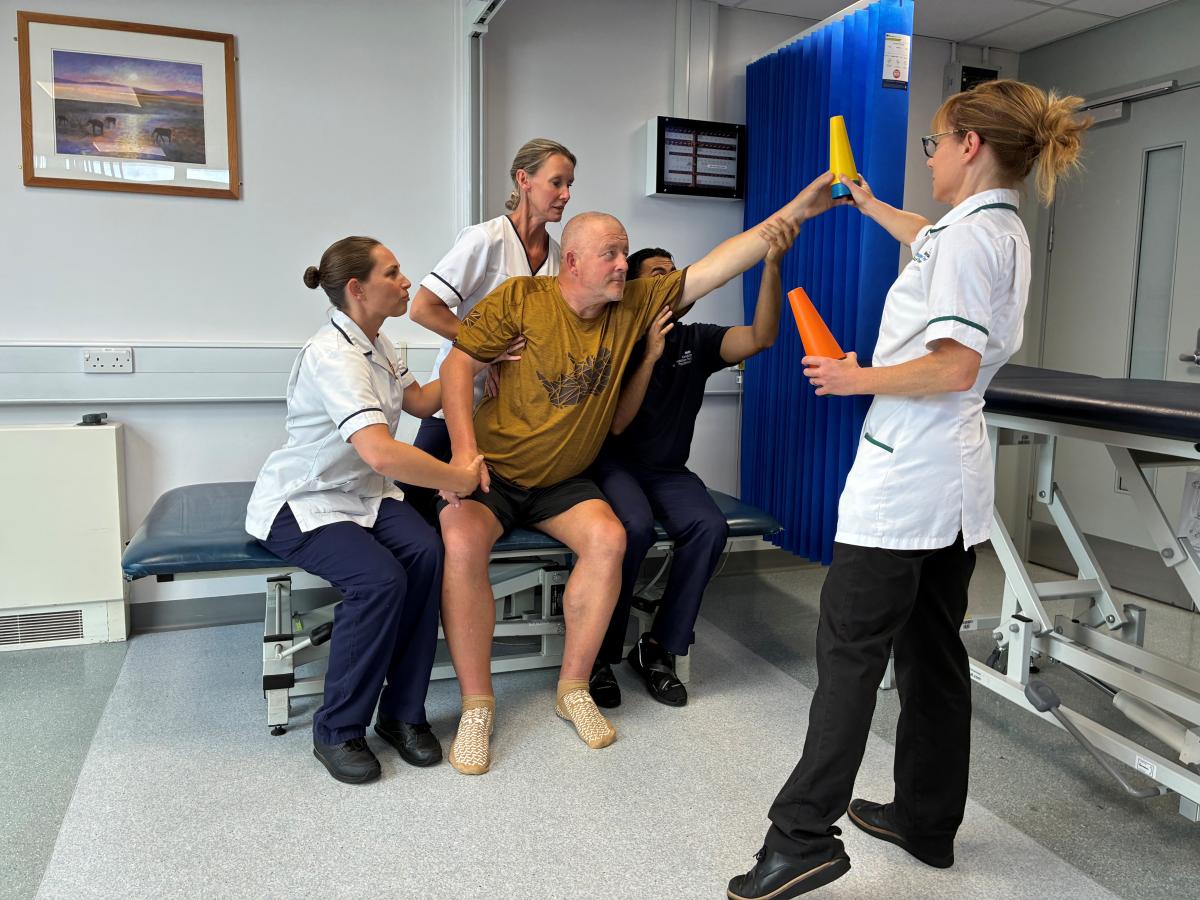
A stroke patient with rehab team

The report raises concerns that the number of stroke survivors is set to increase by 43 per cent over the next decade to 2 million, highlighting that this will have significant implications not only for people affected by stroke, but also for health and social care services. 

The CSP supports the Stroke Association’s recommendation to make rehabilitation and long-term support for stroke survivors a priority. The Stroke Association is a Community Rehabilitation (CRA) partner. 

Sara Hazzard, CSP Leadership Group and co chair of the CRA, said: ‘Early, personalised rehabilitation, combined with the right ongoing support, can make a profound difference to someone’s recovery after stroke, both to help them regain physical function and support their emotional wellbeing.'

Yet despite what we know about the benefits of rehabilitation, access to the right level of support remains far too limited, and progress to improve  availability and intensity is simply not happening quickly enough.

Back in April, the CSP along with ACPIN (Association of Chartered Physiotherapists in Neurology) warned that despite record numbers of registered physiotherapists, stroke services were unable to provide the care(NOT RECOVERY!) patients needed because they were chronically understaffed. (And they don't have the correct rehab protocols for getting survivors recovered fully! The understaffing is NOT the real problem! SOLVE THE CORRECT PROBLEM! )

VR headset invented by Kerala man’s firm set to rewrite stroke therapy

 How does this compare to all the other virtual reality items your doctor has tested?

Can't do that! PURE INCOMPETENCE! Seems like firing is the way to go.

VR headset invented by Kerala man’s firm set to rewrite stroke therapy

 Due to its compact form, physiotherapists can easily carry the headgear to a patient’s home
VR headset invented by Kerala man’s firm set to rewrite stroke therapy
Updated on: 

THIRUVANANTHAPURAM: Rehabilitation after a stroke is often a gruelling journey. Beyond the physical trauma, patients face long recovery timelines requiring endless, repetitive motions. Compounding this challenge is a severe shortage of certified physiotherapists and the high cost of hospital-bound care, which leads many patients to abandon therapy mid-way.

A groundbreaking collaboration between Chennai-based immersive-technology firm Skillveri and Thiruvananthapuram’s Sree Chitra Tirunal Institute for Medical Sciences and Technology (SCTIMST) aims to rewrite this narrative using virtual reality (VR). The partnership, which tapped into Skillveri’s industrial movement-tracking expertise, has translated into XTend, a software as a medical device designed specifically for upper-limb rehabilitation following a stroke. Born out of IIT Madras in 2012, Skillveri secured the exclusive commercialisation licence from SCTIMST following a technology transfer earlier this year. The device itself is a simple, lightweight headgear, a standalone VR headset equipped with built-in optical cameras that track hand movements naturally without requiring any bulky external sensors or wired attachments. Because of its compact form factor, physiotherapists can easily carry the headgear in a small bag to a patient’s home. Unlike conventional therapy, XTend disguises repetitive exercises as interactive games. Wearing the headgear, a patient might find themselves virtually sitting on Kovalam beach, popping balloons arranged precisely to trigger required shoulder, arm, or finger movements.
 The repetitive actions are part of immersive virtual-reality or mixed-reality games,” said Sabarinath Nair, founder and CEO of Skillveri. “The visual elements aid body movements, by acting as subconscious triggers of hand movements. The movements happen naturally, without the patient making a conscious effort.” The technology builds on the concept of hand-eye-mind coordination, developed with SCTIMST’s neurology department led by Dr P N Sylaja, who also serves as the Asia/Oceania member on the board of the World Stroke Organization (WSO). “There is brain damage in the case of a stroke patient. We have to retrain the brain on movements that you have somewhat forgotten,” Sabarinath explained. “We are the technology experts translating the medical knowledge into a gamified equivalent – retraining the brain to do these movements once more.”

While patients can physically operate the device, it is currently deployed in a therapist-supervised mode, where a physiotherapist controls exercise parameters via a smartphone app. Future iterations aim to introduce a fully independent self-mode for home use once patients complete initial guided phases.

Currently awaiting final ethics and regulatory approvals for clinical trials, Skillveri is prioritising affordability for the Indian market. The company is also developing a budget-friendly mobile app version that pairs standard Android phones with home televisions with the aim of further expanding reach.The Rehabilitation Gap

Disability & Economic Impact: Lack of timely post-stroke rehabilitation causes lifelong mobility issues, severely compromising employment chances and self-confidence during a patient’s most productive years (ages 55-60), SCTIMST found.

Treatment Shortfall: Over 70% of stroke survivors seek care at government hospitals, where shortages of occupational therapists and high costs limit long-term therap

Skillveri

  • An immersive technology company founded out of IIT Madras in 2012

  • Well-established in vocational skill simulations (welding, spray painting, HVAC/AC repair) for mechanical and manufacturing sectors

  • Operates as a leading installed building simulator provider with sales primarily across India, Europe, and the United States

  • Founder & CEO Sabarinath Nair, a native of Thiruvananthapuram, completed his BTech in computer science from Kerala University

Doctor reveals '10-year rule' for evaluating brain fog and dementia symptoms

 Can your doctor distinguish between this and dementia from your stroke AND HAVE WAYS TO PREVENT THAT?

Doctor reveals '10-year rule' for evaluating brain fog and dementia symptoms

Losing a train of thought, struggling to recall once-familiar words, or forgetting the reason for walking into a room can leave older adults wondering if they are experiencing early signs of Alzheimer’s disease or dementia.

However, these brain blips can often be tied to hormone shifts later in life, medical experts note — and women who experience a loss of estrogen during perimenopause and menopause are particularly affected.

According to Dr. Rhonda Voskuhl, a professor of neurology at UCLA and co-founder of Cleopatra RX in Los Angeles, women experiencing memory struggles in midlife should compare their cognitive function to their own past performance before assuming something is seriously wrong.

"I think they should listen to themselves," Voskuhl told Fox News Digital at the Livelong Women’s Health Summit in New York City on Friday. "They know. Just ask yourself."

To help women gauge whether their memory lapses are tied to hormonal shifts, Voskuhl recommends using a quick benchmark – comparing their current mental state to where it was a decade ago.

READ ON THE FOX NEWS APP

"The first question is, are you worse than you were 10 years ago?" she said, referencing the Voskuhl Brain Fog Menopause Assessment used at UCLA's Comprehensive Menopausal Program.

Ten years is a pretty good spot. And they think, ‘What was I doing 10 years ago? What was I like? Was I having problems finding the words? Was I at the point where I couldn’t multitask anymore? Was I slow in my processing?’ And they’ll remember."

This comparison can help reveal whether a woman’s cognitive changes may be linked to menopause, while giving patients and physicians a baseline for tracking symptoms over time.

Standard memory tests don’t always catch subtle shifts in everyday cognitive function. In the medical community, this is known as subjective cognitive decline, when patients notice changes even though neurological testing remains normal.

The Centers for Disease Control and Prevention reports that roughly one in nine adults report subjective cognitive decline, yet less than half discuss these memory concerns with a healthcare professional.

To illustrate why routine tests can miss early menopausal brain fog, Voskuhl compared it to a high-performing athlete.

"If somebody is an NFL football player and he says, 'I'm weaker than I used to be,' and I go and do my neurological test, I say, 'Oh no, you're still strong,'" Voskuhl said.

What the player is feeling still matters, even if a standard test shows no obvious decline, she noted.

Women, too, can often recognize when their cognitive abilities have changed from their own baseline and should trust those observations, according to Voskuhl.


Understanding the difference between hormonal brain fog and neurodegenerative disease comes down to how memory loss impacts daily function, experts agree.

Menopausal brain fog typically involves temporary, inconsistent lapses like misplacing items or experiencing slower processing speed, but overall reasoning skills and independence remain intact, according to Mayo Clinic. Symptoms often fluctuate and can be exacerbated by stress or poor sleep.

By contrast, early dementia causes persistent, progressive memory loss that interferes with basic daily tasks like managing finances, following familiar routes while driving or exercising sound judgment.

Amid cognitive concerns, experts advise seeking a full medical evaluation, including brain MRIs or vascular screenings, to rule out underlying issues like blood vessel disease or thyroid imbalances.

CLICK HERE TO DOWNLOAD THE FOX NEWS APP

For women who feel their concerns are being dismissed, Voskuhl offered this advice: "When they have doctors who don't listen to them, they need to find another doctor."

Original article source: Doctor reveals '10-year rule' for evaluating brain fog and dementia symptoms