Changing stroke rehab and research worldwide now.Time is Brain! trillions and trillions of neurons that DIE each day because there are NO effective hyperacute therapies besides tPA(only 12% effective). I have 523 posts on hyperacute therapy, enough for researchers to spend decades proving them out. These are my personal ideas and blog on stroke rehabilitation and stroke research. Do not attempt any of these without checking with your medical provider. Unless you join me in agitating, when you need these therapies they won't be there.

What this blog is for:

My blog is not to help survivors recover, it is to have the 10 million yearly stroke survivors light fires underneath their doctors, stroke hospitals and stroke researchers to get stroke solved. 100% recovery. The stroke medical world is completely failing at that goal, they don't even have it as a goal. Shortly after getting out of the hospital and getting NO information on the process or protocols of stroke rehabilitation and recovery I started searching on the internet and found that no other survivor received useful information. This is an attempt to cover all stroke rehabilitation information that should be readily available to survivors so they can talk with informed knowledge to their medical staff. It lays out what needs to be done to get stroke survivors closer to 100% recovery. It's quite disgusting that this information is not available from every stroke association and doctors group.

Thursday, June 4, 2020

Postural transitions detection and characterization in healthy and patient populations using a single waist sensor

This would seem to be incredibly important to monitor stroke survivors with and set this up so it immediately(text message?) tells them when they are doing these transitions wrong. But alas, nothing will be done with this since there is NO STROKE LEADERSHIP to go to and get this accomplished.  

Postural transitions detection and characterization in healthy and patient populations using a single waist sensor


Abstract

Background

Sit-to-stand and stand-to-sit transitions are frequent daily functional tasks indicative of muscle power and balance performance. Monitoring these postural transitions with inertial sensors provides an objective tool to assess mobility in both the laboratory and home environment. While the measurement depends on the sensor location, the clinical and everyday use requires high compliance and subject adherence. The objective of this study was to propose a sit-to-stand and stand-to-sit transition detection algorithm that works independently of the sensor location.

Methods

For a location-independent algorithm, the vertical acceleration of the lower back in the global frame was used to detect the postural transitions in daily activities. The detection performance of the algorithm was validated against video observations. To investigate the effect of the location on the kinematic parameters, these parameters were extracted during a five-time sit-to-stand test and were compared for different locations of the sensor on the trunk and lower back.

Results

The proposed detection method demonstrates high accuracy in different populations with a mean positive predictive value (and mean sensitivity) of 98% (95%) for healthy individuals and 89% (89%) for participants with diseases.

Conclusions

The sensor location around the waist did not affect the performance of the algorithm in detecting the sit-to-stand and stand-to-sit transitions. However, regarding the accuracy of the kinematic parameters, the sensors located on the sternum and L5 vertebrae demonstrated the highest reliability.

Background

Being able to maintain balance during movements is a prerequisite for an independent life. The inability to do so can lead to an increased risk of falls and consequently a dependent and inactive life [13]. Balance disorders can lead to problems with postural transitions (PTs), such as the sit-to-stand movements [4]. These challenging PTs require complicated coordination of lower and upper limbs [5] and frequently occur during daily living activities [6, 7]. As the sit-to-stand transitions are indicative of lower limb muscle strength and balance control [6, 8, 9], quantifying these movements is key to understand the underlying problem of balance disorders.
Clinicians conventionally assess the sit-to-stand transitions by either diaries [10] and questionnaires [11, 12] or functional tests. Standardized assessment tools can provide here valuable additional information.
The five-time sit-to-stand (5xSTS) test which measures the time to perform five sit-to-stand transitions [13, 14] and thirty-second chair-rise (30SCT) test which includes the numbers of sit-to-stands that can be performed within thirty seconds [8, 15] are standardized functional tests used in clinical routine to assess the ability to perform, and the quality of transitions. Although these methods have been proven to display discriminative properties for balance disorders [16], subtle differences that may provide further relevant information about the movement are not detectable with these tests [17].
For instance, during sit-to-stand transitions, maximum angular velocity has been shown to be associated with inadequate momentum generation and consequently, the success of the PT [18, 19]. Moreover, duration of each phase of sit-to-stand transitions changes between young and old adults [20] and between older adults with a low or a high risk of fall [21]. Peak power of transition has been reported to be associated with muscle power and strength [22, 23]. Therefore, instrumenting these functional tests and extracting meaningful parameters can provide a more in-depth and precise analysis. Sit-to-stand transitions have been studied with optical motion trackers [24] and force plates [25]. Although these methods provide very detailed and granular information about the movements, they are limited to the laboratory environment [7, 17].
The laboratory setting can only assess the performance of the participants in the confined environment (e.g. in-clinic) while individuals demonstrate different behavior in real-life daily activities [26, 27]. For example, sit-to-stand duration has been shown to be higher during daily activities compared to the functional test performed in the clinic in older adults and in patients with idiopathic Parkinson’s disease (IPS) [28]. Thus, it is important to develop methods that can also be used in domestic environments.
Inertial sensors can be applied in almost every environment. Moreover, they have been already used to instrument the 5xSTS [29] or the 30SCT [30] tests. Kinematic parameters extracted from such instrumented assessments have been shown to have greater clinical relevance than the conventional clinical approach [31, 32]. Wearable sensors have provided an objective tool to evaluate PTs during daily activities as well. Barometric pressure sensor within the pendant device was used as a complementary source of data to detect the PTs [3, 33]; however, due to the pressure changes in outdoor environments, the use of the barometric sensor can adversely affect the detection accuracy. For instance, in [3], the sensitivity of the sit-to-stand detection was decreased by 25% in outdoor environments.
There are some studies on monitoring sit-to-stand transitions with a single inertial sensor on either the sternum or on the lower back. In reference [34], the gyroscope and accelerometer signal along with a discrete wavelet transform have been used to obtain the trunk angle and consequently to detect the PTs. A simpler sensor setup with only a tri-axial accelerometer was used in [35]. In this study, the tilt angle of the trunk was estimated by the scalar product of the accelerometer data and gravity vector obtained during a static calibration at the beginning of each measurement. These studies were validated under very controlled conditions that involved sit-to-stand and stand-to-sit movements with a few other activities. More daily activities were included in the measurement protocol used by [36] and to reduce the false positive trunk movements, fuzzy rules have been employed to improve the accuracy of detection based on the previous or next activity. The performance of the PT detection was further improved by employing a template matching technique with dynamic time warping method in [37]. However, the performance of the detection algorithm was still unsatisfying with a positive predictive value and sensitivity of 22% and 50%, respectively. In another study, with a single inertial sensor on the waist, the candidates of the PTs were first detected by detecting the peaks of the tilt angle of the lower back. These were filtered out by double integrating the vertical acceleration and calculating the elevation change of the lower back [38].
The drawback of all of these studies [21, 3440] is that they require the sensor to be attached to a specific and fixed location of the body, which is difficult to maintain during daily activities and may not be achievable by patients themselves without a trained operator, thus limiting its broaden applicability in clinical setting.
This issue has been partially solved through using the signal vector magnitude which is the Euclidean norm of the accelerometer signal [41, 42]. The choice of various wavelets and scale approximations were studied in [42] to detect the PTs in a large group of healthy younger and older adults. However, in both of these studies, no method was suggested to distinguish true PTs from movements that can have similar wavelets to PTs. Their algorithms have been validated in measurements involving only sit-to-stand and stand-to-sit movements with rest periods in-between.
To this end, an algorithm which is robust to sensor placement changes and validated in a range of daily activities is desirable. Furthermore, little is known about the transferability of algorithms developed within a certain cohort, to other cohorts (e.g., with different and without diseases). The goal of this study was therefore to evaluate the performance of a new PT detection algorithm in healthy individuals and patients with different diseases that were all equipped with an inertial sensor on different locations around the waist and on the trunk. The new algorithm was validated in both laboratory and daily activity settings. Finally, the effect of the sensors location on the detection performance and extracted parameters was evaluated.

Shrinking Door-to-Needle Time in Stroke Proves Its Worth

Still useless. WHEN THE HELL WILL YOU DETERMINE THE DELIVERY TIME NEEDED FOR 100% RECOVERY WHEN USING tPA?  Without that knowledge you are just flailing in the dark. We don't want better recovery, we want 100% recovery. GET THERE! This is all still a complete failure because there is no strategy to solve stroke and never will be until we get survivors in charge.

Shrinking Door-to-Needle Time in Stroke Proves Its Worth

Large study affirms better outcomes with quicker start to thrombolysis(Your tyranny of low expectations is completely visible to all. You all need to be  fired.)

Paramedics rush a man on a stretcher from an ambulance into the hospital
Shorter times to thrombolytic therapy in acute stroke led to better long-term survival and lower readmission risk, an analysis of Medicare patients showed.
One year later, ischemic stroke patients treated with intravenous tissue plasminogen activator (tPA) who had door-to-needle times longer than 45 minutes had higher all-cause mortality (35.0% vs 30.8%) and higher all-cause hospital readmission (40.8% vs 38.4%) than patients with shorter times to treatment, reported Gregg Fonarow, MD, of the University of California Los Angeles, and colleagues.
Within a 90-minute window after hospital arrival, every 15-minute increase in door-to-needle time upped the risk of 1-year all-cause death (HR 1.04) or readmission (HR 1.02), they wrote in JAMA.
"Faster treatment translates into better long-term outcomes in patients with stroke," Fonarow said.
"For every 15 minutes improvement in treatment time, improvements in 1-year outcomes were observed," he told MedPage Today.
Faster tPA treatment is associated with better short-term outcomes, but it's less certain whether faster treatment also has long-term benefit, noted Christopher Muth, MD, of Rush University Medical Center in Chicago and senior editor of JAMA, in an accompanying commentary.
"Clinical trial data have not demonstrated a long-term mortality benefit with thrombolytic therapy, perhaps because trials were underpowered for this outcome," Muth wrote. Observational studies have shown better long-term survival in tPA-treated than untreated patients, but have not matched granular treatment data with long-term outcomes, he added.
In this study, Fonarow and colleagues linked detailed information about time to treatment from hospitals participating in the Get With The Guidelines-Stroke (GWTG-Stroke) registry with long-term clinical outcomes from Medicare claims data.
The researchers evaluated associations between door-to-needle times and 1-year outcomes for 61,426 patients with acute ischemic stroke who were treated with intravenous tPA from 2006 to 2016 within 4.5 hours from the time they were last known to be well.
Median age was 80, 43.5% were men, and 82% were white. Median door-to-needle time was 65 minutes.
The 48,666 patients who had a door-to-needle time longer than 45 minutes had significantly higher 1-year all-cause mortality (HR 1.13, 95% CI 1.09-1.18) and higher all-cause hospital readmission (HR 1.08, 95% CI 1.05-1.12) than patients treated within 45 minutes.
They did not, however, have significantly higher recurrent stroke readmission (HR 1.05, 95% CI 0.98-1.12) at 1 year.
A total of 34,367 patients had door-to-needle times longer than 60 minutes. They, too, had significantly higher all-cause mortality (HR 1.11, 95% CI 1.07-1.14) and all-cause readmission (HR 1.07, 95% CI 1.04-1.10) than patients treated within 60 minutes.
Door-to-needle time within 30 minutes was not associated with even better 1-year outcomes, but the analyses may have been underpowered for this group (5.6% of total patients), the researchers noted.
While the study's large sample size is a plus, the dataset and study design have limitations that may affect its generalizability, Muth pointed out. Only older adults were included, as evidenced by the median age of 80. More than 41,000 people in the GWTG-Stroke registry were excluded because their records couldn't be linked with Medicare claims data, including a higher proportion of racial and ethnic minorities. In addition, patients who received concomitant therapy with intra-arterial reperfusion techniques were not part of the study.
  • Judy George covers neurology and neuroscience news for MedPage Today, writing about brain aging, Alzheimer’s, dementia, MS, rare diseases, epilepsy, autism, headache, stroke, Parkinson’s, ALS, concussion, CTE, sleep, pain, and more. Follow
Disclosures
The GWTG-Stroke program is provided by the American Heart Association/American Stroke Association and supported by Novartis, Boehringer Ingelheim, Lilly, Novo Nordisk, Sanofi, AstraZeneca, and Bayer.
Fonarow disclosed support from the Patient-Centered Outcomes Research Institute and the NIH, as well as serving as an associate editor of JAMA Cardiology. Co-authors disclosed multiple relevant relationships with industry. One co-author disclosed serving as an associate editor of JAMA Cardiology.
Muth disclosed no relevant relationships with industry.

Access to rehabilitation for patients with stroke in Australia

Really, you think 'access' is the most important item that survivors care about? NO YOU ARE WRONG, TRY 100% RECOVERY. And I'm sure the goal-directed treatment is goals the therapist and doctor set, NOT THE SURVIVOR.  Because you have to make sure the tyranny of low expectations can be met with the available therapy.  You really would hate for stroke survivors to realize that their stroke medical 'professionals' know absolutely nothing about 100% recovery.

Access to rehabilitation for patients with stroke in Australia

Elizabeth A Lynch, Shylie Mackintosh, Julie A Luker and Susan L Hillier
Med J Aust 2019; 210 (1): 21-26. || doi: 10.5694/mja2.12034
Published online: 14 January 2019


Abstract

Objective: To identify factors associated with receiving acute goal‐directed treatment, being assessed for ongoing rehabilitation, and receiving post‐acute rehabilitation after having a stroke.
Design: Retrospective analysis of National Stroke Audit data for patients with acute stroke treated at Australian hospitals during 1 September 2014 – 28 February 2015.
Setting, participants: 112 Australian hospitals that admit adults with acute stroke.
Main outcomes: Associations between patient‐related and organisational factors and the provision of rehabilitation interventions.
Results: Data for 3462 patients were eligible for analysis; their median age was 74 years, 1962 (57%) were men, and 2470 (71%) had received care(NOT RESULTS?) in a stroke unit. 2505 patients (72%) received goal‐directed treatment during their acute admission; it was not provided to 364 patients (10.5%) who were responsive, had not fully recovered, and did not refuse treatment. Factors associated with higher odds of receiving goal‐directed treatment included goal‐setting with the patient and their family (odds ratio [OR], 6.75; 95% CI, 5.07–8.90) and receiving care in a stroke unit (OR, 2.08; 95% CI, 1.61–2.70). 1358 patients (39%) underwent further rehabilitation after discharge from acute care; factors associated with receiving post‐acute rehabilitation included care in a stroke unit (OR, 1.73; 95% CI, 1.34–2.22) and having an arm or speech deficit. Dementia was associated with lower odds of receiving acute goal‐directed treatment (OR, 0.49; 95%, 0.33–0.73) and post‐acute rehabilitation (OR, 0.43; 95%, 0.30–0.61).
Conclusions: Access to stroke units and to early and ongoing rehabilitation for patients after stroke can be improved in Australia, both to optimise outcomes and to reduce the burden of care on underresourced community and primary care providers.


The dead fairy sign

Does your doctor have a protocol for fairy touch prevention for stroke prevention? It has only been hundreds of years for your doctor to come up with a prevention solution for fairies.  Is hundreds of years enough time for your doctor to do something?

The dead fairy sign

. 1998 Aug 8; 317(7155): 396.
PMCID: PMC1113672
PMID: 9694758

Graham Lewis, general practitioner, Hampton, Middlesex
Insight is reflecting on the words of others. In medicine this means listening to your patients. My memorable patient was a little old lady, who remarked towards the end of a long Monday morning surgery, “Oh, doctor, you have just killed a fairy.” Wondering if my ears needed syringing, I was creating a list of possible psychiatric diagnoses as the patient continued, “Didn’t your mother ever tell you, every time you sigh you kill a fairy?” I was forced to admit that she had not.
This was surprising because a childhood spent in Cornwall had given me a healthy respect for, and knowledge of, the unworldy. I had recently bought my wife, whose family was full of such odd sayings, a copy of A Dictionary of Omens and Superstitions. This book provided no reference to sighs, fairies, and death.
From then on each time I started to sigh mental images of fairies falling from the skies, dying in mid-flight, began to haunt me. These were not the fay, gossamer winged creatures pictured by Arthur Rackham that are again in fashion. My fairies were the original Celtic fairy or piskie. These are amoral creatures, childlike in nature, and capable of great malevolence. In folklore they were held responsible for those otherwise inexplicable episodes of misfortune that afflict us all. Indeed, until recently, being touched by such a fairy was commonly recognised as the cause of a stroke. These were not creatures willfully to destroy, for—like bees—would not the death of one cause the rest of the swarm to fall on you? My sighing days were over and I began to observe others.
Within the practice it struck me that certain patients singlehandedly slay fairies at an almost genocidal rate. On entering the consulting room they have slaughtered several before seating. More lie mortally wounded as the first sentences are uttered. These patients usually present with a list of multiple symptoms or complaints. These patients are difficult to manage, frequently returning with yet more worries or symptoms. My newly discovered mental imagery now revealed a trail of fairies, dying fairies, spiralling down like sycamore seeds in the autumn winds, as these patients left the room.
I now understood why, whatever the advice I proffered, they would soon return with yet more woes. For dogging their steps, growing ever more malicious, were the surviving fairy brethren. Angered by the untimely demise of their kinsmen, they would now ensure misfortune to guarantee the patients’ return. It dawned on me that a chance remark by a little old lady had revealed a new category of patient. These were not just depressed, stressed, or heartsink. They were fairy killers.
Some patients, caught early enough may, like me, benefit from this novel insight. The more Rambo-inspired all seem to have been blinded (by the fairies?) to any possibility of insight. An attempt at explanation only lead to the sort of puzzled look I originally gave the little old lady. Any effort to push the concept is unnecessarily dangerous. The patient could leave the list or, worse still, the local community mental health team might begin to pay me undue attention.
However, now as they leave the room from a supposedly final consultation I can unfailingly spot the patient who will shortly return with yet another tale of woe. For as the surgery door closes, there lying on the floor (visible only to those with eyes to see) and gasping its last breath is a small dying fairy.
And the little old lady? I never saw her again.

Wednesday, June 3, 2020

Stuck on enormity - but what about stroke?

Yep, solving stroke is a BHAG(Big Hairy Audacious Goal.

Which is why you break it down into smaller parts. Choose one of the 5 causes of the neuronal cascade of death in the first week and just focus on that. Or tackle one of these 13 problems in stroke. This is so fucking simple, it is called creating a strategy to solve stroke. We can't allow researchers to willy nilly choose their stroke research, it has to be directed to the goal of 100% recovery. Which is why biomarkers and recovery prediction should never be researched, they assume the status quo will not change.  The status quo in stroke needs to be destroyed. Another great blog post by Seth Godin.

Stuck on enormity 

When a problem appears too large, too intractable and too unspeakable to deal with, it’s easy to give up.
There never seems to be enough time, enough resources or enough money to make the big problems go away.
Perhaps we can start with a very small part of it. One person, one opportunity, one connection.
Drip by drip, with commitment.
Those are the two hard parts. The insight to do it drip by drip and the persistence to commit to it.

Novel complement inhibitor improves ischemic stroke recovery in mice, say researchers

But this is in mice, we don't have ANY STROKE LEADERSHIP WE CAN GO TO TO GET THIS RESEARCHED IN HUMANS. Stroke survivors will continue to be screwed until all stroke associations and hospitals are run by survivors.  They have been incompetent for decades and don't deserve any more time.  I had not heard of the 30 day problem, even more proof that the 'Use it or lose it' statement is wrong.

Novel complement inhibitor improves ischemic stroke recovery in mice, say researchers

Administering a novel complement protein blocker (B4Crry) alongside reperfusion therapy improved cognitive and motor recovery in a model of ischemic stroke.
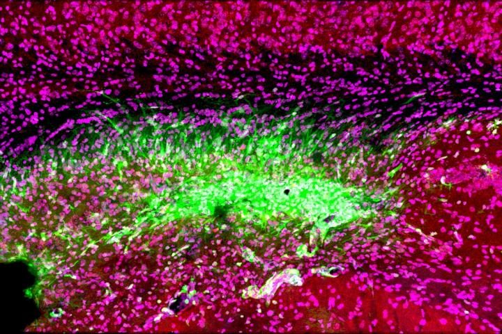
Even after a blocked vessel has been opened, immune cells in the brain (green) continue to attack synapses (red) and neurons (magenta) in the memory center of the brain, the hippocampus, for at least 30 days after stroke.Even after a blocked vessel has been opened, immune cells in the brain (green) continue to attack synapses (red) and neurons (magenta) in the memory center of the brain, the hippocampus, for at least 30 days after stroke [credit: Medical University of South Carolina. Image courtesy of Dr Stephen Tomlinson.].
Researchers have developed a novel complement protein blocker which, when administered alongside reperfusion therapy, improved cognitive and motor recovery in a murine model of ischemic strokes.
Reperfusion therapy restores blood flow to an area of the brain after an ischemic stroke – a stroke caused by a blood clot blocking blood flow to an area of the brain. However, despite being the gold standard of treatment for ischemic strokes, only 10 percent of patients qualify due to its narrow treatment window (ideally given up to four hours after stroke, ineligible after eight hours).
Also, while blood flow is restored in 90 percent of patients, only 40 percent recover their motor and cognitive skills enough to cope with independent living within three months. Researchers at the Medical University of South Carolina (MUSC), US, believe this is because while blood flow is restored, an ongoing inflammatory and phagocytic response to the damaged tissue results in further degeneration and damage.(Yep, call this the correct name: NEURONAL CASCADE OF DEATH)

In an ischemic stroke the lack of oxygen due to a blood clot results in cells becoming stressed and rapidly dying. Even after blood flow is restored, cells which were stressed but did not die give off signal to the immune system, promoting inflammation and repair at the damaged area. The researchers on the latest study have previously shown that the complement system drives this inflammation and its proteins tag both dead tissue and stressed brain cells for removal.
This tagging of stressed neurons results in viable cells being destroyed, further exacerbating the damage to neuronal circuits and so researchers at MUSC sought to salvage them. The team developed a novel complement protein blocker, called B4Crry, which is designed to act only at the site of a stroke injury and blinds the complement proteins to the signals of stressed brain cells, preventing them being tagged.
When B4Crry was administered to a murine model of ischemic stroke alongside reperfusion therapy, the cognitive and motor abilities of mice improved. Evidence of co-ordinated movement recovery was visible even three days after the stroke with B4Crry, where there was no recovery with reperfusion therapy alone. In memory tasks, mice treated with B4Crry made three times fewer errors in a learning and memory task, while their reperfusion therapy only counterparts had the same recovery as mice that went completely untreated.

Complement inhibition and reperfusion safety

The team also demonstrated that after clot removal adding B4Crry to reperfusion therapy reduced the potential for haemorrhage, a complication associated with late administration of reperfusion therapy, even with treatment given up to six hours after the stroke. The team concluded that not only could complement inhibition make reperfusion therapy more effective and safer, but also extend the treatment window.

The team are excited about the possibly using the complement protein blocker in clinic:

Lead author Dr Ali Alawieh, who completed his graduate studies at MUSC and is now a resident in neurosurgery at Emory University School of Medicine, revealed: “Our next step is to see how complement inhibitors work with comorbidities, such as old age, smoking and diabetes, in a preclinical study. Collectively, this information will help us design the best clinical trial when we move to humans.”
Dr Stephen Tomlinson, interim chair of the Department of Microbiology and Immunology at MUSC and senior author of the article, said they are also testing whether the complement inhibitor may be beneficial in treating other brain injuries, such as traumatic brain injury.
“We have shown that we can administer complement inhibitors as far as two months after a traumatic brain injury and see improvements in cognitive recovery,” said Tomlinson. “This is something I’m actually quite excited about. It means that months after an initial event, complement inhibitors could still be beneficial to cognitive recovery after brain injuries, including strokes.”
The paper was published in the Journal of Neuroscience.

Physiological evaluation of gait disturbances post stroke

Maybe there is something in these 8 pages  but that is your doctor's responsibility to know and apply. Only 13 years to accomplish that, how is the competence of your doctor and stroke hospital appearing in light of what should have been done 13 years ago? CREATING PROTOCOLS FOR REHAB BASED ON RESEARCH!

Physiological evaluation of gait disturbances post stroke

 Anouk Lamontagne
*
, Jennifer L. Stephenson, Joyce Fung
School of Physical and Occupational Therapy, McGill University, Montreal, CanadaJewish Rehabilitation Hospital Research Site of the Montreal Centre for Interdisciplinary Research in Rehabilitation (CRIR),3205 Place Alton-Goldbloom, Laval (Quebec), Canada H7V 1R2
Accepted 26 December 2006Available online 16 February 2007

Abstract

A large proportion of stroke survivors have to deal with problems in mobility. Proper evaluations must be undertaken to understand the sensorimotor impairments underlying locomotor disorders post stroke, so that evidence-based interventions can be developed. The current electrophysiological, biomechanical, and imagery evaluations that provide insight into locomotor dysfunction post stroke, as well as their advantages and limitations, are reviewed in this paper. In particular, electrophysiological evaluations focus on the contrast of electromyographic patterns and integrity of spinal reflex pathways during perturbed and unperturbed locomotion between persons with stroke and healthy individuals. At a behavioral level, biomechanical evaluations that include temporal distance factors, kinematic and kinetic analyses, as well as the mechanical energy and metabolic cost, are useful when combined with electrophysiological measures for the interpretation of gait disturbances that are related to the control of the central nervous system or secondary to biomechanical constraints. Finally, current methods in imaging and transcranial magnetic stimulation can provide further insight into cortical control of locomotion and the integrity of the corticospinal pathways.

 2007 International Federation of Clinical Neurophysiology. Published by Elsevier Ireland Ltd. All rights reserved.
Keywords:
 Cerebrovascular accident; Cerebrovascular disease; Locomotion; Electromyography; Kinematics; Kinetics; Electrophysiology; Reflex;Functional imagery
1. Introduction
Gait disorders affect a large proportion of stroke survi-vors and limit their ability to ambulate in the community(Keenan et al., 1984). Walking after stroke is characterizedby slow gait speed (von Schroeder et al., 1995), poor endurance (Dean et al., 2001) and a reduced ability to adapt tothe task and the environmental constraints (Lamontagne et al., 2003; Said et al., 1999). Motor impairments arebelieved to be the primary cause of this poor walking ability, as suggested by the association between muscle weakness of specific muscle groups, such as the plantarflexorson the paretic side, and slow gait speed (Nadeau et al.,1999; Olney et al., 1994). Sensory perception (Lamontagne et al., 2005b) and cognitive factors (Bowen et al., 2001;Haggard et al., 2000; Regnaux et al., 2005), however, alsoinfluence mobility in stroke and are now gaining increased attention. Many electrophysiological, biomechanical and imagery measures have been developed to quantify gait dis-turbances post stroke. This paper aims to review some of the outcomes provided by such measures, emphasizing how the proper evaluation of locomotor disorders will lead to a better understanding of the underlying pathophysiology and hence more effective interventions can be developed. 
5. Conclusions
Gait disturbances post stroke are multi-faceted and hence must be studied from multiple perspectives. Biomechanical measurements such as temporal distance parameters, kinematics, kinetics, mechanical energy and energy costs as well as electromyography can evaluate the behavioral profile of gait and reflect central nervous system adaptation with respect to internal and external demands.Electrophysiological measurements such as stretch reflex,H-reflex and cutaneous reflexes can evaluate the integrity of spinal cord functions during gait and indirectly assess the integrity of the descending control system. Further-more, transcranial magnetic stimulation and advances inimaging techniques have provided new opportunities to examine supraspinal mechanisms and cortical activationsduring gait.

Blood Test May Help Predict Post-Concussion Recovery

Oh god, more laziness and stupidity. Predictions assume the status quo, the status quo is a complete fucking failure. This should be a call to change the status quo by researching solutions. 

Blood Test May Help Predict Post-Concussion Recovery

Exosomal and plasma markers tied to neuropsychiatric symptoms

A healthcare worker draws blood from a man’s arm
Military veterans with a history of three or more concussions were more likely to have high levels of exosomal and plasma levels of neurofilament light (NfL) chain years after injury occurred, a cross-sectional study suggested.
Moreover, elevations of NfL, a measure of axonal damage, were highest in veterans with repetitive traumatic brain injury (TBI) who had symptoms of chronic post-concussive syndrome, post-traumatic stress disorder (PTSD), and depression, reported Kimbra Kenney, MD, of Walter Reed National Military Medical Center in Bethesda, Maryland, and colleagues, in Neurology.
Blood tests that include exosomal and plasma NfL markers may one day help predict which people with mild TBIs will take longer to recover, Kenney noted.
"Our study found there's great potential for this protein to predict the problems people with concussions may experience years after their injuries," she said in a statement.
"While most people with mild concussions recover completely, some never get their lives back fully because of chronic disability," Kenney added. "These people may benefit greatly from a test that could predict those disabilities years ahead of time."(Predicting crap like this does the patient no good at all. We need solutions for recovery.)
Most concussions are single and uncomplicated, but "a subset of individuals experience persistent post-concussive symptoms and substantial disability," noted Silvina Tonarelli, MD, of Texas Tech University in El Paso, and Davin Quinn, MD, of the University of New Mexico in Albuquerque, in an accompanying commentary.
"There is a need for accurate and reliable biomarkers that are tied to the pathophysiologic mechanisms of mild TBI, and that track and predict neuropsychiatric outcomes," they wrote.
"A promising avenue of biomarker development is exosomes, a type of extracellular vesicle released by injured cells, whose contents can be informative about neuronal pathophysiology," Tonarelli and Quinn added.
Exosomes of other proteins like  are being studied as potential biomarkers in Parkinson's and other neurodegenerative diseases. In other disorders, including Alzheimer's disease and multiple sclerosis, blood levels of NfL have been shown to correspond with hallmarks of disease progression.
In this study, Kenney's research team studied 195 military veterans in the Chronic Effects of Neurotrauma Consortium (CENC) cohort. Participants had a history of combat exposure during deployment; their median age was 38, and 85% were men.
Excluded from the study were people with moderate or severe TBI, and people who had coma that lasted more than 30 minutes, amnesia that lasted more than 24 hours, traumatic intracranial lesion on head CT, or a history of major neurologic or psychiatric disorder.
Besides exosomal and plasma levels of NfL, the researchers looked at other candidate biomarkers, including tumor necrosis factor (TNF)-α, interleukin-6 (IL-6), and vascular endothelial growth factor (VEGF).
The researchers divided participants into three groups:
  • 45 people had no history of mild TBI
  • 94 people had one or two mild TBIs
  • 56 people had three or more TBIs
People with one or two mild TBIs had their first concussion a median of 13.53 years ago and their last one 9.53 years ago. People with three or more mild TBIs had their first concussion a median of 22.11 years ago and their last one 6.83 years ago.
Exosomal and plasma levels of NfL were linked to repetitive mild TBIs and correlated with severity of post-concussive, PTSD, and depressive symptoms.
Plasma levels of TNF-α, an inflammatory marker, were tied to post-concussive and PTSD symptoms. The lifetime number of concussions correlated with exosomal and plasma NfL levels and with plasma IL-6, another inflammatory marker.
Moreover, an increased number of years since first concussion correlated with higher levels of exosomal and plasma NfL. Years since first concussion also was linked to plasma VEGF, which may modulate inflammatory responses, and plasma TNF-α.
"These are encouraging findings, suggesting that an ongoing neuroinflammatory process may be an important contributor to persistent post-concussion syndrome in a military population subjected to repetitive mild TBIs," Tonarelli and Quinn pointed out.
Strengths of the study include its "well-characterized cohort and rigorous mild TBI confirmation process," they observed, but "further confirmation of these relationships and clarification of confounding effects of comorbidities is required before these biomarkers can be of use."
The study had several limitations, Kenney and colleagues noted. The sample size was relatively small and there was substantial variability in the number of years in which injury occurred.
There also was variability in mechanisms of injury. "We included participants with deployment-associated TBI, which includes but it is not restricted to blast-related TBI, as well as TBIs sustained before military service," the researchers said.
  • Judy George covers neurology and neuroscience news for MedPage Today, writing about brain aging, Alzheimer’s, dementia, MS, rare diseases, epilepsy, autism, headache, stroke, Parkinson’s, ALS, concussion, CTE, sleep, pain, and more. Follow
Disclosures
The study was supported by the Department of Defense, Department of Veterans Affairs, and NIH.
Kenney and co-authors, as well as Tonarelli and Quinn, disclosed no relevant relationships with industry.

Use of real-time visual feedback during overground walking training on gait symmetry and velocity in patients with post-stroke hemiparesis: randomized controlled, single-blind study.

These are high functioning patients already, stage IV and V and ability to walk 30 minutes at a time.  I have no understanding of real-time visual feedback.  In fact I would assume that the participants are using the Hawthorne effect to please the researchers since they would know which group they were in.

Use of real-time visual feedback during overground walking training on gait symmetry and velocity in patients with post-stroke hemiparesis: randomized controlled, single-blind study.

Abstract 


This study aimed to determine the efficacy of using real-time visual feedback during overground walking training to improve walking function in patients with post-stroke hemiparesis. Twenty-four patients with post-stroke hemiparesis who were able to walk independently under less impact of synergy pattern on the affected lower limbs (Brunnstrom stage IV or V) were randomly assigned to either the experimental group or the control group. All subjects performed overground walking for 30 min, three times a week for 6 weeks, with real-time visual feedback (weight load to the affected lower limb) provided during training for subjects in the experimental group. Outcome measures comprised the timed up-and-go test and gait parameters (step length, stride length, single and double support times, step and stride length ratios, and single support time ratio). In between-group comparison, the changes between pre-test and post-test scores in all parameters were significantly greater in the experimental group than in the control group (P < 0.05), except for double support time and step length ratio. Furthermore, post-test values of all parameters were significantly more improved in the experimental group than in the control group (P < 0.05). Our findings suggest that real-time visual feedback may be an advantageous therapeutic adjunct to reinforce the effects of overground walking training in patients with post-stroke hemiparesis.

Usefulness of Goal Attainment Scaling in Intensive Stroke Rehabilitation During the Subacute Stage

Totally bad research right from the start. Therapist suggested goals;  the tyranny of low expectations in broad daylight. They all need to be fired. 

Usefulness of Goal Attainment Scaling in Intensive Stroke Rehabilitation During the Subacute Stage

Youngsu Jung, MD, Jaehoon Sim, MD, Joonhyun Park, MD, Jongmoon Kim, MD, MinYoung Kim, MD, PhD
Department of Rehabilitation Medicine, CHA Bundang Medical Center, CHA University College of Medicine, Seongnam, Korea
Objective  
To investigate the usefulness of goal attainment scaling (GAS) in intensive stroke rehabilitation during the subacute stage. 
Methods  
Medical records of subacute post-stroke patients who had undergone intensive rehabilitation under hospitalization, before and after the application of GAS, were collected. GAS was conducted at the initial evaluation of each patient. Specific goals were suggested by physical and occupational therapists and were determined by the responsible physiatrist.(This is a fireable offense per Dean.) A 5-point scale was used for the GAS score after 4 weeks of rehabilitation according to the preset criteria of each goal. To evaluate the influence of GAS in rehabilitation practice, functional improvements were compared between two patient groups before (n=121) and after (n=141) GAS. To assess progress in GAS practice, the standard GAS score was calculated, and the changes were observed over a 3-year period. The standard GAS score converged to 50 points when the goal was achieved. The therapists who used GAS also completed a survey regarding its usefulness. Results  There were no statistical differences in the motor and cognitive outcomes of the patient groups before and after applying GAS scoring. Successive yearly changes in the standard GAS scores showed progressive convergence to 50 points, signaling that the patient’s goal-setting abilities improved. According to the survey, most therapists felt that GAS enhanced the quality of therapies (84.6%). Conclusion 
GAS improved goal-setting for the rehabilitation of subacute post-stroke patients and might have a positive effect on rehabilitation.(Yeah, it would show complete failure of all of your rehab interventions since the true goal of all survivors is 100% recovery. NOT THIS CRAP YOU SHOVE DOWN THEIR THROATS.)

An extended stroke rehabilitation service for people who have had a stroke: the EXTRAS RCT

How do you reconcile these two conflicting results?   

did not improve stroke survivors' performance in extended activities of daily living but did improve their overall satisfaction with services. Your stroke team was excellent at getting you to accept the tyranny of low expectations but didn't deliver actual results? Not my idea of excellence.  

An extended stroke rehabilitation service for people who have had a stroke: the EXTRAS RCT

Abstract 


BACKGROUND:
There is limited evidence about the effectiveness of rehabilitation in meeting the longer-term needs of stroke patients and their carers. 
OBJECTIVE:
To determine the clinical effectiveness and cost-effectiveness of an extended stroke rehabilitation service (EXTRAS). DESIGN:A pragmatic, observer-blind, parallel-group, multicentre randomised controlled trial with embedded health economic and process evaluations. Participants were randomised (1 : 1) to receive EXTRAS or usual care. SETTING:Nineteen NHS study centres.  

PARTICIPANTS:
Patients with a new stroke who received early supported discharge and their informal carers.  

INTERVENTIONS:
Five EXTRAS reviews provided by an early supported discharge team member between 1 and 18 months post early supported discharge, usually over the telephone. Reviewers assessed rehabilitation needs, with goal-setting and action-planning. Control treatment was usual care post early supported discharge. 
MAIN OUTCOME MEASURES:
The primary outcome was performance in extended activities of daily living (Nottingham Extended Activities of Daily Living Scale) at 24 months post randomisation. Secondary outcomes at 12 and 24 months included patient mood (Hospital Anxiety and Depression Scale), health status (Oxford Handicap Scale), experience of services and adverse events. For carers, secondary outcomes included carers' strain (Caregiver Strain Index) and experience of services. Cost-effectiveness was estimated using resource utilisation costs (adaptation of the Client Service Receipt Inventory) and quality-adjusted life-years. 
RESULTS:
A total of 573 patients (EXTRAS, n = 285; usual care, n = 288) with 194 carers (EXTRAS, n = 103; usual care, n = 91) were randomised. Mean 24-month Nottingham Extended Activities of Daily Living Scale scores were 40.0 (standard deviation 18.1) for EXTRAS (n = 219) and 37.2 (standard deviation 18.5) for usual care (n = 231), giving an adjusted mean difference of 1.8 (95% confidence interval -0.7 to 4.2). The mean intervention group Hospital Anxiety and Depression Scale scores were not significantly different at 12 and 24 months. The intervention did not improve patient health status or carer strain. EXTRAS patients and carers reported greater satisfaction with some aspects of care. The mean cost of resource utilisation was lower in the intervention group: -£311 (95% confidence interval -£3292 to £2787), with a 68% chance of EXTRAS being cost-saving. EXTRAS was associated with 0.07 (95% confidence interval 0.01 to 0.12) additional quality-adjusted life-years. At current conventional thresholds of willingness to pay for a quality-adjusted life-year, there is a 90% chance that EXTRAS is cost-effective. 
CONCLUSIONS:
EXTRAS did not improve stroke survivors' performance in extended activities of daily living but did improve their overall satisfaction with services. Given the impact on costs and quality-adjusted life-years, there is a high chance that EXTRAS could be considered cost-effective. FUTURE WORK:Further research is required to identify whether or not community-based interventions can improve performance of extended activities of daily living, and to understand the improvements in health-related quality of life and costs seen by provision of intermittent longer-term specialist review. TRIAL REGISTRATION:Current Controlled Trials ISRCTN45203373. FUNDING:This project was funded by the National Institute for Health Research (NIHR) Health Technology Assessment programme and will be published in full in Health Technology Assessment; Vol. 24, No. 24. See the NIHR Journals Library website for further project information.

Tuesday, June 2, 2020

Biomedical Engineering professor and team develop treatment for traumatic brain injury

With ANY BRAINS AT ALL,  our stroke leadership would fire up research on this for stroke. But that will never occur, two problems;

1. We have NO STROKE LEADERSHIP.

2. There are no brains in stroke.  

The minocycline approach was researched/published in October 2009  SO 11 TEARS OF  EPIC FAILURE EVERYWHERE IN STROKE! Proving once again that the existing stroke associations are worthless.  

I put this in my list of 31 things I was going to demand after my next stroke.

The latest here:

Biomedical Engineering professor and team develop treatment for traumatic brain injury

Dr. Teresa Murray, associate professor in Biomedical Engineering and the Center for Biomedical Engineering and Rehabilitation Sciences at Louisiana Tech University, is collaborating with former Louisiana Tech doctoral student Dr. Chelsea Pernici and professors at the University of Arizona to help develop solutions to traumatic brain injury.
Murray and Pernici are working with Dr. Jonathan Lifshitz and Dr. Rachel Rowe, director and research assistant professor, respectively, with the Translational Neurotrauma Research Program at the University of Arizona College of Medicine – Phoenix, to develop a treatment that could prevent memory and emotional problems in patients suffering from axonal injury caused by traumatic brain injuries (TBIs).
The study was led by Murray and Pernici who used a new imaging technology to show how diseases and injuries affect the brain over time and how medicines could reduce brain damage. This novel imaging was created in Murray’s lab. Through this technology, the group of researchers were able to repeatedly image the same axons in the brain before and after brain injury, providing proof of TBI-related axonal pathology.
Using a novel combination of high-resolution imaging and a GRIN lens implanted in the brain of mice, the team documented axonal injury and the recovery process after a blunt injury to the brain. GRIN lenses are microscopic glass lenses that allow scientists to focus closely on the cells for good image quality. These lenses are smaller than Lincoln’s nose on a U.S. penny and were adopted by a handful of biologists to better study the brain.
Murray developed an improved GRIN lens system to see more details of brain cells. Her team also designed 3D-printed hardware to help them find the exact same cells for each imaging session. Her improved GRIN lenses allowed the team to find and evaluate the same injured axons over several weeks post injury.
“The fine communication elements of neurons — axons — are torn, ruptured and damaged, which contributes to a multitude of clinical symptoms,” Lifshitz said. “To date, these processes have been inferred from fixed histological sections, clinical imaging and cells in culture. No one has observed an axon prior to injury, the consequence of injury and the outcome. Here, we show that axons do sustain injury and can either recover or become truncated.”
Tracking the time in which axons become affected, the team employed a clinically approved drug, minocycline, to reduce inflammation, which slowed axon injury, recovered them from injury and preserved function. The team tested the drug’s effectiveness for immediate use within 45 minutes of the incident, compared to delayed use at three days post injury – both time points showed positive effects. The minocycline drug treatment promoted the recovery of injured axons. With treatment, a much larger percentage of the damaged axons healed, compared to those without treatment.
“Most brain injuries cause damage to just a few axons,” Murray said. “These few axons can’t be seen on MRIs and CT scans. So, most TBI patients are sent home with the hope that they will get better over time. However, even a few damaged axons can disrupt communication in the brain and affect neurological function.”
“The novel fluorescence imaging method using GRIN lenses allows researchers to study brain cells in both time and space, offering a unique opportunity to better understand the effect of drugs on brain structure over time,” Pernici added. “It’s exciting to see that although axons become damaged after TBI, the injury can resolve and with this method, we have the opportunity to better understand that timescale.”
The team plans to continue their research by pairing minocycline with another drug to further reduce this early damage. If successful, this treatment could lessen the chances of memory loss and emotional problems for people suffering a TBI.
“This study provides hope that we can use minocycline effectively if the drug can be taken early in the process and not given long-term. This could help reduce the inflammation, and thus some of the permanent damage caused by brain injury,” Murray said.
No drug currently on the market is clinically used to prevent axon injury. The results of their research were published in Scientific Reports on May 8.

Randomized Pragmatic Trial of Stroke Transitional Care

Bad research right from the start. 'Care' in the title. THIS IS WHY WE NEED SURVIVORS IN CHARGE.  We wouldn't take our eyes off the only goal in stroke; 100% recovery. 

Randomized Pragmatic Trial of Stroke Transitional Care

The COMPASS Study
Originally publishedhttps://doi.org/10.1161/CIRCOUTCOMES.119.006285Circulation: Cardiovascular Quality and Outcomes. ;0

Background

The objectives of this study were to develop and test in real-world clinical practice the effectiveness of a comprehensive postacute stroke transitional care (TC) management program.

Methods and Results

The COMPASS study (Comprehensive Post-Acute Stroke Services) was a pragmatic cluster-randomized trial where the hospital was the unit of randomization. The intervention (COMPASS-TC) was initiated at 20 hospitals, and 20 hospitals provided their usual care. Hospital staff enrolled 6024 adult stroke and transient ischemic attack patients discharged home between 2016 and 2018. COMPASS-TC was patient-centered and assessed social and functional determinates of health to inform individualized care plans. Ninety-day outcomes were evaluated by blinded telephone interviewers. The primary outcome was functional status (Stroke Impact Scale-16); secondary outcomes were mortality, disability, medication adherence, depression, cognition, self-rated health, fatigue, care satisfaction, home blood pressure monitoring, and falls. The primary analysis was intention to treat. Of intervention hospitals, 58% had uninterrupted intervention delivery. Thirty-five percent of patients at intervention hospitals attended a COMPASS clinic visit. The primary outcome was measured for 59% of patients and was not significantly influenced by the intervention. Mean Stroke Impact Scale-16 (±SD) was 80.6±21.1 in TC versus 79.9±21.4 in usual care. Home blood pressure monitoring was self-reported by 72% of intervention patients versus 64% of usual care patients (adjusted odds ratio, 1.43 [95% CI, 1.21–1.70]). No other secondary outcomes differed.

Conclusions

Although designed according to the best available evidence with input from various stakeholders and consistent with Centers for Medicare and Medicaid Services TC policies, the COMPASS model of TC was not consistently incorporated into real-world health care. We found no significant effect of the intervention on functional status at 90 days post-discharge.

Registration

URL: https://www.clinicaltrials.gov; Unique identifier: NCT02588664.