Changing stroke rehab and research worldwide now.Time is Brain! trillions and trillions of neurons that DIE each day because there are NO effective hyperacute therapies besides tPA(only 12% effective). I have 523 posts on hyperacute therapy, enough for researchers to spend decades proving them out. These are my personal ideas and blog on stroke rehabilitation and stroke research. Do not attempt any of these without checking with your medical provider. Unless you join me in agitating, when you need these therapies they won't be there.

What this blog is for:

My blog is not to help survivors recover, it is to have the 10 million yearly stroke survivors light fires underneath their doctors, stroke hospitals and stroke researchers to get stroke solved. 100% recovery. The stroke medical world is completely failing at that goal, they don't even have it as a goal. Shortly after getting out of the hospital and getting NO information on the process or protocols of stroke rehabilitation and recovery I started searching on the internet and found that no other survivor received useful information. This is an attempt to cover all stroke rehabilitation information that should be readily available to survivors so they can talk with informed knowledge to their medical staff. It lays out what needs to be done to get stroke survivors closer to 100% recovery. It's quite disgusting that this information is not available from every stroke association and doctors group.

Tuesday, June 2, 2020

Auckland man dies from stroke after being misdiagnosed with a migraine

This is why YOU need to remove subjective determination of stroke, YOU are going to have to get that changed in your hospital.  Your hospital has been doing nothing about this for years.  

For example all the misdiagnosed young adult strokes.

Another reason to switch to artificial intelligence to diagnose strokes. Maybe one of these much faster methods:

Hats off to Helmet of Hope - stroke diagnosis in 30 seconds


Microwave Imaging for Brain Stroke Detection and Monitoring using High Performance Computing in 94 seconds


New Device Quickly Assesses Brain Bleeding in Head Injuries - 5-10 minutes

The latest here:

Auckland man dies from stroke after being misdiagnosed with a migraine

A man died from a stroke days after doctors misdiagnosed him with a migraine, a coroner has found.
Edwin Donald Amundsen, 57, died at Auckland's Middlemore Hospital on October 15, 2015, just two days after he sought treatment for sudden loss of vision and a persistent headache.
According to a coroner's report into his death, the father-of-three may have survived if he had not been misdiagnosed.
The report identified several "missed opportunities" for diagnosis and treatment by multiple medical staff at Counties Manukau and Auckland DHBs – however, the most critical issue was that Amundsen was not seen by a specialist neurologist early enough, it said.
READ MORE:
* Unwell prisoner should have been taken to hospital 'immediately' - coroner
* Doctors misdiagnose woman's strokes as ear infection, send her home 3 times
* In your 40s and think you won't suffer a stroke? It happened to me
Middlemore Hospital does not have an on-site neurology team. However, at the time of Amundsen's death, a neurologist from Auckland City Hospital visited three mornings a week.


Coroner Katharine Greig said the problems were systemic and "no one person is 'to blame'".
The report said Amundsen first realised something was wrong while he was reading the newspaper on October 12, 2015 and noticed words on the pages were "missing".
His vision was blurry and later in the day he developed a constant headache.
The next morning, Amundsen, who lived in Kawakawa Bay, south Auckland, went to his GP.
Believing he may have a brain bleed, the doctor referred Amundsen to Middlemore Hospital, where he was assessed by a sixth-year medical student intern and senior medical officer Dr Michael Rosen.


Middlemore Hospital, where Amundsen was treated, does not have specialist neurology services (file photo).
Stuff-co-nz
Middlemore Hospital, where Amundsen was treated, does not have specialist neurology services (file photo).

Amundsen had blood tests, a CT scan and a chest x-ray before Rosen diagnosed him with a "severe atypical migraine".
After spending the night in Middlemore, Amundsen went to Greenlane Hospital's acute eye clinic where he had further tests, which failed to find a cause for his symptoms.
He was discharged later that afternoon.
About midday on October 15, Rosen phoned Amundsen to check on him.
Amundsen said he still had a headache and his vision was worsening. Rosen told him to return to the hospital immediately.
While Amundsen's wife was driving him back to Middlemore, he started vomiting and became unresponsive.


An ambulance was called and he was rushed to the hospital's emergency department and referred to the stroke team, as his condition was worsening.
Amundsen had a decreased level of consciousness, weakness in his right arm and leg and an unsteady gait. He was also nauseous and slightly disoriented, the report said.
He had an urgent CT scan which was more comprehensive than the first and showed evidence of a stroke, it said.
Staff then called a neurologist at Auckland City Hospital, who recommended Amundsen have another CT scan.
That showed the stroke was worsening. Amundsen died later that night.
Forensic pathologist Dr Deborah Johnson and associate professor and consultant neurologist Annemarei Ranta, an expert on strokes who provided advice to the coroner, both concluded Amundsen's death was caused by a stroke.


A radiologist who assesed a scan of Amundsen's brain failed to pick up changes which indicated he was having a stroke (file photo).
SUPPLIED/123RF
A radiologist who assesed a scan of Amundsen's brain failed to pick up changes which indicated he was having a stroke (file photo).

Ranta reviewed the CT scans and spotted "subtle" changes to Amundsen's brain in the first one which the radiologist failed to pick up.
"A clinical stroke very likely was present and probably missed," she said.
Ranta believed this should have been noticed and likely would have been if Amundsen had been referred to a neurologist earlier.
In her report, Greig said: "Tragically... layers of weakness and holes in the system aligned with the result that harm was caused to Mr Amundsen in that he lost the opportunity for earlier diagnosis and treatment."
In light of the case, the coroner recommended Counties Manukau DHB review its protocols for specialist neurology care.
The DHB did so and found the referral processes adequate.
However, a neurologist from Auckland City Hospital was now at Middlemore every weekday, the report said.

Physical fitness training for stroke patients

OK, nothing useful. But great weasel words used; may, unclear, can improve,
insufficient evidence.

Physical fitness training for stroke patients

David H Saunders1, Mark Sanderson2, Miriam Brazzelli3, Carolyn A Greig4, Gillian E Mead5
1Moray House School of Education, Institute for Sport, Physical Education and Health Sciences (SPEHS), University of Edinburgh, Edinburgh, UK. 2Institute of Clinical Exercise and Health Science, University of the West of Scotland, Hamilton, UK. 3Division of Clinical Neurosciences, University of Edinburgh, Edinburgh, UK. 4School of Sport, Exercise and Rehabilitation Sciences, MRCARUK Centrefor Musculoskeletal Ageing Research,University of Birmingham, Birmingham, UK. 5Centre for Clinical Brain Sciences, University of Edinburgh, Edinburgh, UK
Contact address: David H Saunders, Moray House School of Education, Institute for Sport, Physical Education and Health Sciences (SPEHS), University of Edinburgh, St Leonards Land, Holyrood Road, Edinburgh, Midlothian, EH8 2AZ, UK. Dave.Saunders@ed.ac.uk.
Editorial group: Cochrane Stroke Group. Publication status and date: New search for studies and content updated (conclusions changed), published in Issue 10, 2013. Review content assessed as up-to-date: 31 January 2013.
Citation: Saunders DH, Sanderson M, Brazzelli M, Greig CA, Mead GE. Physical fitness training for stroke patients. Cochrane Database of Systematic Reviews 2013, Issue 10. Art. No.: CD003316. DOI: 10.1002/14651858.CD003316.pub5.
Copyright © 2013 The Cochrane Collaboration. Published by John Wiley & Sons, Ltd.

A B S T R A C T

Background
Levels of physical fitness are low after stroke. It is unknown whether improving physical fitness after stroke reduces disability.
Objectives
To determine whether fitness training after stroke reduces death, dependence, and disability. The secondary aims were to determine the effects of training on physical fitness, mobility, physical function, quality of life, mood, and incidence of adverse events.
Search methods
We searched the Cochrane Stroke Group Trials Register(lastsearchedJanuary2013), the Cochrane Central Register of Controlled Trials (CENTRAL) (The Cochrane Library 2012, Issue 12: searched January 2013), MEDLINE (1966 to January 2013), EMBASE (1980 to January 2013), CINAHL (1982 to January 2013), SPORTDiscus (1949 to January 2013), and five additional databases (January 2013). We also searched ongoing trials registers, handsearched relevant journals and conference proceedings, screened reference lists, and contacted experts in the field.
Selection criteria
Randomised trials comparing either cardiorespiratory training or resistance training, or both, with no intervention, a non-exercise intervention, or usual care in stroke survivors.
Data collection and analysis
Two review authors independently selected trials, assessed quality, and extracted data. We analysed data using random-effects metaanalyses. Diverse outcome measures limited the intended analyses.

Main results
We included 45 trials, involving 2188 participants, which comprised cardiorespiratory (22 trials, 995 participants), resistance (eight trials, 275 participants), and mixed training interventions (15 trials, 918 participants). Nine deaths occurred before the end of the intervention and a further seven at the end of follow-up. No dependence data were reported. Diverse outcome measures made data pooling difficult. Global indices of disability show a tendency to improve after cardiorespiratory training (standardised mean difference (SMD) 0.37, 95% confidence interval (CI) 0.10 to 0.64; P = 0.007); benefits at follow-up and after mixed training were unclear. There were insufficient data to assess the effects of resistance training.
Cardiorespiratory training involving walking improved maximum walking speed (mean difference (MD) 7.37 metresper minute, 95% CI 3.70 to 11.03), preferred gait speed (MD 4.63 metres per minute, 95% CI 1.84 to 7.43), walking capacity (MD 26.99 metres per six minutes, 95% CI 9.13 to 44.84), and Berg Balance scores (MD 3.14, 95% CI 0.56 to 5.73) at the end of the intervention. Mixed training, involving walking, increased preferred walking speed (MD 4.54 metres per minute, 95% CI 0.95 to 8.14), walking capacity (MD 41.60 metres per six minutes, 95% CI 25.25 to 57.95), and also pooled balance scores but the evidence is weaker (SMD 0.26 95% CI 0.04 to, 0.49). Some mobility benefits also persisted at the end of follow-up. The variability and trial quality hampered the assessment of the reliability and generalisability of the observed results.
Authors’ conclusions
The effects of training on death and dependence after stroke are unclear. Cardiorespiratory training reduces disability after stroke and this may be mediated by improved mobility and balance. There is sufficient evidence to incorporate cardiorespiratory and mixed training, involving walking, within post-stroke rehabilitation programs to improve the speed and tolerance of walking; improvement in balance may also occur. There is insufficient evidence to support the use of resistance training. Further well-designed trials are needed to determine the optimal content of the exercise prescription and identify long-term benefits.
PLAIN  LANGUAGE  SUMMARY
Physical fitness training for stroke patients
Physical fitness is important to allow people to carry out everyday activities such as walking and climbing stairs. However, physical fitness is often reduced in stroke patients and may limit their ability to perform everyday activities and also worsen any stroke-related disability. For this reason fitness training has been proposed as a beneficial approach for stroke patients. In January 2013 this review identified 45 trials involving 2188 participants, which tested different forms of fitness training after stroke.
Studies of fitness training can be difficult to carry out. This means most of the studies were small and of moderate quality. However, some consistent findings did emerge. We found that some types of fitness training, particularly those involving walking, can improve exercise ability, walking and balance after stroke. However, there was not enough information to draw reliable conclusions about the impact of fitness training on quality of life or mood.
There was no evidence that any of the different types of fitness training caused injuries or other health problems; exercise appears to be a safe intervention.

Certified Stroke Rehabilitation Specialist (CSRS™)

Absolutely nothing in here tells me that you will get proven results from using persons trained in this.  100% recovery is the goal and nothing in here mentions recovery close to that. But it will be added to your therapist's toolbox.  But since I'm not medically trained I can't comment on this, but you can ask questions on EXACTLY HOW YOUR THERAPIST IS GOING TO GET YOU 100% RECOVERED.

Certified Stroke Rehabilitation Specialist (CSRS™)

The CSRS™ is the field's only stroke certification for occupational and physical therapists.

About CSRS™ Seminars

We offer a rigorous series of live seminars culminating in an online certification examination. Successful participants list the CSRS™ designation behind their other professional credentials. The CSRS™ is the field’s only stroke certification for occupational and physical therapists.
Want to be notified when future courses are launched? Contact our business administratorand write, “add me to the mailing list” in the subject line. We’ll email you when registration is launched for a new course.

Ageing without dementia: can stimulating psychosocial and lifestyle experiences make a difference?

Now you just need your doctor to have EXACT STROKE PROTOCOLS ON THIS.  Since none exist you'll have to guess.

Mine are here, don't follow me:

The following two quotes are going to be my life: 

"Your body is not a temple: It's an amusement park. Enjoy the ride." Anthony Bourdain


Part of my Hunter S. Thompson journey;
“Life should not be a journey to the grave with the intention of arriving safely in a pretty and well preserved body, but rather to skid in broadside in a cloud of smoke, thoroughly used up, totally worn out, and loudly proclaiming "Wow! What a Ride!”

Men must drink with male friends twice a week to stay healthy, study finds

Drinking within recommended limits not tied to dementia

And look at that, it gets you to age 107:

Man Dies Aged 107, Thanked Red Wine For Long Life - 3 liters a day, no water

When I finally do settle down it will be 1-3 months in a different country moving constantly. Living out of two suitcases as a friend does.


Ageing without dementia: can stimulating psychosocial and lifestyle experiences make a difference?


Summary

In a world with an ageing population, dementia has become an urgent threat to global health and wellbeing. Psychosocial and lifestyle factors, such as higher socioeconomic positions, longer times spent in education, greater occupational complexity, reduced stress at work, and engagement in mental, physical, and social activities, have been hypothesised to supply resilience against dementia. Although questions remain surrounding the role of these factors in the development of dementia, scientific advancements have considerably expanded our understanding of modifiable psychosocial and lifestyle factors and their neuroprotective and compensatory influences over a life course. Evidence from observational studies is robust enough to suggest that stimulating psychosocial and lifestyle factors are protective against dementia. And, although the corresponding evidence from intervention studies is still scarce, public health campaigns promoting psychosocial and lifestyle factors might improve the health and wellbeing of people aged 60 years and older.


To read this article in full you will need to make a payment.

Magnetic Resonance Imaging Alteration of the Brain in a Patient With Coronavirus Disease 2019 (COVID-19) and Anosmia

You better hope doctors figure this out by the time you get COVID-19.  I however will be demanding massive anti-coagulation therapies because of the clotting seen in autopsies. I'm not medically trained so don't listen to me.


Magnetic Resonance Imaging Alteration of the Brain in a Patient With Coronavirus Disease 2019 (COVID-19) and Anosmia

JAMA Neurol. Published online May 29, 2020. doi:10.1001/jamaneurol.2020.2125
The neurotropism of human coronaviruses has already been demonstrated in small animals, and in autoptic studies the severe acute respiratory syndrome coronavirus (SARS-CoV), which was responsible for the SARS outbreak during 2002 to 2003, was found in the brains of patients with infection.1 It has been proposed that the neuroinvasive potential of the novel SARS-CoV-2, responsible for coronavirus disease 2019 (COVID-19), may be at least partially responsible for the respiratory failure of patients with COVID-19.2 In this article, we share the magnetic resonance imaging (MRI) evidence of in vivo brain alteration presumably due to SARS-CoV-2 and demonstrate that anosmia can represent the predominant symptom in COVID-19.
A 25-year-old female radiographer with no significant medical history who had been working in a COVID-19 ward presented with a mild dry cough that lasted for 1 day, followed by persistent severe anosmia and dysgeusia. She did not have a fever. She had no trauma, seizure, or hypoglycemic event. Three days later, nasal fibroscopic evaluation results were unremarkable, and noncontrast chest and maxillofacial computed tomography results were negative. On the same day, a brain MRI was also performed. On 3-dimensional and 2-dimensional fluid-attenuated inversion recovery images, a cortical hyperintensity was evident in the right gyrus rectus (Figure 1) and a subtle hyperintensity was present in the olfactory bulbs (Figure 1). Because many patients in Italy are experiencing anosmia3 and the cortical signal alteration was suggestive of viral infection, a swab test was performed and reverse transcription–polymerase chain reaction analysis yielded positive results for SARS-CoV-2. During a follow-up MRI performed 28 days later, the signal alteration in the cortex completely disappeared and the olfactory bulbs were thinner and slightly less hyperintense (Figure 24). The patient recovered from anosmia. No brain abnormalities were seen in 2 other patients with COVID-19 presenting anosmia who underwent brain MRI 12 and 25 days from symptom onset.
Figure 1.  Brain Magnetic Resonance Imaging Alterations in a Patient With Coronavirus Disease 2019 (COVID-19) Presenting With Anosmia 4 Days From Symptom Onset

Brain Magnetic Resonance Imaging Alterations in a Patient With Coronavirus Disease 2019 (COVID-19) Presenting With Anosmia 4 Days From Symptom Onset
Coronal (A) and axial (B) reformatted 3-dimensional fluid-attenuated inversion recovery (FLAIR) images showing cortical hyperintensity in the right gyrus rectus (yellow arrowheads in A and B). In the inset in A, a coronal 2-dimensional FLAIR image shows subtle hyperintensity in the bilateral olfactory bulbs (white arrowheads). The cortical hyperintensity is present only in the posterior portion of the right gyrus rectus (B). Accordingly, the cortical hyperintensity of the right gyrus rectus is evident in the more posterior coronal image (A) and not in the anterior coronal one (inset).
Figure 2.  Follow-up Magnetic Resonance Imaging Study in the Same Patient 28 Days From Symptom Onset

Follow-up Magnetic Resonance Imaging Study in the Same Patient 28 Days From Symptom Onset
Coronal (A) and axial (B) reformatted 3-dimensional fluid-attenuated inversion recover (FLAIR) images showing complete resolution of the previously seen signal alteration within the cortex of the right gyrus rectus. In the inset, a coronal 2-dimensional FLAIR image shows a slight reduction of the hyperintensity and the thickness of the olfactory bulbs, suggesting a postinfection olfactory loss.4
To our knowledge, this is the first report of in vivo human brain involvement in a patient with COVID-19 showing a signal alteration compatible with viral brain invasion in a cortical region (ie, posterior gyrus rectus) that is associated with olfaction. Alternative diagnoses (eg, status epilepticus, posterior reversible encephalopathy syndrome–like alterations, other viral infections, and anti–N-methyl-d-aspartate receptor encephalitis) are unlikely given the clinical context. Based on the MRI findings, including the slight olfactory bulb changes, we can speculate that SARS-CoV-2 might invade the brain through the olfactory pathway and cause an olfactory dysfunction of sensorineural origin; cerebrospinal fluid and pathology studies are required to confirm this hypothesis. Ours and others’ observations of normal brain imaging in other patients with COVID-19–associated olfactory dysfunctions4 and the disappearance of the cortical MRI abnormalities in the follow-up study of this patient suggest that imaging changes are not always present in COVID-19 or might be limited to the very early phase of the infection. Further, anosmia can be the predominant COVID-19 manifestation, and this should be considered for the identification and isolation of patients with infection to avoid disease spread.
Back to top
Article Information
Corresponding Author: Letterio S. Politi, MD, IRCCS Istituto Clinico Humanitas, via Alessandro Manzoni 56, Rozzano 20089, Italy (letterio.politi@hunimed.eu).
Published Online: May 29, 2020. doi:10.1001/jamaneurol.2020.2125

Treadmill walking with partial body weight support versus floor walking in hemiparetic subjects

It has only been 21 years, how fucking long will you allow your stroke doctors and hospital to stay incompetent by not having protocols on this? Is another 50 years good? So your grandchildren have the same incompetent and ineffective stroke rehab you had? It is up to you to change the trajectory of stoke rehab. Your stroke medical professionals have failed for decades. I suggest firings start at the top with the board of directors.


Treadmill walking with partial body weight support versus floor walking in hemiparetic subjects

 Stefan Hesse, MD, Matthias Konrad, MD, Dietmar Uhlenbrock, MPhil

ABSTRACT. 

Hesse S, Konrad M, Uhlenbrock D. Treadmill walking with partial body weight support versus floor walking in hemiparetic subjects. Arch Phys Med Rehabil 1999;80: 421-7. 
Objective: 
To compare the gait of hemiparetic subjects walking on a treadmill with various body weight supports and walking on the floor. Design: Hemiparetic subjects walked on a treadmill, secured in a harness, with no body weight support and with 15% and 30% body weight relief, and walked on a floor. 
Setting: 
Kinematic laboratory of a department of rehabilitation. 
Subjects: 
Eighteen hemiparetic stroke patients. 
Main Outcome Measures: 
Gait cycle parameters and kinesiologic electromyogram of six muscles of the affected side and of two muscles of the nonaffected side. 
Results: 
On the treadmill, patients walked more slowly because of a reduced cadence, with a longer single stance period of the paretic limb, more symmetrically, and with a larger hip extension (multivariate profile analysis, p < .05). The mean functional activities of the gastrocnemius muscle and of the first crest of the erector spinae of the paretic side were smaller on the treadmill (univariate test, p < .05). Further, the premature activity of the gastrocnemius muscle, indicating spasticity, was less on the treadmill (univariate test, p < .05); correspondingly the qualitative muscle pattern analysis re- vealed less co-contraction between the gastrocnemius and tibialis anterior muscles in 11 of the 18 subjects. 
Conclusions: 
Treadmill training with partial body weight support in hemiparetic subjects allows them to practice a favorable gait characterized by a greater stimulus for balance training because of the prolonged single stance period of the affected limb, a higher symmetry, less plantar flexor spasticity, and a more regular activation pattern of the shank muscles as compared with floor walking.
1999 by the American Congress of Rehabilitation Medicine and the American Academy of Physical Medicine and Rehabilitation

Consuming high-fat dairy tied to less metabolic syndrome, diabetes, hypertension

But this can't be true. This line from this research come to the opposite conclusion. So go ask your doctor for clarification. You do need a diet protocol, WHEN THE HELL WILL YOUR DOCTOR PROVIDE ONE?

For each 1 percentage point increase in milk fat consumed (e.g., 1% to 2%), adults had more than 4 years of additional biological aging

From here: 

Milk Fat Intake and Telomere Length in U.S. Women and Men: The Role of the Milk Fat Fraction

March 2020

Or maybe this one?

Higher dairy intake may lower CVD, mortality risks

September 2018

Confusion reigns.

 The latest here:

Consuming high-fat dairy tied to less metabolic syndrome, diabetes, hypertension 

Higher intake of whole-fat — but not low-fat — dairy is associated with a lower prevalence of metabolic syndrome, as well as lower incidence of hypertension and diabetes, researchers reported.
Higher intake of dairy foods, such as milk, yogurt and cheese, especially from whole-fat dairy rather than low-fat dairy, is associated with a lower prevalence of metabolic syndrome and with a lower risk for developing hypertension and diabetes,” Andrew Mente, PhD, associate professor, Population Health Research Institute, McMaster University, Ontario, told Healio. “Consumption of two to three servings of dairy, especially from whole-fat dairy foods, may represent a feasible and low-cost approach to reducing hypertension, diabetes, and ultimately cardiovascular disease events worldwide.”
Global analysis
In a cross-sectional study published in BMJ Open Diabetes Research & Care, Mente and colleagues analyzed data from 112,922 participants in the PURE study, a prospective, epidemiological study of adults aged 35 to 70 years from 21 countries on five continents, with a median follow-up of 9.1 years. Participants completed country-specific validated food frequency questionnaires. Total dairy was defined as milk, yogurt, yogurt drink, cheese, and mixed dishes prepared with dairy; mixed dishes prepared with dairy were disaggregated into their constituents and a proportional weight was assigned to each component. Researchers grouped foods as whole-fat dairy and low-fat dairy. Follow-up occurred at least once every 3 years by telephone or in-person visits by local research teams; participants were asked if they had been diagnosed with hypertension of diabetes.
Andrew Mente
Andrew Mente
Researchers assessed the association of dairy intake with prevalent metabolic syndrome, defined as having at least three of five components: systolic BP of at least 130 mm Hg or diastolic BP of at least 85 mm Hg; waist circumference greater than 80 cm for women and at least 94 cm for men (except among Asians or South Americans); HDL cholesterol of 40 mg/dL or lower for men and 50 mg/dL or lower for women; triglyceride level of at least 150 mg/dL; and a fasting blood glucose of at least 100 mg/dL or use of diabetes drugs.
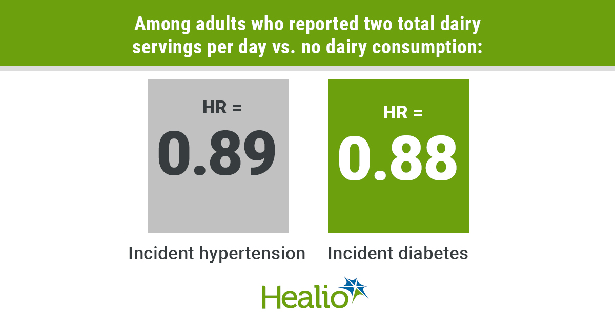
Within the cohort, mean total dairy intake was 179 g per day, with whole-fat dairy consumption almost twice as high as low-fat dairy consumption (mean, 124.6 g per day vs. 65 g per day). Researchers observed the highest intakes of total dairy in Europe, North America, the Middle East and South America; lowest intakes were observed in South Asia, China, Africa and Southeast Asia.
Among adults who reported two total dairy servings per day vs. no dairy consumption: H

Monday, June 1, 2020

The Effect of Priming on Outcomes of Task-Oriented Training for the Upper Extremity in Chronic Stroke: A Systematic Review and Meta-analysis

You can try to understand what the hell priming is by looking at these 602 thousand results on a Google search. I'm not doing it.

priming in stroke rehabilitation

The latest here:

The Effect of Priming on Outcomes of Task-Oriented Training for the Upper Extremity in Chronic Stroke: A Systematic Review and Meta-analysis

First Published May 26, 2020 Review Article


Background. 
Priming results in a type of implicit memory that prepares the brain for a more plastic response, thereby changing behavior. New evidence in neurorehabilitation points to the use of priming interventions to optimize functional gains of the upper extremity in poststroke individuals. Objective. To determine the effects of priming on task-oriented training on upper extremity outcomes (body function and activity) in chronic stroke.  
Methods.
The PubMed, CINAHL, Web of Science, EMBASE, and PEDro databases were searched in October 2019. Outcome data were pooled into categories of measures considering the International Classification Functional (ICF) classifications of body function and activity. Means and standard deviations for each group were used to determine group effect sizes by calculating mean differences (MDs) and 95% confidence intervals via a fixed effects model. Heterogeneity among the included studies for each factor evaluated was measured using the I2 statistic.  
Results. 
Thirty-six studies with 814 patients undergoing various types of task-oriented training were included in the analysis. Of these studies, 17 were associated with stimulation priming, 12 with sensory priming, 4 with movement priming, and 3 with action observation priming. Stimulation priming showed moderate-quality evidence of body function. Only the Wolf Motor Function Test (time) in the activity domain showed low-quality evidence. However, gains in motor function and in use of extremity members were measured by the Fugl-Meyer Assessment (UE-FMA). Regarding sensory priming, we found moderate-quality evidence and effect size for UE-FMA, corresponding to the body function domain (MD 4.77, 95% CI 3.25-6.29, Z = 6.15, P < .0001), and for the Action Research Arm Test, corresponding to the activity domain (MD 7.47, 95% CI 4.52-10.42, Z = 4.96, P < .0001). Despite the low-quality evidence, we found an effect size (MD 8.64, 95% CI 10.85-16.43, Z = 2.17, P = .003) in movement priming. Evidence for action observation priming was inconclusive. Conclusion.
 Combining priming and task-oriented training for the upper extremities of chronic stroke patients can be a promising intervention strategy. Studies that identify which priming techniques combined with task-oriented training for upper extremity function in chronic stroke yield effective outcomes in each ICF domain are needed and may be beneficial for the recovery of upper extremities poststroke.

A Novel Wearable Device for Motor Recovery of Hand Function in Chronic Stroke Survivors

Well then, write this up as a protocol and deliver it to ALL PREVIOUS STROKE SURVIVORS. 

6.4 million stroke survivors living in the U.S.

There are over 1.2 million stroke survivors in the UK.
 

A Novel Wearable Device for Motor Recovery of Hand Function in Chronic Stroke Survivors 

First Published May 26, 2020 Research Article


Background.
In monkey, reticulospinal connections to hand and forearm muscles are spontaneously strengthened following corticospinal lesions, likely contributing to recovery of function. In healthy humans, pairing auditory clicks with electrical stimulation of a muscle induces plastic changes in motor pathways (probably including the reticulospinal tract), with features reminiscent of spike-timing dependent plasticity. In this study, we tested whether pairing clicks with muscle stimulation could improve hand function in chronic stroke survivors.  
Methods.
Clicks were delivered via a miniature earpiece; transcutaneous electrical stimuli at motor threshold targeted forearm extensor muscles. A wearable electronic device (WD) allowed patients to receive stimulation at home while performing normal daily activities. A total of 95 patients >6 months poststroke were randomized to 3 groups: WD with shock paired 12 ms before click; WD with clicks and shocks delivered independently; standard care. Those allocated to the device used it for at least 4 h/d, every day for 4 weeks. Upper-limb function was assessed at baseline and weeks 2, 4, and 8 using the Action Research Arm Test (ARAT), which has 4 subdomains (Grasp, Grip, Pinch, and Gross). Results.
Severity across the 3 groups was comparable at baseline. Only the paired stimulation group showed significant improvement in total ARAT (median baseline: 7.5; week 8: 11.5; P = .019) and the Grasp subscore (median baseline: 1; week 8: 4; P = .004).  
Conclusion.
A wearable device delivering paired clicks and shocks over 4 weeks can produce a small but significant improvement in upper-limb function in stroke survivors.

Inhibition versus facilitation of contralesional motor cortices in stroke: Deriving a model to tailor brain stimulation

I don't understand. 

Inhibition versus facilitation of contralesional motor cortices in stroke: Deriving a model to tailor brain stimulation

 Vishwanath Sankarasubramanian a, 
Andre G. Machado b, 
Adriana B. Conforto c,d, 
Kelsey A. Potter-Baker a,
David A. Cunningham a,e, 
Nicole M. Varnerin a, 
Xiaofeng Wang f, 
Ken Sakaie g, 
Ela B. Plow a,b,h,

a Department of Biomedical Engineering, Lerner Research Institute, Cleveland Clinic, Cleveland, OH 44195, USA
b Center for Neurological Restoration, Neurological Institute, Cleveland Clinic, Cleveland, OH 44195, USA
c Neurology Clinical Division, Neurology Department, Hospital das Clinicas, São Paulo University, 05508-090 São Paulo, SP, Brazil
d Hospital Israelita Albert Einstein, 05652-900 São Paulo, SP, Brazil
e School of Biomedical Sciences, Kent State University, Kent, OH 44242, USA
f  Department of Quantitative Health Sciences, Lerner Research Institute, Cleveland Clinic, Cleveland, OH 44195, USA
g Department of Diagnostic Radiology, Imaging Institute, Cleveland Clinic, Cleveland, OH 44195, USA
h Department of Physical Medicine and Rehabilitation, Neurological Institute, Cleveland Clinic, Cleveland, OH 44195, USA
a r t i c l e i n f o
 Article history:
Accepted 14 March 2017Available online 21 March 2017
Keywords:
Diffusion tensor imagingMotor cortexPremotor cortexNeuronal plasticityRehabilitationStrokeTranscranial magnetic stimulation
h i g h l i g h t s

 Mildly affected chronic stroke patients improved upon paretic upper limb reaching with standardinhibitory 1Hz rTMS of contralesional motor cortex.

 Severely affected patients improved with a new method involving facilitatory 5Hz rTMS of contrale-sional dorsal premotor cortex.

 A preliminary cut-off level of damage/impairment separated responders to each form of stimulation.

a b s t r a c t

Objective:
 The standard approach to brain stimulation in stroke is based on the premise that ipsilesionalM1(iM1)is important for motor function of the paretic upper limb, while contralesional cortices compete with iM1. Therefore, the approach typically advocates facilitating iM1 and/or inhibiting contralesionalM1 (cM1). But, this approach fails to elicit much improvement in severely affected patients, who on account of extensive damage to ipsilesional pathways, cannot rely on iM1. These patients are believed to instead rely on the undamaged cortices, especially the contralesional dorsal premotor cortex(cPMd), for support of function of the paretic limb. Here, we tested for the first time whether facilitation of cPMd could improve paretic limb function in severely affected patients, and if a cut-off could be identified to separate responders to cPMd from responders to the standard approach to stimulation.
Methods:
 In a randomized, sham-controlled crossover study, fifteen patients received the standard approach of stimulation involving inhibition of cM1 and a new approach involving facilitation of cPMdusing repetitive transcranial magnetic stimulation (rTMS). Patients also received rTMS to control areas.At baseline, impairment [Upper Extremity Fugl-Meyer (UEFM PROXIMAL, max=36)] and damage to path-ways [fractional anisotropy (FA)] was measured. We measured changes in time to perform proximal paretic limb reaching, and neurophysiology using TMS.
Results:
 Facilitation of cPMd generated more improvement in severely affected patients, who had expe-rienced greater damage and impairment than a cut-off value of FA (0.5) and UEFM PROXIMAL
 (26–28). The standard approach instead generated more improvement in mildly affected patients. Responders to cPMd showed alleviation of interhemispheric competition imposed on iM1, while responders to the standard approach showed gains in ipsilesional excitability in association with improvement.
Conclusions:
A preliminary cut-off level of severity separated responders for standard approach vs. facilitation of cPMd

Microstroke Recovery Restores Blood Flow Before Brain Tissue

What this means is that there are NEVER strokes too good/small to treat. So your doctor has a lot of research to get accomplished to see what interventions are needed to recover from this problem.  COVID-19 has this problem on a massive scale, thrombi all over the place.

Your action point on this is to make damn sure you have all the classic symptoms, lost sensation, slurred speech, lack of movement on one side, dropped mouth. You don't want to be classified as too good to treat.

Every single stroke coming into your stroke hospital should have a protocol to follow. There is never a stroke that is too good to treat. You never magically recover from a stroke. Your doctor should never have to make a subjective decision. You have an objective damage diagnosis(The NIH Stroke Scale is not objective so we have a problem right from the start.). What should follow directly from that is a stroke protocol to remove the clot or stop the bleeding and then a protocol to stop the neuronal cascade of death or the hemorrhage cascade of death.  This is so fucking simple, why can't it be done?  Laziness? Incompetence? Or just don't care? No leadership? No strategy? Not my job?


Study: Even the smallest stroke can damage brain tissue, impair cognitive function

December 2012

Microstroke Recovery Restores Blood Flow Before Brain Tissue

Increased blood flow to the brain after a microscopic stroke doesn’t mean that part of the brain has recovered. At least not yet.
A study in Science Advances by Rice University neuroengineer Lan Luan and her colleagues used advanced neural monitoring technology to discover a significant disconnect between how long it takes blood flow and brain function to recover in the region of a microinfarct, a tiny stroke in tissue less than 1 millimeter in size.

The study led by Luan, a core faculty member of Rice’s Neuroengineering Initiative, shows “a pronounced neurovascular dissociation that occurs immediately after small-scale strokes, becomes the most severe a few days after, lasts into chronic periods and varies with the level of ischemia,” the researchers wrote.

The study in rodent models revealed the restoration of blood flow in the brain occurs first, followed by restoration of neuronal electrical activity. They observed that neuronal recovery could take weeks even for small strokes, and possibly longer for larger strokes.

The study required implants and instrumentation designed to monitor both blood flow and brain activity simultaneously before, during and after the onset of strokes.

“This started with the device,” said Luan, an assistant professor of electrical and computer engineering at Rice’s Brown School of Engineering, who developed a flexible neural electrode with co-author Chong Xie while both were at the University of Texas at Austin. “That was my transition from being trained as a material physicist to neuroengineering.

“As soon as we had the electrodes, I wanted to use them to understand brain functions and dysfunctions in a domain that was difficult to probe with previous technology,” she said. “The electrodes are extremely flexible and well suited to be combined with optical imaging in exactly the same brain regions.”

The electrodes were combined with optical lines able to measure blood flow by recording laser speckle patterns. The combined data, gathered for as long as eight weeks, gave the researchers an accurate comparison between blood flow and electrical activity.

“The strokes we focus on are so small that when they happen, it’s very hard to detect them from behavioral measures,” Luan said. “We would not easily see impairment in animal locomotion, meaning the animal could walk away just fine, from a lay perspective.

“The implications in humans are similar,” she said. “These microinfarcts can occur spontaneously, especially in aged populations. Because they’re so tiny, it’s not like you’re having a stroke. You will not notice it at all. But it has been long hypothesized that it’s related to vascular dementia.”

Luan said the neurological impact of individual microinfarcts is largely unknown. “That’s what motivated us to set up a series of experiments to really directly measure the impacts of those extremely small-scale injuries,” she said.

While the study would be hard to replicate in humans, the implications could improve diagnoses of patients who suffer microinfarcts.

“There are a lot of similarities in neurovascular coupling in rodent models and in humans,” she said. “What we observed in rodents likely has a similar signature in humans, and I hope that can be of use to clinicians.”

Luan said she is continuing her research at Rice, supported by a five-year R01 grant from the National Institute of Neurological Disorders and Stroke.

“We’re interested in knowing not just how a single microinfarct would alter neural activity but also, cumulatively, whether the effect of multiple microinfarcts that occur at different times would be stronger or weaker than the sum of the individuals,” she said.

Reference

He et al. (2020). Multimodal mapping of neural activity and cerebral blood flow reveals long-lasting neurovascular dissociations after small-scale strokes. Science Advances. DOI: https://doi.org/10.1126/sciadv.aba1933

This article has been republished from the following materials. Note: material may have been edited for length and content. For further information, please contact the cited source.

Mesenchymal Stem Cell–Derived Exosomes Improve Functional Recovery in Rats After Traumatic Brain Injury: A Dose-Response and Therapeutic Window Study


It is your doctor and hospital responsibility to get this followed up by testing for stroke AND THEN HUMAN PATIENTS.  Or you could let incompetence reign and allow them to DO NOTHING.  1, 4, 7 days after injury, so no need for speed, although 1 day was quite a bit better.

Mesenchymal Stem Cell–Derived Exosomes Improve Functional Recovery in Rats After Traumatic Brain Injury: A Dose-Response and Therapeutic Window Study

Here is how to act F.A.S.T. to prevent a deadly stroke

Completely and totally wrong title. F.A.S.T. does nothing to prevent a stroke, it is used to recognize a stroke is occurring and get yourself to a stroke hospital. 

Here is how to act F.A.S.T. to prevent a deadly stroke

Did you know that 80 percent of strokes are preventable?
According to the Centers for Disease Control and Prevention, stroke is the fifth leading cause of death in the United States and a major cause of long-term disability affecting about 795,000 individuals each year.
In observance of National Stroke Awareness Month, Dr. Sumathi Venkatesh, a Health Specialist with Texas A&M AgriLife Extension Service, offers some insight into stroke risk factors and prevention measures.
Regular blood supply is vital for a healthy brain — an important organ in our body that regulates our thoughts and actions. A stroke occurs when oxygen and blood flow to the brain is prevented either by a blood clot that is blocking an artery (ischemic stroke) or by the rupturing of a weakened blood vessel (hemorrhagic stroke).
When blood flow to a part of the brain is obstructed, it could affect specific functions corresponding to that region, resulting in cognitive impairment, physical disability, or loss of major body functions. In more severe cases such as a brain stem stroke, paralysis or even death could occur.
One of the main risk factors for a stroke, especially a hemorrhagic stroke, is high blood pressure. When blood pushes the arteries with high force for a prolonged period, it can damage the walls of the arteries, causing them to rupture. Likewise, high levels of blood cholesterol and blood sugar could damage the blood vessels.
Therefore, routine medical monitoring and the maintenance of an ideal weight through physical activity and a healthy diet are critical measures to prevent some of the most important stroke-related comorbidities such as obesity, hypertension, diabetes, and heart disease.
You could save a life by learning the F.A.S.T. warning signs of a stroke. If you experience or witness Face drooping, Arm weakness, and Slurred speech, then it is Time to call 911.
Other signs of concern are sudden numbness or weakness, confusion or trouble understanding, vision problems, loss of balance and coordination, and sudden severe headaches without any known problems.
If you notice one or more of the warning signs, seek immediate medical attention to detect the underlying causes and begin a treatment plan.
Visit the American Stroke Association at https://www.stroke.org/ for more information and resources on stroke awareness and management. For programs on healthy cooking, physical activity, heart health, diabetes, and blood pressure management, contact your Brown County Extension Office at 325-646-0386.
Sumathi Venkatesh is an Extension Program Specialist. She can reached by email at Sumathi.venkatesh@ag.tamu.edu

Feasibility of combining multi-channel functional neuromuscular stimulation with weight-supported treadmill training

You can ask your doctor where the protocol for this is from 16 years ago. 

Feasibility of combining multi-channel functional neuromuscular stimulation with weight-supported treadmill training


Janis J. Daly*, Robert L. Ruff
 Department of Neurology, Case Western Reserve University School of Medicine, United States Louis Stokes Cleveland VA Medical Center, Research Service 151-W 10701 E. Blvd., Cleveland, OH 44106, United States
Received 13 April 2004; received in revised form 30 June 2004; accepted 2 July 2004Available online 27 August 2004

Abstract

More than 3 million stroke survivors live with residual disabilities and mobility deficits even after rehabilitation. Therefore, it is important to develop new, more effective, gait training methods. The purpose of this study was twofold: (1) testing the feasibility of combining multi-channel functional neuromuscular stimulation (FNS) using intramuscular (IM) electrodes and body weight supported treadmill training(BWSTT) for gait training; and (2) documenting the potential gait practice advantages afforded by combining FNS-IM and BWSTT. Eight subjects with gait deficits in the chronic phase (
N
12 months) were enrolled. Intramuscular electrodes were placed in the paretic hip abductors,knee flexors and extensors, and ankle dorsiflexors,  plantar flexors, and evertors. Subjects were treated with exercise and gait training using the combined technologies 1 1/2 h/week, four times/week, for 12 weeks. Feasibility was tested according to performance of the technologies,clinician skill factors, and subject satisfaction. Impairment, function, and quality of life were measured. Provision of practice for eight gait characteristics was catalogued. We found the following results for the combined technologies: (1) the combined technologies were safe and feasible; (2) clinicians required five training sessions to reach proficiency; (3) subjects were satisfied; (4) there were significant gains inimpairment and functional measures; (5) a greater number of gait practice characteristics were provided with the combined technologies than with either alone.
D
 2004 Elsevier B.V. All rights reserved.