Use the labels in the right column to find what you want. Or you can go thru them one by one, there are only 33,849 posts. Searching is done in the search box in upper left corner. I blog on anything to do with stroke. DO NOT DO ANYTHING SUGGESTED HERE AS I AM NOT MEDICALLY TRAINED, YOUR DOCTOR IS, LISTEN TO THEM. BUT I BET THEY DON'T KNOW HOW TO GET YOU 100% RECOVERED. I DON'T EITHER BUT HAVE PLENTY OF QUESTIONS FOR YOUR DOCTOR TO ANSWER.
Changing stroke rehab and research worldwide now.Time is Brain!trillions and trillions of neuronsthatDIEeach day because there areNOeffective hyperacute therapies besides tPA(only 12% effective). I have 523 posts on hyperacute therapy, enough for researchers to spend decades proving them out. These are my personal ideas and blog on stroke rehabilitation and stroke research. Do not attempt any of these without checking with your medical provider. Unless you join me in agitating, when you need these therapies they won't be there.
What this blog is for:
My blog is not to help survivors recover, it is to have the 10 million yearly stroke survivors light fires underneath their doctors, stroke hospitals and stroke researchers to get stroke solved. 100% recovery. The stroke medical world is completely failing at that goal, they don't even have it as a goal. Shortly after getting out of the hospital and getting NO information on the process or protocols of stroke rehabilitation and recovery I started searching on the internet and found that no other survivor received useful information. This is an attempt to cover all stroke rehabilitation information that should be readily available to survivors so they can talk with informed knowledge to their medical staff. It lays out what needs to be done to get stroke survivors closer to 100% recovery. It's quite disgusting that this information is not available from every stroke association and doctors group.
Isn't your competent? doctor already testing you with the 2 Finger Test In Dementia? and you correctly declined it since your stroke deficits bias the test. Declining it proves you don't have dementia since you know of the bias.
A mail-in pinprick blood test accurately detects markers of Alzheimer’s disease
The test’s results mirrored those from standard blood and spinal fluid tests
The test evaluates dried blood samples dripped onto a card
TUESDAY, Jan. 6, 2026 (HealthDay News) — A mail-in blood test accurately detects markers linked to Alzheimer’s disease, potentially making the degenerative brain condition easier to diagnose and research, a new study says.
The
finger-prick test accurately measures blood levels of tau proteins,
glial fibrillary acidic proteins and neurofilament light fragments,
researchers reported Jan. 5 in the journal Nature Medicine.
All are hallmarks of brain damage associated with Alzheimer’s, researchers said.
The
test is still years away from clinical use among regular patients, but
currently could help fuel research efforts into Alzheimer’s, researchers
said.
“Ultimately, we are moving toward a pathway of treating
people for Alzheimer’s disease before symptoms emerge,” said senior
researcher Nicholas Ashton, senior director of Banner Health’s Fluid Biomarker Program in Sun City, Arizona.
“If
this trajectory continues, we will need innovative ways to identify
eligible individuals who are not routinely presenting in clinical
settings,” Ashton said in a news release. “This work represents one such
approach in that direction and further validation remains.”
For
the study, researchers analyzed dried blood samples provided by 337
people. Participants provided a few drops of blood, which were dried on a
card before lab analysis.
The lab work looked at blood levels of:
Phosphorylated tau proteins, which form toxic clumps in the brains of Alzheimer’s patients
Protein fragments of neurofilament light chain (NfL), which are released from damaged or dying brain cells
Glial Fibrillary Acidic Protein (GFAP), a protein produced by cells that heal and protect neurons in the brain and spinal cord
The pin-prick samples showed levels of tau proteins that closely matched those found in standard blood and spinal fluid tests.
The dried blood samples also accurately captured people’s levels of GFAP and NfL, researchers said.
This
simple technique could make it easier to perform large-scale
Alzheimer’s studies by enabling remote participation, researchers said.
“What excites me most is the democratization of biomarker research this enables,” Anne Corbett,
a professor of dementia research at the University of Exeter in the
U.K., said in a news release. “We’re moving toward a future where
anyone, anywhere, can contribute to advancing our understanding of brain
diseases. This isn’t just a technical advancement — it’s a paradigm
shift in how we conduct neuroscience research.”
This method also could help research efforts associated with other brain diseases, including Parkinson’s disease, multiple sclerosis, Amyotrophic Lateral Sclerosis (ALS) and brain injuries, researchers said.
So, you described something and incompetently provided NO EXACT NEXT STEPS TO SOLVE THE PROBLEM! In the business world that would be grounds for immediate firing. Aren't you glad you're in the absolutely incompetent stroke medical world?
The systematic review included 24 studies and 4,688 adults with hemorrhagic stroke.
There were no significant associations between headache and diabetes mellitus, hypertension, alcoholism or previous headache.
Nearly half of all patients with hemorrhagic stroke also experience
headache across its acute and chronic phases that could contribute to
long-term morbidity, according to a review published in Headache.
Yet the prevalence of headache varied substantially across populations and clinical settings, Bradley Ong, MD, adult neurology resident at Neurological Institute, Cleveland Clinic, and colleagues wrote.
Data derived from Ong B, et al. Headache. 2025; doi:10.1111/head.70008.
“In clinical practice, headaches after hemorrhagic stroke came up
quite often in our clinical practice, but they were rarely addressed,”
Ong told Healio.
Most treatment after stroke focuses on motor recovery and preventing
its recurrence, he said, with headache treated as an incidental or
transient symptom.
Bradley Ong
“When we looked at the literature, there was no clear, consolidated
picture of how common these headaches are or how long they last,” Ong
said. “That gap is what motivated this study.”
Ong and colleagues conducted a systematic review and meta-analysis
that included 24 peer-reviewed, observational studies from Medline,
Embase and CENTRAL with 4,688 adults (mean age, 56.9 years; weighted
mean, 58.2% women) with hemorrhagic stroke.
“The most striking finding was how common headaches are,” Ong said.
“Nearly half of patients with hemorrhagic stroke experience headache,
and more than one-third go on to have persistent headaches months or
years later.”
Overall, 46.1% (95% CI, 36.3% to 56.1%) of these patients experienced
headache after their stroke. Eleven studies (n = 2,481) found that
55.9% of patients (95% CI, 41.1% to 70.1%) experienced acute headache.
Thirteen studies (n = 2,207) found that 36.7% of patients (95% CI, 25.6%
to 48.5%) had persistent headache.
“This challenges the assumption that headache is mainly an ‘acute’ symptom, especially in hemorrhagic stroke,” Ong said.
Specific prevalences of headache included 58.3% (95% CI, 44.4% to
71.6%) for those with subarachnoid hemorrhage (SAH) and 36.1% (95% CI,
26.7% to 46%) for those with intracerebral hemorrhage (ICH).
Prevalence of severe headaches included 42.7% (95% CI, 15.8% to
72.1%) among those whose headaches were acute/subacute and 14.3% (95%
CI, 10.4% to 18.7%) among those whose headaches were persistent.
With an overall I2 of 96.7%, the researchers said their findings
indicated substantial heterogeneity in these pooled prevalence
estimates, with no statistically significant differences based on study
design, population, geography, Human Developmental Index or risk for
bias.
Further, Ong and colleagues said there were no significant
associations between risk for headache and female sex, nor were there
any significant associations with history of diabetes mellitus,
hypertension, alcoholism or previous headache.
“Another important finding was that headache at stroke onset strongly
predicted chronic headache, which gives us an early clinical signal we
can actually act on,” Ong said.
The odds ratio for post-stroke headache among patients with headache
at stroke onset was 1.7 (OR = 1.7; 95% CI, 1.4-2.05). Also, the odds
ratio for post-stroke headache among patients with lobar ICH was 1.93
(95% CI, 1.08-3.44).
There were no significant associations between headache risk and
cortical ICH or delayed cerebral ischemia. Also, there were no
significant associations between headache risk and the presence of an
anterior circulation aneurysm among patients with SAH.
Patients with atrial fibrillation had less risk for headache (OR =
0.59; 95% CI, 0.37-0.95), which the researchers attributed to
differences in stroke severity and symptom reporting and not to any
direct protective effect.
Noting that the prevalence of headache among patients with
hemorrhagic stroke exceeds the prevalence of other primary headache
disorders among the general population, with substantial variations by
population and clinical settings, the researchers called these headaches
“common” as well as “persistent and disabling.”
Ong said that clinicians can use these findings to improve outcomes for patients with stroke.
“Clinicians should ask about headache routinely, both in the hospital
and during follow-up. Headache should be treated as a meaningful
post-stroke complication,” he said.
“Patients who report headache early may benefit from closer
monitoring and earlier referral to headache care,” he continued. “Even
simple steps like education and avoiding unnecessary opioid exposure can
improve quality of life.
Looking ahead, the researchers called for studies with standardized
diagnostic criteria, clearly defined populations and detailed headache
characteristics into protective therapies and secondary prevention
strategies.
“The next step is prospective, longitudinal studies using
standardized headache definitions and patient-reported outcomes,” Ong
said.
“We also need clinical trials focused specifically on post-stroke
headache treatment, rather than extrapolating from primary headache
disorders,” he added. “Ultimately, the goal is to integrate headache
care into routine stroke recovery.”
Women experienced worse functional outcomes up to 12 months after a first ischemic stroke compared to men, despite their improvements in activities of daily living (ADLs) from 3 to 6 months, a new study showed. Although both sexes showed improvement in neurologic outcomes, only men had cognitive gains within a year following the stroke.
METHODOLOGY:
Researchers analyzed data from the Brain Attack Surveillance in Corpus Christi Project for more than 1000 patients in Texas with first-ever ischemic stroke (mean age, 66 years; 52% men; 58% Mexican American; 34% non-Hispanic White American) between 2014 and 2019.
Participants completed structured interviews at baseline after the onset of stroke (median time post-stroke, 7 days) and follow-up assessments at 3, 6, and 12 months, which were conducted in English or Spanish.
Researchers analyzed functional difficulties with ADL/instrumental activities of daily living (IADL), neurologic outcomes with the National Institutes of Health Stroke Scale (NIHSS), and cognition with a modified Mini-Mental State Examination (3MSE).
TAKEAWAY:
Women had worse functional status compared to men, as shown with higher adjusted mean ADL/ IADL scores at 3, 6, and 12 months (mean difference [MD] for all time points, 0.1).These sex differences were observed among patients with less severe stroke at baseline (initial NIHSS score of 5 or less) but not among those with moderate or severe strokes.
Women had small but significant overall functional improvement from 3 to 12 months (adjusted MD in ADL/ IADL scores, -0.08), whereas scores for men did not change significantly.
Neurologic outcomes improved in both sexes from 3 to 12 months, with women having a greater decrease in NIHSS scores compared to men (adjusted MDs, -0.8 vs -0.2, respectively). Only men showed cognitive improvement (adjusted MD in 3MSE scores, 0.97).
IN PRACTICE:
"Since many people live with physical, cognitive and emotional challenges after stroke, it is important to find ways to improve recovery(Your tyranny of low expectations is showing! SURVIVORS WANT 100% RECOVERY! Why aren't you delivering that?
Laziness? Incompetence? Or just don't care? NO leadership? NO strategy? Not my job? Not my Problem!
). Our study provides a better understanding of sex differences during stroke recovery," lead author Chen Chen, PhD, University of Michigan School of Public Health, Ann Arbor, said in a press release.
My right carotid artery was at 80% blockage at time of stroke and then thankfully fully closed up 3 years later. Remained closed for 10 years
and I cognitively functioned quite well with no episodes of
fainting or poor executive functioning. Eventually collaterals grew around the blockage. Since my Circle of Willis is complete, I still had 3 fully functioning arteries supplying blood to the brain, obviously enough to keep me highly functioning. I'm glad that my doctors were so incompetent they never found that 80% blockage, otherwise they probably would have insisted I undergo either stenting or endarterectomy, both of which they couldn't guarantee no problems. And I didn't find out about those problems until years later researching for this blog.
Increased severity of carotid stenosis is significantly associated with poorer executive function and slower processing speed, independent of traditional vascular risk factors and history of stroke. Patients with greater carotid stenosis demonstrate significantly worse processing speed and executive function performance compared with those who have less severe disease, according to results of a study published in Alzheimer’s & DementiaAsymptomatic extracranial carotid atherosclerotic disease (aECAD) has been associated with a 22% increased risk for Alzheimer disease, but previous studies evaluating the effect of aECAD on cognitive outcomes have been limited by methodological biases.To better characterize early cognitive changes associated with aECAD, researchers from the University of Arizona examined data from the Carotid and Mind (CAM) clinical study. The researchers evaluated data from the first 182 individuals enrolled in CAM between 2022 and 2024, of whom 167 were included in the final analysis. Participants were aged 50 to 85 years and were recruited from vascular surgery and cardiology clinics. All participants underwent magnetic resonance imaging (MRI), carotid stenosis assessment using North American Symptomatic Carotid Endarterectomy Trial criteria, and comprehensive neurocognitive testing. The researchers defined aECAD as greater than 50% carotid stenosis without a history of stroke or transient ischemic attack in the previous 6 months. This work builds momentum for clinical management changes and future studies with a long-term goal of dementia prevention.
The study population had a mean (SD) age of 72 (7) years, 57% were men, 96% were White, 49% had 13 to 16 years of education, 36% had less than 50% carotid stenosis, and 26% carried the apolipoprotein E (APOE) ε4 allele.
After adjusting for age, sex, race, and ethnicity, increasing carotid stenosis severity was significantly associated with slower processing speed (adjusted b [ab], -0.20; 95% CI, -0.34 to -0.07; P =.004) and poorer executive function (ab, -0.19; 95% CI, -0.33 to -0.05; P =.009).
At the individual test level, greater carotid stenosis was associated with worse performance on the Wechsler Adult Intelligence Scale (ab, -0.26; 95% CI, -0.40 to -0.12; P <.001), Stroop Color-Word Interference (ab, -0.21; 95% CI, -0.35 to -0.07; P =.003), and Trail Making Test part A (ab, -0.21; 95% CI, -0.36 to -0.06; P =.007). Using these 3 tests, a composite Carotid Cognitive Index (CCI) was formulated.
The CCI was also inversely related with phosphorylated tau217 (p-tau217; r, -0.33; P <.001). This relationship was independent of white matter lesion volume (ab, -0.27; P =.006); demographic factors of age, gender, race, ethnicity, and education (ab, -0.24; P =.005); vascular risk factors (ab, -0.22; P =.01); APOE ε4 status (ab, -0.21; P =.01); and vascular disease history (ab, -0.21; P =.02). Higher p-tau217 levels were independently associated with APOE ε4 carriership (b, 0.57; P <.001).
Study limitations include a lack of racial and ethnic diversity.
The study authors concluded, “Although patients with carotid disease are not regularly evaluated clinically for cognitive impairment or dementia, this work builds momentum for clinical management changes and future studies with a long-term goal of dementia prevention.”
Disclosures: One study author declared affiliations with biotech, pharmaceutical, and/or device companies. Please see the original reference for a full list of authors’ disclosures.
So, we now can exactly identify the signals between neurons that tell one neuron to drop their use and take on a neighboring neuron's use! That could then make neuroplasticity repeatable on demand. If your doctor and hospital aren't pushing for further research on this; THEY ARE COMPLETELY FUCKING INCOMPENT!
Do you prefer your doctor, hospital and board of director's incompetence NOT KNOWING? OR NOT DOING?
Researchers validated a wireless EEG method to measure neuroplasticity, offering a non-invasive way to track how the brain responds to new treatments.
The approach could provide an objective readout of brain response to therapies for psychiatric and neurodegenerative conditions.
Neuroplasticity is the brain’s ability to reorganise its connections after experience, injury or disease, for example by forming new pathways to aid recovery.
The research, conducted by Cumulus Neuroscience, used visual evoked potential (VEP) modulation to mark neuroplasticity.
VEPs are brain signals produced in response to visual patterns.
The method was tested in two US-based clinical trials involving 50 healthy participants.
Brian Murphy, co-founder and chief scientific officer at Cumulus Neuroscience, said: “This study demonstrates that it is possible to measure neuroplasticity reliably in real-world clinical environments using a non-invasive, low-burden, scalable approach.
“Our findings open the door to incorporating objective plasticity measures into early-phase CNS drug development, which could accelerate progress in treating neuropsychiatric and neurodegenerative disorders.”
Conventional EEG assessments are time-consuming and burdensome, which has limited their use in trials.
The new approach uses a quick set-up dry EEG headset with frequency-domain analyses to extract precise measures.
Dr David Walling, chief clinical officer for CenExel-CNS and principal investigator for the study, said: “The ability to capture valid VEPs in clinical studies with easy-to-deploy technology and short sessions has the potential to transform how we assess the efficacy of new therapies in this space.
“Historically, we have not had a way to directly measure neuroplasticity outside of animal models which require invasive techniques.
“We can now integrate non-invasive VEP measures into clinical study workflows, providing sponsors with objective biomarkers of target engagement and treatment effects early in development.”
The specialist shortage is not the problem! LACK OF 100% RECOVERY PROTOCOLS IS THE PROBLEM! Are you that blitheringly stupid? These specialists have nothing that will get survivors 100% recovered! That is the only goal in stroke for survivors!
Thousands of people in the UK are at risk death or severe disability due to a lack of NHS stroke specialists, senior doctors have warned.
A chronic shortage of stroke consultants means patients face delays to clot-busting drugs and surgery, with horrendous consequences, they said.
Prof David Werring, past president of the British and Irish Association of Stroke Physicians (BIASP), said: “People are either dying or living with disability unnecessarily because they’re not getting the correct evaluation and treatment by the right expert at the right time.”
About 100,000 people across the UK have a stroke each year.
Between 10,000 and 20,000 of them died or sustained a serious disability because of treatment delays linked to staff shortages, said Dr Sanjeev Nayak, a senior stroke specialist at Royal Stoke hospital.
“It is heartbreaking to see the real and avoidable impact that workforce shortages have on patient outcomes.
In my experience workforce shortages directly lead to avoidable disability and, in some cases, avoidable death,” said Nayak, a consultant interventional radiologist.
“It is reasonable to estimate that around 10-20 per cent of stroke patients each year are left avoidably dead or more disabled than they otherwise would have been because of delays in the system.
“Those delays are multifactorial, but workforce shortages are a major, repeatedly identified contributor.”
The most recent Sentinel Stroke national audit programme report found it took four hours and 11 minutes to get someone who had had a stroke to hospital in 2024-25, more than 90 minutes longer than a decade ago.
Just 46.5 per cent of stroke patients last year were admitted to a specialist stroke unit within four hours of arrival, down more than 10 percentage points on a decade ago.
New research by BIASP surveyed 100 hospitals in England providing acute stroke care and found:
70 per cent of stroke units are short of at least one consultant, and many are two down
53 of 84 hospitals that responded had vacancies for a total of 96 consultants
10 per cent of the NHS’s 423 permanent consultants are due to retire in the next five years
Dr Louise Shaw, BIASP’s current president, said some smaller hospitals did not have a senior specialist on duty around the clock.
“That’s very unacceptable,” Shaw said.
“All patients admitted to a hospital with an acute stroke should have access to an immediate stroke consultant opinion and advice on their care. And at the moment that’s not available.”
Key treatments such as thrombolysis, which uses drugs to dissolve a clot, and mechanical thrombectomy, surgery to remove a blood clot from the brain, are extremely time-critical.
Nayak said: “When services are understaffed, patients miss treatment windows altogether or are treated too late, resulting in far worse neurological outcomes that could have been prevented.
“Delays in specialist assessment or transfer to a thrombectomy centre can mean the difference between independent recovery and devastating, lifelong disability, or not surviving at all.”
The Stroke Association said patients were being denied “time-critical, life-changing” treatment because of staff shortages.
The situation threatens Labour’s pledge to cut deaths from heart disease and stroke by 25 per cent by 2035.
A Department of Health and Social Care spokesperson said the NHS had 7,000 more doctors than the same time last year. “And our upcoming workforce plan will set out how we ensure the NHS has the right people in the right places, with the right skills to care for patients when they need it.”
The number of people in the UK who have a stroke is expected to rise from 100,000 to 151,000 a year by 2035, according to Stroke Association analysis.
Have your competent? doctor verify that your body clock is still functioning post stroke and have EXACT PROTOCOLS to fix it if needed! A competent doctor would have these interventions already on hand! Your doctor should have the Body Clock test already in hand, IF COMPETENT AT ALL!
Small asymmetries (leg-length differences, collapsed arches, shoulders) can snowball over time, changing how you move and loading knees, hips, and low back. Super Age Advisor Michelle MacDonald says you should book a quick movement screen or gait/posture check with a trainer or physical therapist, (My physical therapists DID NOTHING to objectively identify my walking problems and correct them. As a result, my left bad knee and my good right knee are slowly being destroyed along with my left hip.)
“Check yourself before you wreck yourself,” she says. “Check your leg length. Look at your pelvis. Look at your foot pressure.” Getting a clear view of how your body is moving and how to correct imbalances will help you keep moving, she says. Do a monthly self-audit: stand barefoot and check arches, balance on one leg for 30 seconds, film a squat from the side, and note what drifts. Adjust your plan before pain shows up. Here’s how to build strength that lasts.
Puncture to recanalization time was significantly associated with discharge outcome, wherein each 15-minute increase was linked to a higher likelihood of death or hospice discharge. Patients with acute ischemic stroke who experience longer puncture to recanalization (PTR) times during thrombectomy have worse outcomes and greater acute care costs. These findings were published in Stroke: Vascular and Interventional Neurology. Researchers conducted a retrospective analysis of 721 patients who underwent mechanical thrombectomy for large vessel occlusion at a high-volume neuroendovascular practice between January 2011 and June 2020. The researchers examined the effect of PTR time on immediate poststroke disposition and associated costs. The analysis included patients with successful reperfusion (modified Thrombolysis in Cerebral Infarction≥2B) and excluded those with missing data or those with extreme time outliers. Patients were categorized into 4 discharge groups: transfer to acute or subacute rehabilitation, home or home with physical therapy (PT), in-hospital death or hospice, and long-term care placement. The researchers employed a multinomial logistic regression model to assess how incremental increases in PTR time influenced these outcomes. Reducing procedural time should be prioritized not only to improve individual patient outcomes but also to support institutional cost efficiency and inform national stroke care policies. The median PTR time was 34.7 minutes, and the cohort was 50% women, with a mean age of 67.0 years. Most patients (95%) presented with anterior circulation occlusions. Common comorbidities included hypertension (78.8%), hyperlipidemia (51.5%), atrial fibrillation (38.4%), and diabetes (29.3%). At discharge, 54.2% of patients were transferred to rehabilitation, 21.9% were discharged home or home with PT, 18.7% died or were transitioned to hospice, and 5.1% required long-term care placement. There was a significant association between PTR and discharge disposition (P= .003). Each 15-minute increase in PTR was linked to a 2% to 4.6% higher likelihood of death or hospice discharge and a 1.5% to 2.5% lower likelihood of being discharged home or home with PT. In adjusted analyses, longer PTR was independently associated with greater odds of death or hospice disposition (odds ratio [OR], 1.020; 95% CI, 1.008-1.032; From an economic standpoint, every 15-minute increase in PTR was associated with an average increase in direct acute care costs of $190.04 per stroke episode (95% CI, $184.74-$196.20;P<.001). When extrapolated to an estimated 39,000 annual thrombectomies nationwide, the researchers estimated a $7.4 million annual increase in acute stroke care costs for each 15-minute increase in mean PTR (95% CI, $7.2–$7.7 million). Study limitations include a single-center design, exclusion of patients with incomplete data, and reliance on modeled cost estimates that did not capture postacute or indirect costs. “These results suggest that procedural speed remains a crucial determinant of clinical outcome, and reducing procedural time should be prioritized not only to improve individual patient outcomes but also to support institutional cost efficiency and inform national stroke care policies,” the study authors concluded. Disclosures: This research was supported by Microvention. Multiple study authors declared affiliations with biotech, pharmaceutical, and/or device companies. Please see the original reference for a full list of disclosures.
The human brain, with its intricate neural networks, continues to fascinate and baffle researchers, particularly in the realm of recovery following adverse events such as strokes. A recent scoping review highlighted in BMC Neuroscience delves into the role of the contralesional primary motor cortex in aiding upper limb recovery after a stroke. This development is particularly pivotal, given that strokes significantly impair motor functions, leading to long-term disability in numerous individuals. Studying the contralesional primary motor cortex offers a new lens through which we can understand post-stroke rehabilitation.
Strokes occur when the blood supply to part of the brain is interrupted or reduced, preventing brain tissue from getting oxygen and nutrients. The consequences can be devastating, often resulting in the loss of motor functions, particularly in the limbs. Upper limb recovery becomes a critical goal in rehabilitation, as it heavily influences a person’s ability to carry out daily activities and ultimately impacts their quality of life. The contralesional primary motor cortex—the part of the brain that processes motor functions for the limbs opposite the side of body affected—holds promise in facilitating recovery from such debilitating conditions.
Researchers Hernan Fregni, Pattharawadee Suputtitada, and Victor Costa conducted this comprehensive review as part of their efforts to elucidate how the contralesional primary motor cortex contributes to functional recovery. Through the meticulous application of PRISMA-ScR guidelines—an established framework ensuring transparency and reproducibility in scoping reviews—they meticulously sifted through varied studies to extract pertinent findings. The synthesis of these studies offers critical insights into how contralesional regions can be harnessed to enhance rehabilitation strategies.
One of the most striking findings from this review is how the brain exhibits remarkable plasticity. Even after significant injury, the brain can adapt and reorganize itself to compensate for lost functions. This plasticity is particularly pronounced in the contralesional hemisphere, which, following the injury of the ipsilesional hemisphere—typically where the stroke occurs—can take over some motor tasks. This neural adaptation widens the horizon for therapeutic interventions, suggesting that targeted stimulation of the contralesional motor cortex could engender recovery pathways that were previously thought unattainable.
Moreover, the review meticulously highlights various therapeutic strategies aiming to exploit this contralesional connectivity. Rehabilitation techniques including transcranial magnetic stimulation (TMS) have emerged as frontrunners in modulating activity within the contralesional primary motor cortex. By using non-invasive brain stimulation techniques, therapists can enhance excitability in this area, thereby improving motor function. Such efficient stimulation protocols could provide a similar stimulus to the impaired areas of the brain, catalyzing the recovery process.
Additionally, the involvement of augmented feedback mechanisms in upper limb rehabilitation is worth noting. Studies included in the review reflect how feedback mechanisms, whether intrinsic or extrinsic, can significantly influence motor relearning and recovery. The contralesional primary motor cortex, capable of modifying its functional representation based on feedback from the environment, indicates that we might not only be able to recover lost motor functions but also optimize existing ones. Harnessing this feedback in therapeutic practices could lead to profound improvements in recovery trajectories.
Interestingly, the review also emphasizes the role of engaging patients in active rehabilitation practices. Motor imagery and mental practice, where patients visualize themselves performing movements, have been shown to engage the contralesional motor cortex, further underscoring the power of mental processes in recovery. These findings support a broader paradigm shift where cognitive engagement becomes a central tenet in rehabilitation, integrating both mental and physical stages in recovery protocols.
The clinical implications of this research are profound. With a clearer understanding of how the contralesional primary motor cortex facilitates recovery, therapists can tailor individualized rehabilitation protocols. These tailored approaches pivot from traditional methods, incorporating new dimensions such as virtual reality or gamified platforms that directly stimulate contralesional pathways, which can engage patients more effectively and promote better recovery outcomes.
A dynamic interplay between clinical techniques and neuroscience is evident, where researchers and practicing clinicians must collaborate closely. This scoping review nostalgically harkens to previous studies that highlighted the potential of the contralesional cortex, yet it provides a panoptic view of contemporary knowledge and outlines future directions for research. It poses essential questions regarding optimal stimulation parameters and the timing of interventions that are ripe for exploration.
As we look to the future, this research serves as an impetus for further studies aimed at unlocking the full potential of the contralesional primary motor cortex. Larger randomized controlled trials will likely refine the role of various rehabilitation strategies in exploiting this brain area effectively. The collective goal remains to enhance the quality of recovery for stroke patients, ultimately helping them regain independence and improve their quality of life.
In conclusion, the exploration of the contralesional primary motor cortex in relation to recovery from stroke represents an exciting frontier in neuroscience. The implications not only provide hope for individuals affected by strokes but also highlight a crucial intersection of clinical application and theory. Such advancements reinforce the necessity for continuous research and innovation, ensuring that recovery techniques remain ahead of the curve, aligning with our growing understanding of neuroplasticity and motor learning processes.
Subject of Research: The role of the contralesional primary motor cortex in upper limb recovery after stroke.
Article Title: The role of the contralesional primary motor cortex in upper limb recovery after stroke: a scoping review following PRISMA-ScR guidelines.
Article References: Suputtitada, P., Costa, V. & Fregni, F. The role of the contralesional primary motor cortex in upper limb recovery after stroke: a scoping review following PRISMA-ScR guidelines. BMC Neurosci 26, 31 (2025). https://doi.org/10.1186/s12868-025-00950-y
Image Credits: AI Generated
DOI: https://doi.org/10.1186/s12868-025-00950-y
Keywords: Stroke recovery, contralesional primary motor cortex, motor cortex plasticity, rehabilitation techniques, transcranial magnetic stimulation, motor imagery, neuroplasticity.
Tags: BMC Neuroscience scoping reviewbrain plasticity in stroke recoverycontralesional primary motor cortexenhancing quality of life post-strokeimpact of stroke on daily activitiesmotor function impairment due to strokeneural networks in stroke recoverypost-stroke rehabilitation strategiesrole of motor cortex in recoverystroke recovery mechanismsstroke-related long-term disabilitiesupper limb rehabilitation after stroke
Welcome to Impact Factor, your weekly dose of commentary on a new medical study. I’m Dr F. Perry Wilson from the Yale School of Medicine.
I was lucky enough to have lived in Paris as a young man, spending a year between college and medical school working as a singing waiter in a restaurant called the Hollywood Savoy in the 2nd arrondissement. It’s still open, but it looks like the waiters don’t sing anymore. Tant pis.
In any case, living on a French waiter’s salary back then wasn’t easy. There were no tips. So I subsisted, as I imagined Hemingway did in his Moveable Feast days, on a diet composed almost entirely of wine, baguettes, and goat cheese. This might not have been the best for my GI health, but according to a new study at least, all that cheese intake might serve to protect my brain in the long term.
That’s right. This week, nutritional epidemiology rears its delicious head again as we discuss whether high-fat-cheese-eating prevents dementia.
The public gets understandably frustrated with new studies that suggest that a particular food or ingredient is healthy, or unhealthy, or should be avoided at all costs. And this paper will have the same effect. Think of all the “balanced diet” advice you’ve ever heard: Eat a mix of fruits and vegetables, grains and low-fat dairy — you know: yogurt, cottage cheese, and so on.
And yet here we have a study, “High- and Low-Fat Dairy Consumption and Long-Term Risk of Dementia,” appearing in the journal Neurology, that turns that dairy part on its head. It’s the high-fat cheese that is good for you after all. Throw out your part-skim mozzarella and pass the Stilton. For someone like me, this sounds too gouda to be true.
Let me walk you through how the study worked.
Researchers used the Malmö Diet and Cancer cohort, a long-running population cohort study out of Sweden. From 1991 to 1996, as new participants were brought into the study, a fairly detailed dietary profile was created. This included data from food diaries, a dietary interview, and, of course, my old nemesis: the food frequency questionnaire, which I take to task in my book about medical research. Still, let’s take it on faith that, broadly, researchers captured how much dairy, and how much high-fat dairy, people were eating.
After the initial collection of data, the participants were followed for around 25 years through the national electronic health record system of Sweden. Researchers used those records to identify new cases of dementia and to subset those cases into vascular causes of dementia and Alzheimer's disease. These diagnoses were based on administrative codes but validated in a subset with direct review, which revealed pretty high fidelity.
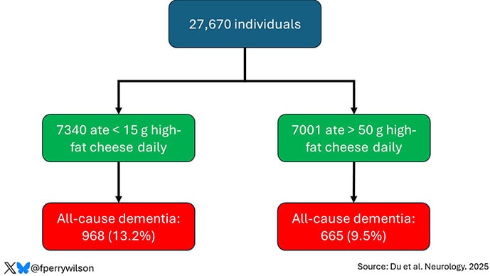
All told, 27,670 individuals were analyzed. Of those, 3208 (11.6%) developed dementia over those 25 years of follow-up.
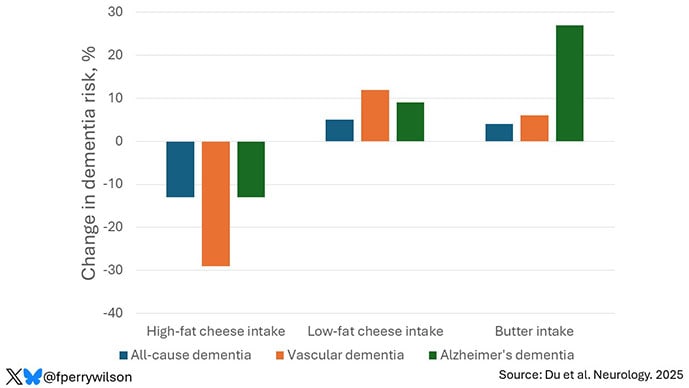
The people who ate more high-fat cheese were less likely to be in that 11.6% group. According to the study, they had about a 13% lower risk for all-cause dementia and a 29% lower risk for dementia than those who didn’t eat much high-fat cheese. There was no significant relationship between cheese intake and Alzheimer's disease.
OK. The first thing we need to remember when we are thinking about a nutrition study is that food is complicated. Cheese, even high-fat cheese, is not one thing, one chemical, the way a medication or vitamin or even amino acid is. It’s an incredibly complex mixture of fats, proteins, enzymes, bacterial cultures, molds, salt, vitamins, and minerals. Finding a signal of benefit from a foodstuff like cheese is problematic, because even if it is real, we may never discover what thing in the cheese is driving that benefit.
The study looked at other dairy intake as well and, frankly, that made me more confused about these cheese results. If it’s the protein content in the cheese that is beneficial, then we should see a protective signal from low-fat cheese intake, but we don’t. If it’s the fat that is protective, you’d think we’d see a protective signal from butter, which is about 80% milkfat, but we don’t. So, we’re left having to wave our hands about some complex interaction of various brain-protecting substances that exist only in high-fat cheeses. Possible? Maybe. Likely? I don’t think so.
But beyond that, food is a cultural phenomenon. What you eat varies dramatically based on who you are and, dare I say it, what you can afford. It has not escaped my notice that high-fat cheeses — your mascarpones and aged cheddars — tend to be more expensive than their lower-fat counterparts. They therefore may be consumed by people of higher socioeconomic status.
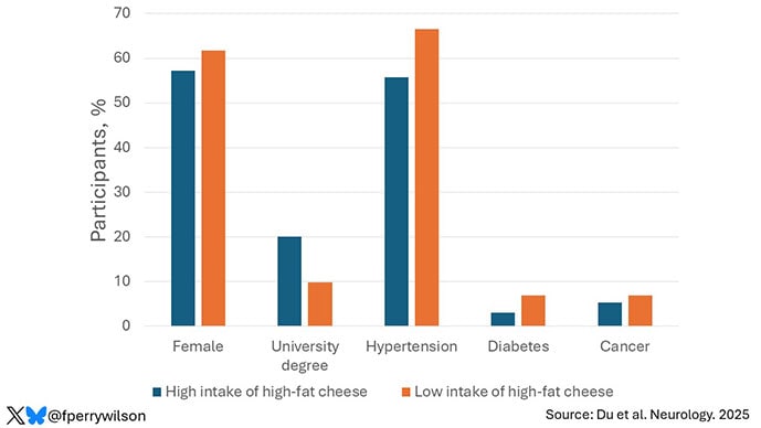
There’s direct support for this hypothesis in the study, by the way. High-fat cheese eaters were younger, less likely to be female, had a lower BMI, and were twice as likely to have a university degree. They were also less likely to have hypertension, diabetes, or a cancer history.
The authors adjusted for these factors as best they could, of course, but adjustment is virtually never perfect and relies on how precisely you can measure the cultural factors that might associate with high-fat cheese intake. That’s not easy. So, I am always inherently skeptical of findings of health benefits of foods that are a little more costly than their alternatives. I always believed that red wine was firmly in this category, and although no one has studied it, I suspect that those who eat more foie gras also have better health outcomes.
One mechanism that I do find plausible for the protective effect of high-fat cheese is via food substitution. The idea is that when you eat certain high-fat foods, you may eat less of other foods that scratch that same itch; think red meat for the umami-ness of it all, or processed meats for the saltiness. The authors analyzed the data with this in mind and indeed showed that replacing the high-fat cheeses with some of these other foods increased dementia risk. This suggests that there may not be anything magic in cheese itself; instead, cheese helps us resist the temptations of the foods that are really bad for our brains. Could be.
Still, most people who see headlines about this study are likely to end up confused. What am I supposed to do: High-fat dairy or low-fat dairy? Gruyère or ricotta? We are all looking for that “one simple thing” we can do to change the trajectory of our health. But the truth is that there is no “one simple thing.” No one food will prevent dementia. No one exercise will give you six-pack abs. No one supplement will keep you young and full of energy as time goes on. To really change the trajectory of our health, we have to be willing to change our lifestyles in pretty substantial ways. That’s never going to be one simple thing, it won’t be easy, and it definitely won’t be delicious.
With that said, though cheeses may not protect your brain over time, many will absolutely delight your senses in the moment. And if this paper, or my commentary on it, gives you a bit more permission to enjoy that baguette and chèvre, well, I think you should do so. Bon appétit.
F. Perry Wilson, MD, MSCE, is an associate professor of medicine and public health and director of Yale’s Clinical and Translational Research Accelerator. His science communication work can be found in the Huffington Post, on NPR, and here on Medscape. He posts at@fperrywilsonand his book, How Medicine Works and When It Doesn’t, is available now.
Sponsor National Institute of Neurological Disorders and Stroke (NINDS) Information Provided by Lawrence L Latour, Ph.D. ,Study Start (Actual) 2001-01-26 Enrollment (Estimated) 4000 Study Type Observational Last Update Posted 2025-12-19
Study Overview The purpose of this study is to learn more about stroke and obtain information that may serve as the basis for future investigations. It will 1) establish a registry of patients with cerebrovascular disease (stroke); 2) characterize the natural history of acute stroke and transient ischemic attacks (TIA)-an interruption of blood flow to the brain that causes stroke symptoms for a short period of time); and 3) evaluate the data to generate ideas for future studies.(ARE YOU THAT FUCKING INCOMPETENT YOU HAVEN'T BEEN KEEPING TRACK OF RESEARCH THAT LOOKS PROMISING?)
(Here for your perusal: See how fucking easy it is, if a stroke survivor can do that, WHY THE FUCK DO YOU NEED THESE Ph. D's? Just start running research, needing to do this research just proves your COMPLETE FUCKING INCOMPETENCE! I'd have you all keel hauled!)
Patients 18 years of age or older with suspected acute stroke or TIA may be eligible for this study. Subjects will be recruited from patients who present with stroke at the emergency department of Suburban Hospital in Bethesda, Maryland. The study will gather data collected from diagnostic and laboratory tests the patient undergoes as part of standard medical care, including findings of medical and neurological examinations and other tests. In addition, studies will be done for research purposes only to gather data about stroke and TIA. These may include the following: * Blood and urine tests not more than 2 tablespoons of blood will be drawn for various tests. * Electrocardiogram (EKG) (heart tracing)-electrodes placed on the chest wall detect the heartbeat and heart rhythm. * Computed tomography (CT) scan of the head-specialized X-rays are used to obtain images of the brain. * Magnetic resonance imaging (MRI) of the brain-a strong magnetic field and radio waves are used to produce images that provide information about the brain tissue and blood vessels. * Transcranial Doppler (TCD)-sound waves are used to image the arteries of the brain and neck. * Echocardiogram-sound waves are used to image the heart and evaluate heart function. Patients may be asked to return to Suburban Hospital for follow-up testing in 1, 3, and/or 12 months, when some of these tests may be repeated to assess changes over time ... To learn more, visitClinicalTrials.gov
Contacts and Locations Contact Information Study Contact Name:Nicole L Peterkin Email nicole.peterkin@nih.gov Study Contact Backup Name Lawrence L Latour, Ph.D. Phone (301) 435-2395 Email latourl@ninds.nih.gov United States Locations District of Columbia Washington D.C., District of Columbia, United States 20010 Medstar Washington Hospital Center Maryland Bethesda, Maryland, United States 20814 Suburban Hospital - Johns Hopkins Medicine Bethesda, Maryland, United States 20892 National Institutes of Health Clinical Center Participation Criteria INCLUSION CRITERIA: In order to be eligible to participate in this study, an individual must meet all of the following criteria: Aged >=18 Presented to participating study site (ED, ICU, or inpatient unit) with or at risk of acute stroke, TIA, or other disturbances of cerebrovascular circulation7nbsp;An individual who meets any of the following criteria will be excluded from participation in this study: Subjects with contraindication to MRI scanning will be excluded from any testing which involves the use of MRI. The contraindications include subjects with the following devices or conditions: Implanted neural stimulator Implanted cardiac pacemaker or defibrillator Ocular foreign body (e.g. metal shavings) Metal shrapnel or bullet Any implanted device that is incompatible with MRI Subjects with a condition precluding entry in the scanner (e.g. morbid obesity, Claustrophobia, etc.) will not be included in the MRI portion of this study. Pregnancy Ages Eligible for Study Minimum Eligible Age Not available Maximum Eligible Age Elderly Age Groups adult, older_adult Eligible Sexes all Accepts Healthy Volunteers:No Sampling Method Non-Probability Sample Study Population Subjects are recruited from the collaborative stroke programs between NINDS and affiliated hospitals (Suburban Hospital and MedStar Washington Hospital Center) and will be followed for the duration of their hospitalization. Select subjects will then be seen for follow up for up to one year.