Changing stroke rehab and research worldwide now.Time is Brain! trillions and trillions of neurons that DIE each day because there are NO effective hyperacute therapies besides tPA(only 12% effective). I have 523 posts on hyperacute therapy, enough for researchers to spend decades proving them out. These are my personal ideas and blog on stroke rehabilitation and stroke research. Do not attempt any of these without checking with your medical provider. Unless you join me in agitating, when you need these therapies they won't be there.

What this blog is for:

My blog is not to help survivors recover, it is to have the 10 million yearly stroke survivors light fires underneath their doctors, stroke hospitals and stroke researchers to get stroke solved. 100% recovery. The stroke medical world is completely failing at that goal, they don't even have it as a goal. Shortly after getting out of the hospital and getting NO information on the process or protocols of stroke rehabilitation and recovery I started searching on the internet and found that no other survivor received useful information. This is an attempt to cover all stroke rehabilitation information that should be readily available to survivors so they can talk with informed knowledge to their medical staff. It lays out what needs to be done to get stroke survivors closer to 100% recovery. It's quite disgusting that this information is not available from every stroke association and doctors group.

Thursday, September 29, 2022

Increased myelination plays a central role in white matter neuroplasticity

 What are your doctor's and hospital's EXACT PROTOCOLS for rebuilding your myelination post stroke? Don't have any? Then you don't have a functioning stroke doctor or hospital.

Increased myelination plays a central role in white matter neuroplasticity


https://doi.org/10.1016/j.neuroimage.2022.119644Get rights and content
Under a Creative Commons license
Open access

Abstract

White matter (WM) neuroplasticity in the human brain has been tracked non-invasively using advanced magnetic resonance imaging techniques, with increasing evidence for improved axonal transmission efficiency as a central mechanism. The current study is the culmination of a series of studies, which characterized the structure-function relationship of WM transmission efficiency in the cortico-spinal tract (CST) during motor learning. Here, we test the hypothesis that increased transmission efficiency is linked directly to increased myelination using myelin water imaging (MWI). MWI was used to evaluate neuroplasticity-related improvements in the CST. The MWI findings were then compared to diffusion tensor imaging (DTI) results, with the secondary hypothesis that radial diffusivity (RD) would have a stronger relationship than axial diffusivity (AD) if the changes were due to increased myelination. Both MWI and RD data showed the predicted pattern of significant results, strongly supporting that increased myelination plays a central role in WM neuroplasticity.

Keywords

Transmission efficiency
motor learning
cortico-spinal tract
neuroimaging
tractography
myelin water imaging (MWI)

Abbreviations

WM
white matter
CST
cortico-spinal tract
MWI
myelin water imaging
DTI
diffusion-tensor imaging
RD
radial diffusivity
AD
axial diffusivity
MRI
magnetic resonance imaging
FA
fractional anisotropy
BOLD
blood-oxygen level dependent
LFOs
low-frequency oscillations
fMRI
functional MRI
MWF
myelin water fraction
BUAN
bundle analytics
ROI
region of interest
GRASE
gradient and spin echo
FSL
FMRIB's software library

1. Introduction

White matter (WM) neuroplasticity in human motor learning has been previously tracked using multimodal magnetic resonance imaging (MRI) across both structural and functional levels (Frizzell et al., 2021, 2020; Reid et al., 2017; Sale et al., 2017). In general, neuroplasticity involves restructuring and reshaping neural networks as a result of experience, injury, learning, and healing (Sampaio-Baptista and Johansen-Berg, 2017). WM is composed of axons and associated glial cells (e.g., oligodendrocytes that produce myelin). Possible mechanisms for WM neuroplasticity include increased myelination, axon diameter, internode length, and ion channel density – all of which are thought to improve transmission efficiency of action potentials (Sampaio-Baptista and Johansen-Berg, 2017). Improved axonal transmission efficiency has recently been investigated in MRI studies and shown to account for WM neuroplasticity improvements during motor learning (Frizzell et al., 2021, 2020; Reid et al., 2017; Sale et al., 2017). Specifically, when participants underwent motor skill training, there were significantly larger motor learning effects for their non-dominant relative to dominant hands, which matched structural and functional MRI changes detected in the contralateral cortico-spinal tracts (CST) (Frizzell et al., 2021, 2020).

In a recent series of studies, we evaluated structural and functional MRI changes using diffusion tensor imaging (DTI) and functional MRI (fMRI), to non-invasively investigate WM neuroplasticity (Frizzell et al., 2021, 2020). DTI analyses examined fractional anisotropy (FA) as a measure of changes in WM structure. As expected, a significant increase in FA was detected in the right CST (i.e., non-dominant left hand). No such effect was detected in the left CST (i.e., dominant right hand). This series of studies had the primary aim of investigating the full structural-functional relationship in white matter tracts, therefore, only the CST was focused on in the current study. Emerging from the early demonstration studies (Courtemanche et al., 2018; D'Arcy et al., 2006; Gawryluk et al., 2011a; Mazerolle et al., 2008, 2010; Omura et al., 2004; Tettamanti et al., 2002; Weber et al., 2005), white matter fMRI is increasingly being reported across expanded applications (Abramian et al., 2021; Frizzell et al., 2021, 2020; Gawryluk et al., 2011b; Grajauskas et al., 2019; Huang et al., 2018; Li et al., 2019). When examining functional MRI changes, right CST increases were similarly detected for both blood-oxygen level-dependent (BOLD) contrast responses and low-frequency oscillations (LFOs). Taken together, the findings suggested improved axonal transmission efficiency. The fMRI results were particularly noteworthy, as fMRI activation in white matter has historically been thought to be undetectable (Gawryluk et al., 2014; Grajauskas et al., 2019; Li et al., 2019) and therefore provide novel non-invasive imaging window into potential WM neuroplasticity mechanisms in the human brain.

The current study represents the culmination of the planned experimental series, designed to identify the central mechanism(s) for WM neuroplasticity. The evidence from fMRI and DTI for increased axonal transmission efficiency suggested that, out of the possible mechanisms for neuroplasticity, increased myelination likely plays a central role. A recent MRI-based technique of measuring myelin levels is myelin water imaging (MWI). Different water environments in the brain can be organized based on the differing T2 relaxation properties. MWI uses multicomponent T2 relaxation from the proton signal of water in central nervous system tissue. Water between the myelin bilayers (myelin water) can be separated from intra- and extracellular water and cerebrospinal fluid water to give the myelin water fraction (MWF) (MacKay et al., 1994).

The results from MWF can be subsequently compared to DTI. DTI measurements of FA, radial diffusivity (RD) and axial diffusivity (AD) have been used to characterize microstructural changes during motor learning (Frizzell et al., 2021; Reid et al., 2017; Sale et al., 2017; Scholz et al., 2009; Taubert et al., 2010; Wang et al., 2014). RD measures the amount of water traveling perpendicular to the tract, making it more sensitive to the cross-sectional extent of myelination (i.e., increased myelination leads to reduction of RD) (Winklewski et al., 2018). By comparison, AD measures the amount of water diffusing along the tracts in a voxel, which makes it more sensitive to factors effecting axonal tract integrity (i.e., axonal injury or reduced axonal caliber may lead to a decrease of AD) (Winklewski et al., 2018). While both are expected to be sensitive to myelin levels, RD is proposed to have a greater sensitivity (Winklewski et al., 2018). Therefore, RD should have a greater correlation with MWI results than FA or AD. While DTI measurements have been proved useful for analyzing microstructural changes, they are an indirect measure of myelin levels, as they compare all water in the brain and do not specify different water environments. Specifying different environments of water, as MWI does, results in a more direct measurement of myelin levels and can be more sensitive to a change in myelination.

Using MRI to measure myelin is not necessarily a new idea as several reviews break down its different techniques. More specifically, there are several reviews on MWI that speak to its validity as a measure of myelin levels (Alonso-Ortiz et al., 2015; MacKay et al., 2006; MacKay and Laule, 2016). MacKay and Laule, 2016 review the imaging acquisition, analysis, findings in animal and human work, and importantly, post-mortem validation work. More recently, Lee et al, 2021 created an extensive review and practical guide for MWI, which concluded that even with the technical and physiological limitations, MWI is an effective stand-in biomarker of myelination. MWF has been shown to correlate strongly with the histological staining of myelin (Laule et al., 2006; Moore et al., 2000). Therefore, MWF can quantify increased myelination in the CST and evaluate whether this plays a central role in WM neuroplasticity.

Numerous studies have demonstrated a measurable change of FA or MWF during longitudinal monitoring of motor training tasks, as well as correlation of FA and its components to MWI results (Baumeister et al., 2020; Frizzell et al., 2021; Lakhani et al., 2016; Mädler et al., 2008; Scholz et al., 2009; Song et al., 2005; Taubert et al., 2010; Tu et al., 2016; Wang et al., 2014). Notably similar to this study, Lakhani, et al., 2016 used MWI to conclude that motor task acquisition led to myelination in the contralateral brain. Mädler, et al., 2008 and Baumeister et al., 2020, found correlation of MWI results to FA. Deeper correlation studies by Song et al., 2005 and Tu et al., 2016, were able to find myelin compactness in rats correlated stronger with the RD, than FA or AD. Across different regions of interests, Kiely et al, 2022 showed that across the adult lifespan, RD shows the strongest correlation with MWF over any other DTI-derived measurement (Kiely et al., 2022). The current study is able to expand measurable MWF, FA, and RD changes along the segmented CST during motor learning, as well as measure the relationship of these changes with each other.

The current study built upon prior investigations (Frizzell et al., 2021, 2020), which localized WM neuroplasticity changes to the internal capsule of the CST using DTI FA, BOLD fMRI, and LFOs. The current study included myelin water-based analyses along the entire CST. Specifically, the MWF, FA, RD, and AD mean value at 15 nodes along the CST was determined and compared between baseline and endpoint scans following motor control training. As in previous studies, the analyses focused on right greater than left CST comparisons in correspondence with the functional motor learning results showing significant improvement for non-dominant (left) greater than dominant (right) hand performance.

The primary hypothesis tested whether increased axonal transmission efficiency was due to increased myelination. Myelination was measured with MWI, which directly evaluated the CST during neuroplasticity-related improvements in motor control. To further connect myelin levels to DTI FA, the secondary hypothesis predicted that RD, rather than AD, would show a strong relationship with MWF as a function of increased myelination. Specifically, RD would decrease with increased myelination in the right CST, with no change in the left CST. MWI and RD changes taken together, would demonstrate that directly increased myelination plays a central role in WM neuroplasticity.

More at link.

The effectiveness of immersive virtual reality in physical recovery of stroke patients: A systematic review

 What are the chances your hospital will bring this in?

The effectiveness of immersive virtual reality in physical recovery of stroke patients: A systematic review

Irini Patsaki1*, Nefeli Dimitriadi2, Akylina Despoti1, Dimitra Tzoumi1, Nikolaos Leventakis1, Georgia Roussou1, Argyro Papathanasiou3, Serafeim Nanas1 and Eleftherios Karatzanos1
  • 1Clinical Ergospirometry, Exercise and Rehabilitation Laboratory, 1st Critical Care Department, Evangelismos Hospital, School of Medicine, National and Kapodistrian University of Athens, Athens, Greece
  • 2Department of Film Studies, Aristotle University of Thessaloniki, Thessaloniki, Greece
  • 3Virtual Reality Applications (ViRA), Athens, Greece

Background: Over the past few years, technological innovations have been increasingly employed to augment the rehabilitation of stroke patients. Virtual reality (VR) has gained attention through its ability to deliver a customized training session and to increase patients’ engagement. Virtual reality rehabilitation programs allow the patient to perform a therapeutic program tailored to his/her needs while interacting with a computer-simulated environment.

Purpose: This study aims to investigate the effectiveness of a fully immersive rehabilitation program using a commercially available head-mounted display in stroke patients.

Methods: A systematic search was conducted in three databases, namely, PubMed, Google Scholar, and PEDro. Four hundred thirty-two references were identified. The keywords used for the literature search were in English, which are given as follows: immersive, virtual reality, neurorehabilitation, stroke, and head-mounted display. Additionally, applicable articles were identified through screening reference lists of relevant articles.

Results: Only 12 studies used head-mounted display for immersing the patient into the virtual world. Apart from the feasibility of this new technology, a range of benefits were identified, especially in terms of functional ability as measured by FIM or Barthel, the Action Research arm Test, Box and Block Test, Fugl-Meyer assessment of physical performance, strength, and balance outcomes.

Conclusion: The results from this review support the potential beneficial effect of fully immersive virtual reality in the rehabilitation of stroke patients, maximizing recovery through increased motivation and adherence.

More at link.

Effect of head-down tilt on clinical outcome and cerebral perfusion in ischemic stroke patients: A case series

 Since this is not ready for prime time what methods are your doctors and stroke hospital using NOW to increase cerebral blood flow?

No protocol, you need to fire the complete stroke department, starting with the board of directors.  I would suggest one of these:

Possible solutions: Obviously not vetted coming from me. Don't do them.

You can look at the years these were reported on and tell how long your hospital has been incompetent.


How to Improve Your Brain Function with An Oxygen Concentrator April 2018 

Or is it more important to increase the loading ability of red blood cells to carry more oxygen? 

Like this?

University of Glasgow Study Demonstrates the Ability of Oxycyte® to Supply Oxygen to Critical Penumbral Tissue in Acute Ischemic Stroke  August 2012

Or like this?

chronic cannabis users have higher cerebral blood flow and extract more oxygen from brain blood flow than nonusers. August 2017   

Vinpocetine increases cerebral blood flow and oxygenation in stroke patients: a near infrared spectroscopy and transcranial Doppler study May 2015 

Or this? having red blood cells release more oxygen.

Methylene blue shows promise for improving short-term memory


HOW FUCKING LONG WILL YOU LET YOUR INCOMPETENT STROKE HOSPITAL STILL TREAT PATIENTS LIKE NOTHING NEW HAS OCCURRED IN THE PAST 50 YEARS?


Or maybe this newest one I found about on Shark Tank, what is the downside? You can't listen to anything I have to say, I'm not medically trained, is your doctor?

Boost Oxygen on Shark Tank highlights

The latest here:

Effect of head-down tilt on clinical outcome and cerebral perfusion in ischemic stroke patients: A case series

Zi-Ai Zhao1†, Nan-Nan Zhang1†, Lin Tao1, Yu Cui1, Meng Li2, Shou-Liang Qi2 and Hui-Sheng Chen1*
  • 1Department of Neurology, General Hospital of Northern Theater Command, Shenyang, China
  • 2School of Sino-Dutch Biomedical and Information Engineering, Northeastern University, Shenyang, China

Background: The effect of head position on stroke is not clear. The current study aimed to observe the effect of head-down tilt on acute ischemic stroke (AIS) patients with large vessel occlusion.

Methods: We observed the influence of head-down tilt position on clinical outcomes, myocardial enzymogram and N-terminal pro b-type Natriuretic Peptide in 4 AIS patients who suffered early neurological deterioration (END). Cerebral perfusion imaging was performed in 3 patients using arterial spin labeling.

Results: In series of AIS patients with END, head down tilt (-20°) prevented further neurological deterioration and improved clinical outcomes. An increase in cerebral blood flow was observed by arterial spin labeling after head down tilt treatment. No obvious adverse events occurred.

Conclusion: The case series suggest that head-down tilt may improve clinical outcome in AIS patients through increasing the cerebral perfusion with no obvious adverse events. The finding needs to be confirmed in future clinical trials.

Introduction

The prognosis in patients with acute ischemic stroke (AIS) depend on the location and size of the occluded cerebral vessel (1), the extent of collateral blood flow (2, 3) and the time to reperfusion therapy (4). Head position after ischemic stroke affects cerebral blood flow (CBF), intracranial pressure, blood flow velocity, cerebral perfusion pressure, oxygenation, as well as other factors which are closely related to the prognosis after ischemic stroke (5, 6). Promoting blood flow during the acute phase of ischemic stroke may directly influence the development of brain infarct and clinical deficit (7). Until now, for the acute ischemic stroke patients, the question of optimal body position has not been fully addressed (4).

The previous studies about the influence of lying-flat vs. sitting-up position on clinical outcome after stroke were controversial. Comparing with an upright position, lying flat induces a significant increase in intracranial pressure in patients with brain injury (8, 9). Moderate head elevation is a standard practice in the management of intracranial pressure (10). Conversely, in AIS patients, some studies indicated that an increase in blood flow velocity and cerebral perfusion pressure could be achieved after changing the upright position to lying flat (11–14), which will promote residual blood flow to ischemic brain tissue (15). However, the HeadPoST (Head Position in Acute Stroke Trial) study did not find the difference in disability outcomes between AIS patients assigned to a lying-flat position and sitting-up position with the head elevated to at least 30° for 24 h (6). We contend that the aggressive head position such as head-down tilt can exert neuroprotective effect in particular AIS patients such as those with large vessel occlusion (LVO). To the best of knowledge, there was no study to investigate the effect of head down tilt position on neurological outcome of ischemic stroke patients.

Head-down tilt may improve clinical outcome of AIS patients through increasing collateral blood flow and CBF. CBF refers to perfusion per unit of tissue, and is optimally measured with a diffusible tracer that can exchange between the blood and the brain. In arterial spin labeling (ASL), the diffusible tracer is a magnetic label applied to blood water molecules, produced by saturating or inverting the longitudinal component of the magnetic resonance signal (16). ASL perfusion magnetic resonance imaging (MRI) sequences are increasingly being used to provide non-invasive MR-based CBF quantification. Clinically, ASL has mainly been used in cerebrovascular disease including stroke (17), steno-occlusive disease (18), arteriovenous malformation (19), and Moyamoya disease (20). Comparing with transcranial Doppler, which only provided indirect information of CBF, ASL perfusion imaging provides quantification of CBF. Additionally, ASL can be performed routinely and repeatedly without contrast administration or ionizing radiation (21).

In this study, we observed the influence of head down position on clinical outcomes and CBF in a small group of AIS patients who suffered neurological deterioration.

Methods

Participants

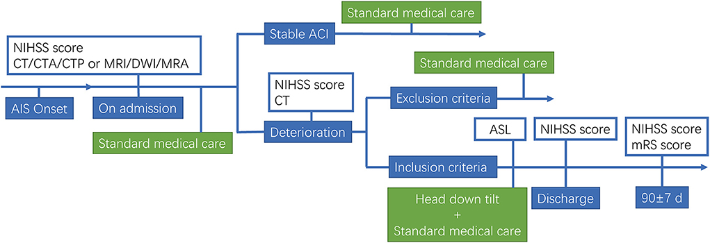
We performed a pilot trial in AIS patients. This study was approved by the Ethics Committee of former General Hospital of Shenyang Military Region (No. k2017-10). Informed consent was obtained from all study subjects, and the study was performed in accordance with the ethical standards as laid down in the 1964 Declaration of Helsinki and its later amendments or comparable ethical standards. Main inclusion Criteria: (1) age ≥18 years; (2) AIS with large vessel occlusion; (3) the patients occurred neurological deterioration within 7 days of onset; (4) 6–16 NIHSS after deterioration; (5) first stroke onset or past stroke without neurological deficit (modified Rankin Scale, mRS = 0); (6) fully understand and cooperate with the doctor's instructions; (7) the availability of informed consent. Exclusion Criteria: (1) Hemorrhagic stroke or mixed stroke; (2) combined with severe organ dysfunction; (3) a history of hemorrhagic stroke; (4) a history of stroke with severe sequelae; (5) planning revascularization in 3 months; (6) ischemic stroke due to surgical intervention; (7) participating in other clinical trials within 3 months; (8) pregnant or lactating women. Early neurological deterioration (END) was defined as an increase of 2 points or more on the NIHSS, except for cerebral hemorrhage. Schematic for head down tilt treatment of AIS patients is shown in Figure 1.

FIGURE 1
www.frontiersin.org

Figure 1. Flow diagram of the study.

Head down tilt treatment in AIS patients

After the informed consent was signed, the patient was treated with head down tilt as follows. The patients lay supine on tilt platform bed, which was adjusted to −20° for about 60 min for one time. During the treatment, a 3–5 min break to lay flat was allowed for patients who cannot tolerate head down tilt position. Head down tilt treatment was performed 3 times daily for 7 days. During the treatments, the patients were continuously monitored by ECG and blood oxygen saturation, and the side-lying head down position with −20° was allowed if high risk of aspiration was suspected.

Arterial spin labeling

After finishing the first treatment of head down tilt, ASL was performed to determine the influence of head down tilt on cerebral blood flow. In the ASL examination, nodes can be ideally described as regions which have coherent patterns of connections in anatomical structure or function in brain. Different parcellation methods of brain regions caused distinct brain network constructions, usually the entire brain surface would be covered completely by the parcellation method. The procedure to determine nodes in the study is as follows. First, the PdWI image of every subject was linearly registered to T1-weighted image and the result matrix was obtained. The T1-weighted image was normalized into MNI space, using both linear registration and non-linear registration. The transformation field derived from T1 to MNI normalization warped the co-registered PdWI image in structural space. Both of the warp-fields and transformation matrix were inverted by the command of convert_xfm and invwarp respectively, and then they were applied to the Human Brainnetome Atlas which is based on connectional architecture. At last, the atlas registered to each subject was resampled to CBF pcolor image, then 246 regions were obtained by using this procedure and each region represents a node in the brain network. Formula for the calculation is as follow.

Statistical analysis

Results were presented as mean values ± standard deviation (SD). The comparisons of the changes in myocardial enzymogram and NT-proBNP before and after head down tilt position were analyzed by Paired t- test. A value of p < 0.05 was considered statistically significant.

Data availability

The data that support the findings of this study are available from the corresponding author upon reasonable request.

Results

Head down tilt position alleviates clinical outcomes in acute ischemic stroke patients with LVO

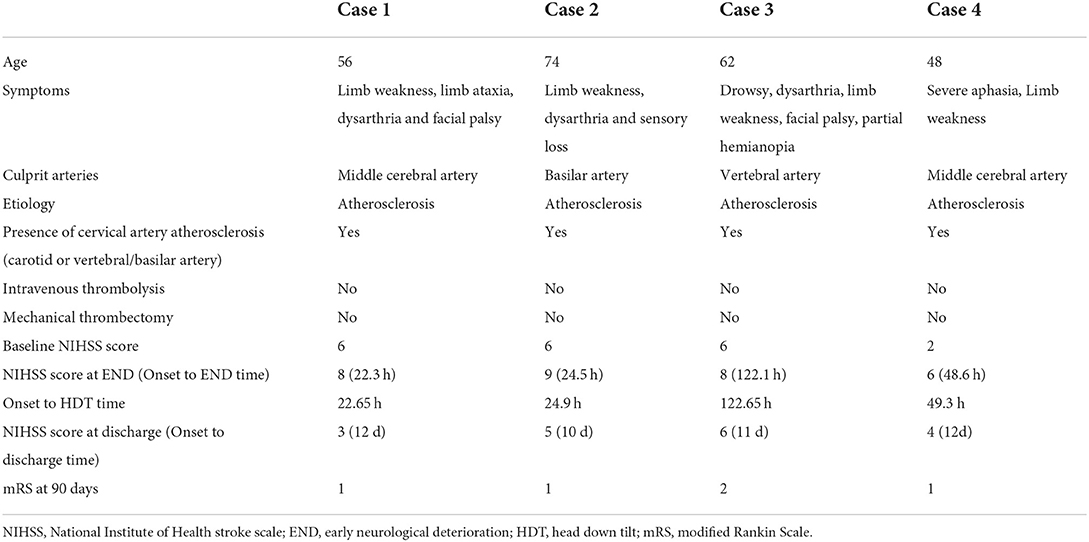
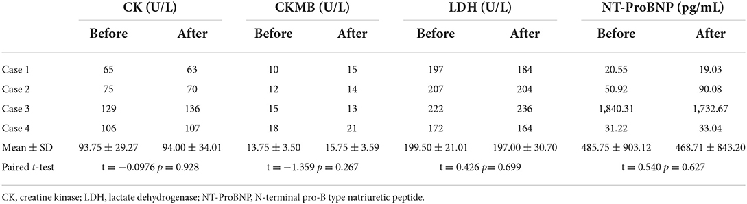
In the present study, 4 eligible AIS patients who occurred END within 7 days after onset were treated with head down tilt position (ages 48–74, average 60 years). Baseline characteristics of patients, symptoms, culprit arteries, etiology, presence of cervical artery atherosclerosis, onset to head down tilt time, prior intravenous thrombosis, prior mechanical thrombectomy, NIHSS score at different stages, and adverse events were listed in Table 1. The results showed that neurological function did not further deteriorate after head down tilt, and neurological deficit at discharge and neurological outcome 90 days after onset were significantly improved. No patients reported obvious uncomfortable events. The electrocardiographic monitoring did not show visible change during head down tilt. There were no changes in myocardial enzymogram and N-terminal pro b-type Natriuretic Peptide (NT-proBNP) before and after head down tilt in all patients (Table 2).

TABLE 1
www.frontiersin.org

Table 1. Characteristics of patients.

TABLE 2
www.frontiersin.org

Table 2. Changes in myocardial enzymogram and NT-proBNP before and after head down tilt position.

Head down tilt increases cerebral blood flow in patients with AIS

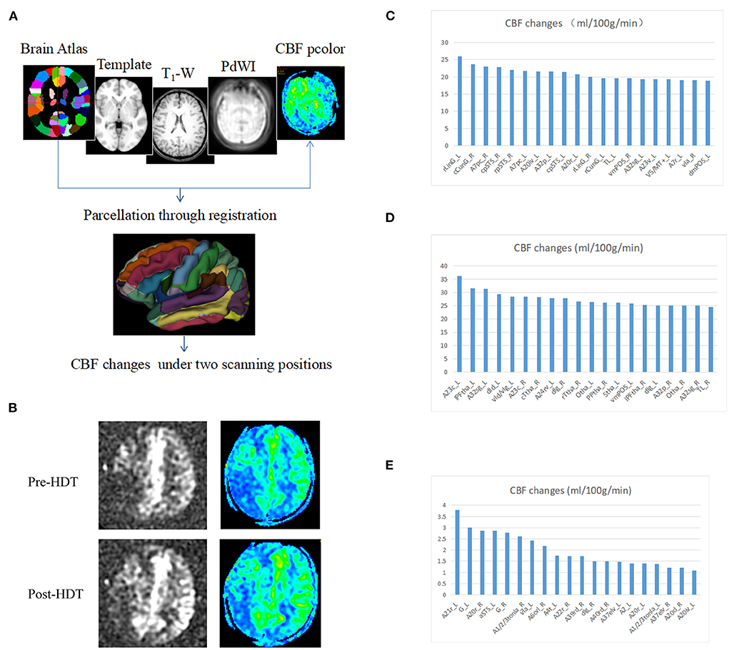
To explore the differences in CBF changes between head down tilt position and lying flat position, 3 anterior circulation ischemic stroke patients (Case 1, 3 and 4) underwent arterial spin labeling examination. The CBF value under lying flat and head down tilt positions were subtracted and ranked. In most regions of Case 1 and Case 3, the CBF value under head down tilt position increased significantly compared to that under lying flat position (Figure 2). The CBF value in Case 4 also increased although not significant. The top 20 brain regions changed the most in each subject were shown in Figures 2C–E.

FIGURE 2
www.frontiersin.org

Figure 2. Changes in cerebral blood flow after head down tilt position. (A) Pipeline for the registration and determination of regions. The Brainnetome Atlas, template, T1-weighted, PdWI and CBF pcolor were used to co-registration and 246 regions of interest were obtained in every subject. In addition, the CBF changes were calculated under two scanning positions (head down tilt position and lying flat position). (B) Representative ASL and CBF pcolor images from Case 1. (C) CBF changes in Case 1. (D) CBF changes in Case 3. (E) CBF changes in Case 4. ASL, arterial spin labeling; CBF, cerebral blood flow; HDT, head down tilt.

Discussion

In AIS patients, various interventions were performed to improve CBF through collateral arteries, leptomeningeal recruitment, and increasing residual blood flow, aiming to improve cerebral perfusion and decrease injury within the ischemic penumbra region. But none so far has been demonstrated effective except reperfusion strategy. HeadPoST study demonstrated no difference in the functional recovery of patients assigned to flatting or sitting up position (6), which may be due to the broad inclusion of the patients (14). Head positioning trials should be performed in discrete patient cohorts with endpoints supported by pilot data (15). The present study first showed that the head down tilt position improved neurological function in acute ischemic stroke patients by possibly increasing the CBF of affected side.

We found that head down tilt treatment could prevent the further deterioration of clinical symptoms in AIS patients with END, because symptoms in all patients stop progression after the treatment. Meanwhile, improvements in NIHSS scoring were detected after head down tilt treatment and an increase in CBF of the ischemic area was induced in patients with anterior circulation infarction by head down tilt position. As we know, ischemic stroke is a potentially reversible process that is dependent on restoration of CBF within a time window of cellular viability that varies according to the severity and duration of the flow deficit. The prognosis of acute ischemic stroke was determined according to the location and size of the occluded cerebral vessel (1), the extent of collateral blood flow (1, 2) and the time to reperfusion therapy (22). Previous studies indicated that changes in head position could influence the prognosis of AIS patients through regulating cerebral perfusion pressure, intracranial pressure, cortical oxygenation (23), systolic and diastolic blood pressure, residual CBF to the ischemic brain tissue (24), collateral blood flow (25, 26), and mean velocity of the residual arterial blood flow (7). A study on “collateral therapeutics” in a rat stroke model of transient MCAO evaluated the effect of different strategies including induced hypertension, intravascular volume load, cerebral arteriolar vasodilation, and head down tilt on stroke outcome, and indicated that treatment with collateral therapeutics was associated with lower infarct volumes and higher chance of good functional outcome. Notably, the highest efficacy and safety profile was observed for head down tilt treatment (27). In addition, cerebral autoregulation ability was impaired in stroke patients and could lead to a decrease in CBF velocity during orthostatic stress with head-up tilt. Therefore, we argue that positioning AIS patients with head down tilt may exert neuroprotective effect through increasing CBF due to collateral circulation improvement by gravitational force (28). Furthermore, we argue that this benefit may be more obvious in the moderate stroke patients with LVO in the acute phase (within 24 h). For acute mild stroke, intensive antithrombosis should be best strategy to prevent the deterioration or reoccurrence of stroke (29). For severe ischemic stroke patients, recanalization as soon as possible may be the best way to acquire good outcome due to limited ischemic penumbra that can be salvaged. In addition, head down tilt treatment may further increase intracranial pressure for severe stroke. In the present study, 2 of the 4 patients had a posterior circulation stroke, which is expected to have a smaller response to collateral therapeutics due to less developed collaterals, compared to anterior circulation stroke. Taken together, we argue that the best target population should be acute moderate stroke patients with LVO, which is being determined by our ongoing trial (NCT03744533).

In previous studies, transcranial laser Doppler-recorded mean flow velocity (MFV) was usually used to reflect the changes in CBF after head positioning treatment. However, Transcranial Doppler only provided indirect information of CBF, due to measuring the flow velocity and not the volume flow. Therefore, changes of blood flow velocity could correspond to the change of CBF only if the vessel diameter was constant, which cannot be assumed a priori. Moreover, even very small changes in the position of the transcranial Doppler probe could alter the blood flow velocity readings. Furthermore, the relevance of changes in MFV to any improvement in clinical outcomes after AIS is uncertain (30). In our study, CBF was evaluated by arterial spin labeling, which steadily reflect the volume flow.

It was reported that serious adverse events of head down tilt treatment included visual field loss, cognitive aberrations, potentially life-threatening complications of the respiratory and cardiovascular systems (31). In our study, no serious adverse events, including aspiration pneumonia, were observed in patients treated with head down tilt position, which may be due to short-term and moderate tilt of head down position. Another important concern was about its effect on CBF, because decreased CBF may occur due to increased intracranial pressure (ICP) induced by head down tilt. The mechanism appeared to be related to the changes in venous outflow through the valueless jugular veins and vertebral venous plexuses. Cerebral venous and jugular venous pressures increase with head position lowering, leading to an increase in the cerebral venous blood volume and a subsequent increase in ICP. The cerebral perfusion pressure (CPP) was calculated as the difference between mean arterial pressure and ICP. The CPP may increase when head position was under the level of heart since the mean arterial pressure increased in this head down tilt position. We argued that in certain angle and duration time, head down tilt position exerted neuroprotective effect through increasing CPP. However, ICP increased due to the impairment of venous outflow in a greater minus angle, leading to the decrease in CPP and the deterioration of neurological function. The main limitation of the clinical study is the small sample, no control design.

Conclusion

For the first time, the case series study suggests that head down tilt may exert neuroprotective effect in AIS patients possibly through increasing the cerebral perfusion. Due to its low cost and easy operation, the efficacy and safety of head down tilt position in acute ischemic patients are urgently needed to be confirmed in further clinical trials.