Based on this, what will your competent? doctor do to cure spasticity?
Nothing like usual? PURE INCOMPETENCE THEN!
A fireable offence for the past two years!
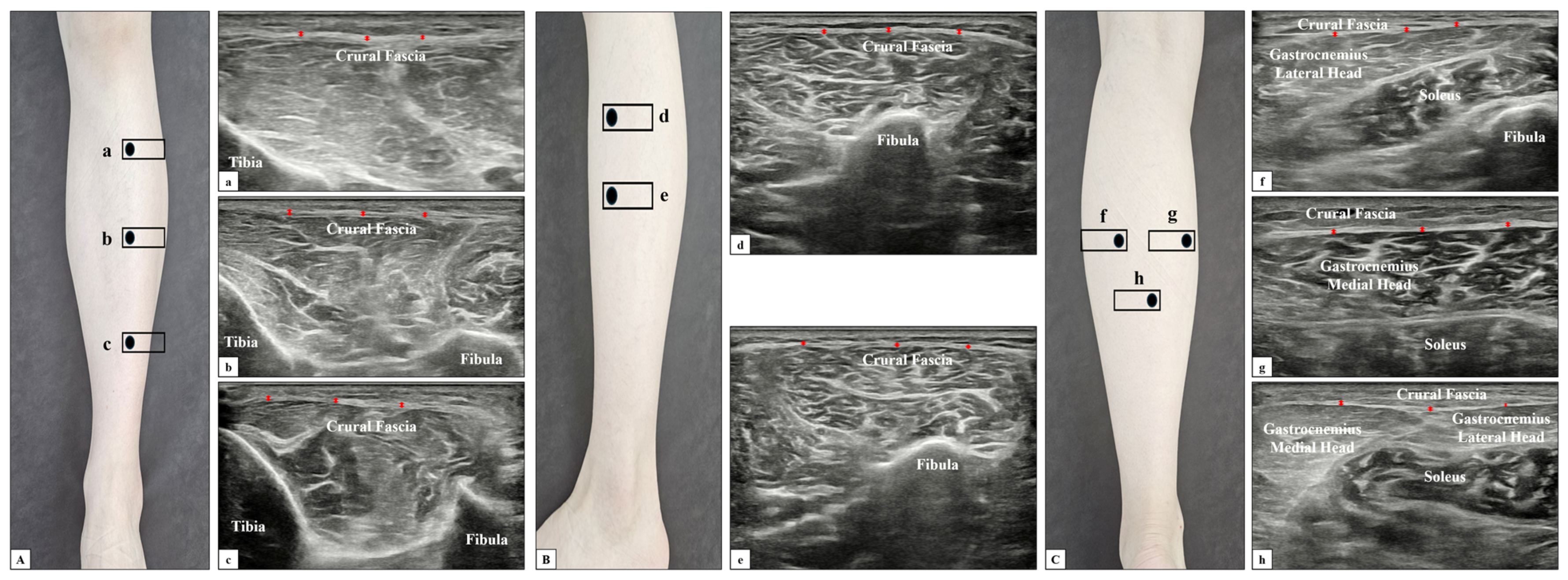
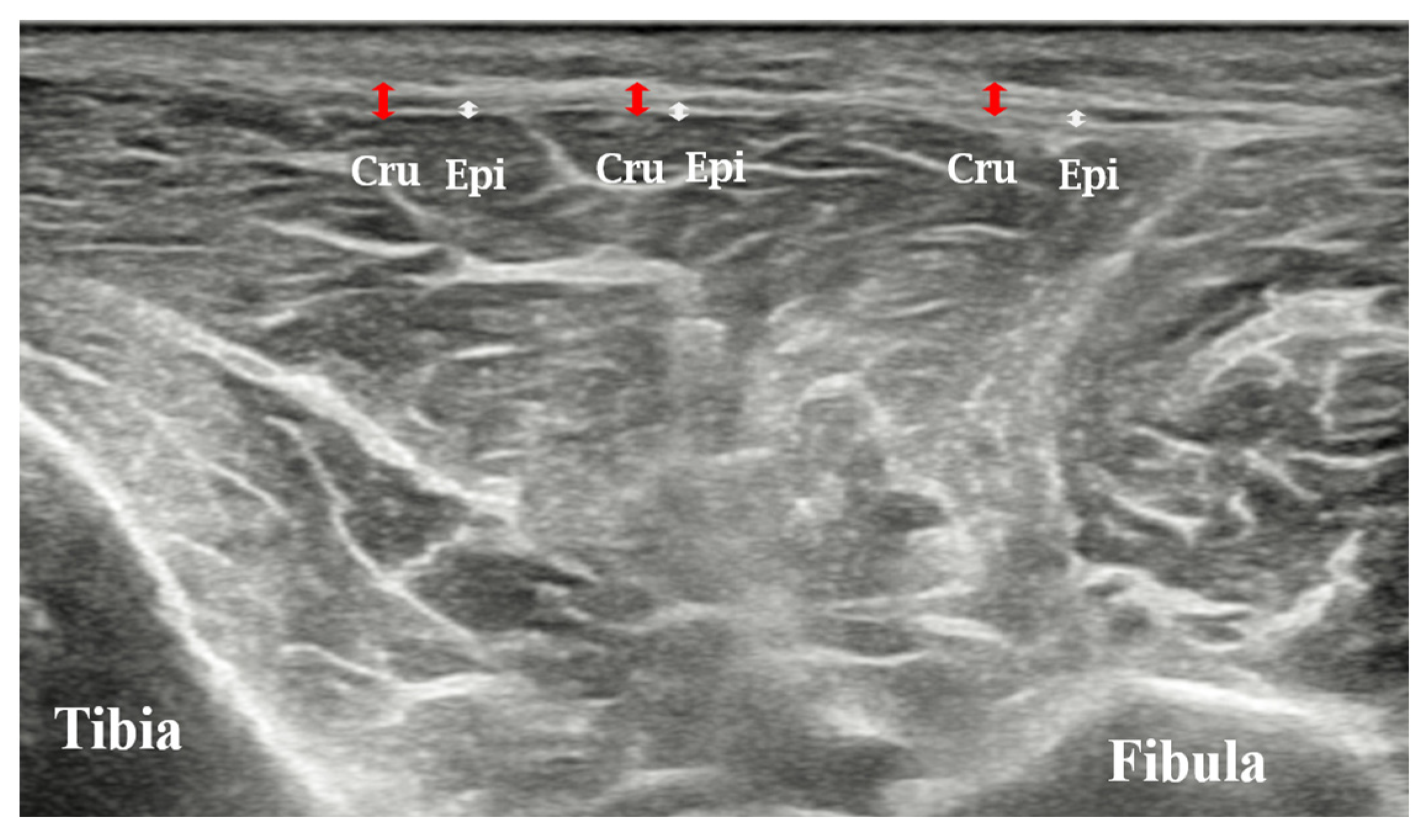
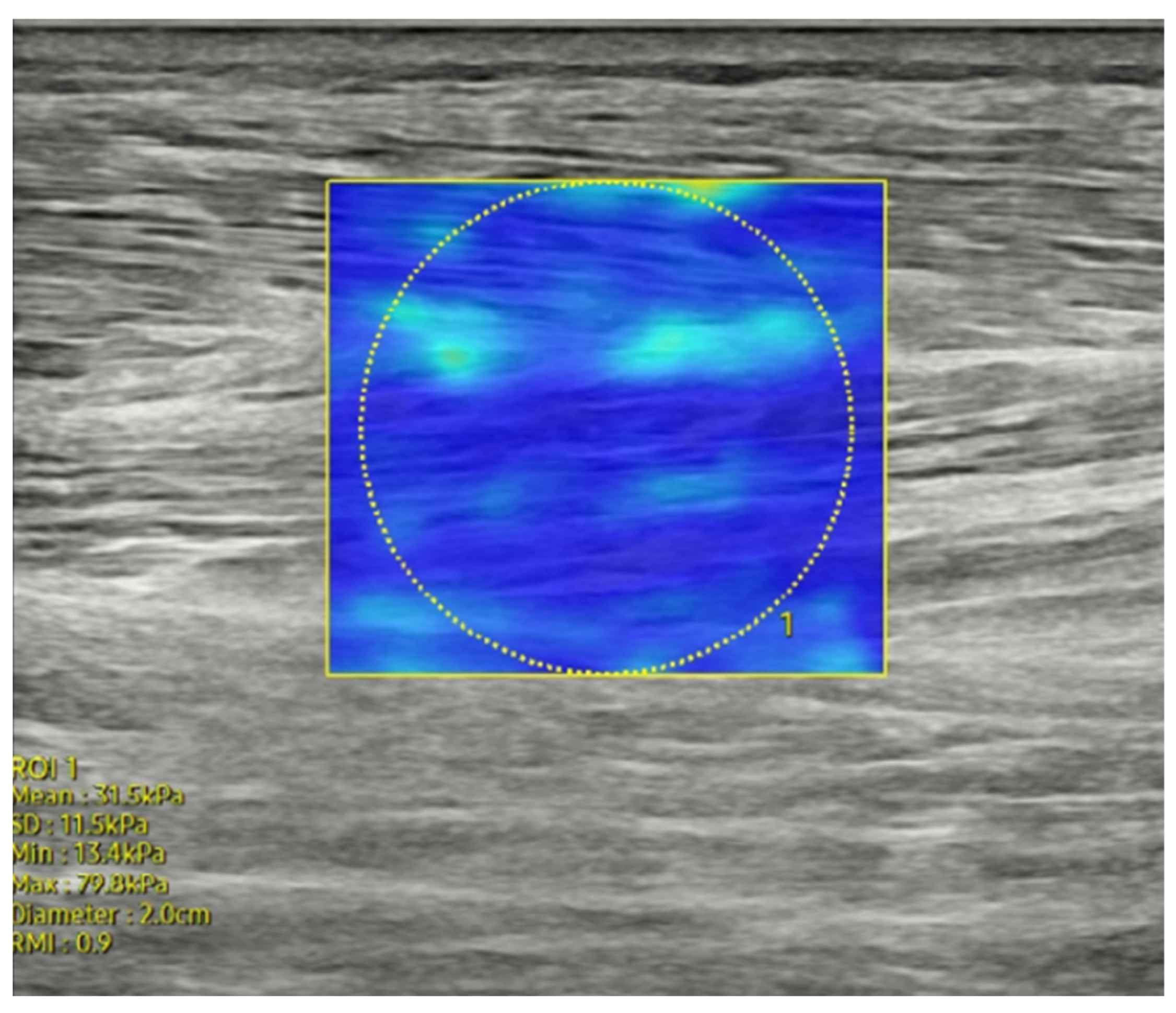
Ultrasound Imaging Comparison of Crural Fascia Thickness and Muscle Stiffness in Stroke Patients with Spasticity

1
Fascia Research Institute, Myofascial Release Korea, Seoul 06136, Republic of Korea
2
Department of Physical Therapy, The Graduate School, Gachon University, Incheon 21936, Republic of Korea
3
Department and Research Institute of Rehabilitation Medicine, College of Medicine, Yonsei University, Seoul 03722, Republic of Korea
4
Department of Physical Therapy, College of Medical Science, Gachon University, Incheon 21936, Republic of Korea
*
Author to whom correspondence should be addressed.
Diagnostics 2024, 14(22), 2606; https://doi.org/10.3390/diagnostics14222606
Submission received: 13 October 2024
/
Revised: 14 November 2024
/
Accepted: 17 November 2024
/
Published: 20 November 2024
(This article belongs to the Special Issue Advances in Ultrasound Imaging for Musculoskeletal Diseases)
Abstract
Background/Objective:
Spasticity following stroke causes structural changes in the muscles and fascia, affecting the mobility and functional recovery of patients. Understanding these structural changes is critical to optimizing the rehabilitation strategies for patients. Therefore, in this study, we aimed to investigate the differences in crural and epimysial fascia thickness and muscle stiffness in the affected and unaffected lower limbs of chronic stroke patients with spasticity.
Methods:
A total of 88 patients with chronic stroke (mean age: 62.7 ± 10.2 years) were included in this study. Ankle range of motion, crural fascial thickness, and muscle stiffness in affected and unaffected lower limbs were assessed using ultrasound. Results: For the affected lower limbs, 59 patients (67.1%) exhibited a modified Ashworth scale score of 2, whereas 29 patients (32.9%) exhibited a score of 3. Ankle range of motion, fascia thickness, and muscle stiffness were also measured. The range of motion in ankle dorsiflexion and plantar flexion was significantly reduced on the affected side (p < 0.05). Crural fascia thickness was significantly greater in all regions of the affected side (anterior: 0.96 ± 0.14 vs. 0.72 ± 0.08 mm [p < 0.001]; lateral: 1.01 ± 0.14 vs. 0.75 ± 0.14 mm [p < 0.001]), and the epimysial fascia of the tibialis anterior muscle was similarly greater in the affected side (0.46 ± 0.07 vs. 0.34 ± 0.03 mm [p < 0.001]). However, no significant differences in muscle stiffness were observed between the affected and unaffected sides (p > 0.05).
Conclusions:
Overall, these findings revealed significant fascial thickening with only minimal changes in muscle stiffness on the affected side, highlighting the importance of controlling fascial changes for post-stroke spasticity management.


No comments:
Post a Comment