Changing stroke rehab and research worldwide now.Time is Brain! trillions and trillions of neurons that DIE each day because there are NO effective hyperacute therapies besides tPA(only 12% effective). I have 523 posts on hyperacute therapy, enough for researchers to spend decades proving them out. These are my personal ideas and blog on stroke rehabilitation and stroke research. Do not attempt any of these without checking with your medical provider. Unless you join me in agitating, when you need these therapies they won't be there.

What this blog is for:

My blog is not to help survivors recover, it is to have the 10 million yearly stroke survivors light fires underneath their doctors, stroke hospitals and stroke researchers to get stroke solved. 100% recovery. The stroke medical world is completely failing at that goal, they don't even have it as a goal. Shortly after getting out of the hospital and getting NO information on the process or protocols of stroke rehabilitation and recovery I started searching on the internet and found that no other survivor received useful information. This is an attempt to cover all stroke rehabilitation information that should be readily available to survivors so they can talk with informed knowledge to their medical staff. It lays out what needs to be done to get stroke survivors closer to 100% recovery. It's quite disgusting that this information is not available from every stroke association and doctors group.

Thursday, July 28, 2016

Astrocytes found to transfer mitochondria to neurons after stroke

Never mind, more study is needed which will never occur under the current fucking failures of stroke associations. Unless you can personally fund researchers to find the answer All these possibilities and NO ONE is following up them to actually help stroke survivors. 
http://medicalxpress.com/news/2016-07-astrocytes-mitochondria-neurons.html
A combined team of researchers from Massachusetts General Hospital/Harvard Medical School in the U.S. and Xuanwu Hospital, Capital Medical University in China has found that when neurons in the mouse brain suffer mitochondrial damage astrocytes donate some of their own to help repair them. In their paper published in the journal Nature, the team describes how they conducted a series of tests designed to find out whether astrocytes donate mitochondria material and if so, whether it helps to restore health to damaged neurons.
Astrocytes are star-shaped glial cells that surround neurons, providing insulation and support—prior studies have shown that they are involved in carrying out removal of dead material. In this new effort, the researchers started with the results of experiments conducted by a team at Columbia University four years ago that showed that bone marrow stem cells provided mitochondria to damaged to help them recover—they wanted to know if the same might be true for astrocytes and neurons.
To find out, the researchers engineered mice to produce extra amounts of a signaling enzyme called CD38. They then found that when rodent astrocytes were mixed with them, they expelled some degree of mitochondrial material—neurons added to the mix were then found to absorb some of the mitochondrial material.
The next step was to find out if the same process actually happened in a living animal. They found that it did by causing brain injuries to mice and then injecting the sites with mitochondria they had retrieved from —microscopic analysis showed the neurons had, indeed, absorbed the material and that as a result, the neurons were healthier than were injured cells that had not received injections.
The researchers also wanted to know if CD38 signaling was necessary for the process to work—to find out, they injected material that interfered with its function into test mice—those with such injections were found to have less astrocyte-donated mitochondrial material in their than did those that did not receive such injections, which suggest that it is a necessary part of the process.
The overall results by the team suggest that human stroke patients might benefit from CD38 injections or drugs that cause the body to produce it, but the researchers are quick to point out that the protein is very active throughout the body, which means such therapies could cause a large number of unknown side effects. More study is needed, but the findings do offer hope for treating such injuries and perhaps maladies such as Parkinson's disease.
Journal reference: Nature search and more info website

A minute of secondhand marijuana smoke may damage blood vessels

But do the benefits outweigh the harm? Or does this mean we should get it in foods? Your doctors thoughts on this?
My 13 reasons for marijuana use post-stroke.  
Seems like trying to demonize marijuana any way possible. 
http://www.alphagalileo.org/ViewItem.aspx?ItemId=166350&CultureCode=en
Rats’ blood vessels took at least three times longer to recover function after only a minute of breathing secondhand marijuana smoke, compared to recovery after a minute of breathing secondhand tobacco smoke, according to new research in Journal of the American Heart Association, the Open Access Journal of the American Heart Association/American Stroke Association.
When rats inhaled secondhand marijuana smoke for one minute, their arteries carried blood less efficiently for at least 90 minutes, whereas similar exposure to secondhand tobacco smoke caused blood vessel impairment that recovered within 30 minutes.
“While the effect is temporary for both cigarette and marijuana smoke, these temporary problems can turn into long-term problems if exposures occur often enough and may increase the chances of developing hardened and clogged arteries,” said Matthew Springer, Ph.D., study senior author and professor of medicine at the University of California, San Francisco’s Division of Cardiology.
Blood vessel function was examined in rats before and after exposure to secondhand marijuana smoke at levels similar to real-world secondhand tobacco smoke.
“Arteries of rats and humans are similar in how they respond to secondhand tobacco smoke, so the response of rat arteries to secondhand marijuana smoke is likely to reflect how human arteries might respond,” Springer said.
Researchers also found the mere burning of the plant material appears responsible for the impaired blood vessels, not chemicals like nicotine and tetrahydrocannabinol (THC), the psychoactive ingredient in marijuana, nor rolling paper.
“There is widespread belief that, unlike tobacco smoke, marijuana smoke is benign,” Springer said.  “We in public health have been telling the public to avoid secondhand tobacco smoke for years, but we don't tell them to avoid secondhand marijuana smoke, because until now we haven’t had evidence that it can be harmful.”
Springer also noted that the increasing number of states legalizing medicinal and recreational marijuana, along with increasing potential for corporate expansion within the cannabis industry, makes it important to understand the health consequences of secondhand marijuana smoke exposure.
The inhalation of smoke should be avoided, regardless of whether it comes from tobacco, marijuana, or other sources. Inhaling smoke is bad for you – period, researchers said.
Co-authors are Xiaoyin Wang, M.D.; Ronak Derakhshandeh, M.S.; Jiangtao Liu, M.D.; Shilpa Narayan, B.S.; Pooneh Nabavizadeh, M.D.; Stephanie Le, B.A.; Olivia Danforth, B.S.; Kranthi Pinnamaneni, M.D.; Hilda Rodriguez, A.S.; Emmy Luu, B.S.; Richard Sievers, B.S.; Suzaynn Schick, Ph.D.; and Stanton Glantz, Ph.D. Author disclosures are on the manuscript.
The National Institutes of Health’s National Institute on Drug Abuse and a generous grant from Elfenworks Foundation funded the study.
http://newsroom.heart.org/news/a-minute-of-secondhand-marijuana-smoke-may-damage-blood-vessels?preview=d01b7b4584badcd39cedb6903e591302

Catheter-Based Closure Not Recommended for Patients With Heart Defect, Stroke

Popular news here, dumping all the risk on the patient.If you want the closure it means you will have to out argue your medical staff and insurance company. Be prepared.
http://dgnews.docguide.com/catheter-based-closure-not-recommended-patients-heart-defect-stroke?
MINNEAPOLIS, Minn -- July 27, 2016 -- An updated recommendation from the American Academy of Neurology (AAN) states that catheter-based closure should not be routinely recommended for people who have had a stroke and also have a patent foramen ovale (PFO).
The practice advisory, which updates a previous AAN guideline, is published in the July 27, 2016, online issue of the journal Neurology.
To develop the advisory, researchers reviewed all available scientific studies on people with PFO who also had an ischemic stroke or a transient ischemic attack (TIA).
“Compared with other ways to prevent a second stroke, such as medications to reduce blood clots, the devices used to close a patent foramen ovale have limited evidence to support their use,” said practice advisory author Steven R. Messé, MD, University of Pennsylvania Perelman School of Medicine, Philadelphia, Pennsylvania. “It’s still uncertain how effective these devices are in reducing stroke risk, and the procedure is associated with uncommon but potentially serious complications.”
In addition, Dr. Messé noted that the devices used for PFO closure are not available for routine use in the United States, so the procedure must be done off-label with a device approved for treating a similar heart defect or with another device that does not have strong evidence regarding its use. At the time of publication, the US Food and Drug Administration (FDA) is currently reviewing the one device that has the best evidence regarding closure.
“People should know that having a PFO is common -- 1 in 4 people have one -- and the risk of having a second stroke is low,” said Dr. Messé. (Well shit, Dr. Messé you are not the person at risk for a stroke.You can blithely talk about it without consequence.)
When the AAN developed the earlier guideline on this topic in 2004, not enough evidence was available to make a recommendation on whether closing a PFO was effective in reducing stroke risk.
The advisory also recommends that aspirin or other antiplatelet drugs be used to prevent blood clots instead of blood thinners unless there is another reason to use blood thinners, such as a person with a history of blood clots in the legs or lungs.
SOURCE: American Academy of Neurology

Improving Patient Outcomes in Preventing Atrial Fibrillation-related Stroke with Non-Vitamin K Antagonist Oral Anticoagulants

You might want to read if you have atrial fibrillation, unless you think your doctor will tell you this in the next two weeks.
http://www.touchneurology.com/articles/improving-patient-outcomes-preventing-atrial-fibrillation-related-stroke-non-vitamin-k
European Neurological Review, 2016;11(1):27–35 DOI: http://doi.org/10.17925/ENR.2016.11.01.1a

Abstract:

The rising incidence of atrial fibrillation (AF) is increasingly resulting in a substantial worldwide increase in AF-related stroke, particularly in elderly patients and this is creating an increasingly serious healthcare burden. Guidelines recommend the use of AF-related stroke prophylaxis but adherence to these remains poor. Studies conducted in the 1990s showed that warfarin reduced the risk of AF-related stroke by an overall 64% compared with placebo. Subsequently, prophylactic treatment was further improved with the development of non-vitamin K antagonist oral anticoagulants (NOACs). More recently, a meta-analysis of four large clinical trials on NOACs (dabigatran, rivaroxaban, apixaban, and edoxaban) showed there was a relative risk reduction of 0.81 (p<0.0001) favouring NOAC treatment over warfarin for stroke or systemic embolic events in patients with AF. The largest trial of NOACs in AF-related stroke, to date, was the ENGAGE AF-TIMI 48 study (n=21,105) which showed that edoxaban was non-inferior to warfarin for ischaemic stroke reduction but significantly reduced bleeding and cardiovascular mortality. A recent subgroup analysis of this study showed that with edoxaban the incidences of intracranial haemorrhage (ICH) subtypes (all ICH, fatal ICH, fatal, subdural and epidural bleed) were significantly lower with 60 mg of edoxaban (p=0.013–<0.001). Edoxaban was also shown to be an effective option in patients with prior stroke. In addition edoxaban was shown to reduce deaths due to fatal bleeds compared with warfarin. The results of current studies, especially the ENGAGE AF-TIMI 48 subgroup analysis therefore, show that the benefits of anticoagulation therapy in patients with AF substantially outweigh the risks
Keywords: Atrial fibrillation-related stroke, outcomes, non-vitamin K oral anticoagulants (NOACs)
Disclosure: Peter Kelly has served on advisory boards or received speakers fees or benefits from the American Stroke Association, Bayer and Daiichi Sankyo, and has received research unit grants from the Health Research Board of Ireland, Irish Heart Foundation and Bayer. Carlos Molina has nothing to declare in relation to this article. Christian T. Ruff has received research support from GlaxoSmithKline, Daiichi Sankyo, Intarcia and AstraZeneca, and serves as a consultant and on the advisory boards for Boehringer Ingelheim, Bayer, Daiichi Sankyo, Portola and DrugDev. Roland Veltkamp has received speaker fees, consulting honoraria and research support from Bayer, Boehringer Ingelheim, BMS, Pfizer, Daiichi Sankyo, CSL Behring, Apoplex Medical Technologies, Morphosys, Biogen, Medtronic.
Acknowledgments: Editorial assistance was provided by James Gilbart at Touch Medical Media, London, this was supported by an unrestricted grant from Daiichi Sankyo Europe GmbH. This article reports the proceedings of a sponsored satellite symposium and as such has not been subject to the journal’s usual peer-review process
Received: October 16, 2015 Accepted February 19, 2016
Correspondence: Peter Kelly, Stroke Service and NeuroVascular Unit for Translational and Therapeutics Research, University College Dublin, Ireland E: pjkelly@mater.ie.
Support: The publication of this article was supported by Daiichi Sankyo Europe GmbH. The views and opinions expressed are those of the authors and not necessarily those of Daiichi Sankyo Europe GmbH.
Open Access: This article is published under the Creative Commons Attribution Noncommercial License, which permits any non-commercial use, distribution, adaptation and reproduction provided the original author(s) and source are given appropriate credit
In atrial fibrillation (AF), considerable harm can result from the lack of appropriate preventive therapy, and optimal prevention is critical, especially in vulnerable elderly or frail patients. AF markedly increases the risk of stroke and this condition must be monitored and potentially treated wherever it is detected.1–4 AF is an increasing concern for physicians worldwide as populations age and more people are at risk.5–7 Although guidelines for stroke prevention in AF that recommend anticoagulation have been established for many years, many at-risk patients receive inadequate anticoagulation or none at all.8–11 This ‘reluctance to treat’ stems largely from a fear of inducing intracranial haemorrhage (ICH) and other serious bleeding types that are associated with warfarin and the non–vitamin K antagonist oral anticoagulants (NOACs). This risk, however, is often over-stated and substantially less than the risks that are associated with the lack of stroke prevention treatment in AF. This review discusses the burden of AF-related stroke and evidence that supports current treatments, and considers novel insights on the use of edoxaban as provided by recent subgroup analyses of the ENGAGE AF-TIMI 48 trial results (see end of article for trial name definitions). These topics were presented at a satellite symposium convened at the European Stroke Organisation Annual Meeting in Glasgow, UK, in April 2015.
Preventing the Rise of AF-related Stroke– A Call to Action
Large-scale population-based observational studies have shown AF to be a serious factor increasing the likelihood of strokes and substantially worsening mortality and morbidity after a stroke.12 Various studies have predicted increasing incidence and prevalence of AF-related stroke and the associated heavy burden this will place on healthcare authorities worldwide. Professor Peter Kelly assessed the history and rising incidence of AF-related stroke. His message constitutes a call to action, encouraging physicians to treat all patients with AF to help stem the burgeoning number of ischaemic strokes and reduce the burden strokes impose on healthcare services.

7 more pages at link.

AAN Nixes Routine PFO Closure

So what is the consensus? Let the patient worry and hope the warfarin or aspirin works well enough not to have another stroke? What do our fucking failures of stroke associations have to say about this? Or will they once again hide and say this is a medical decision that your doctors should handle?
http://www.medpagetoday.com/Neurology/Strokes/59347?xid=nl_mpt_DHE_2016-07-28&eun=g424561d0r&pos=1

Questions definition of 'routine'

  • by Nicole Lou
    Reporter, MedPage Today/CRTonline.org

Patent foramen ovale (PFO) closure is not recommended as a routine therapy for patients with cryptogenic ischemic stroke, according to the American Academy of Neurology (AAN).
A systemic review of the literature for the Academy's stroke care guideline update published in Neurology turned up no stroke prevention benefit with the STARFlex PFO occluder compared with medical therapy alone (risk difference [RD] 0.13%, 95% CI -2.2% to 2.0%).
With the Amplatzer, however, there was a decreased risk of recurrent stroke (RD -1.68%, 95% CI -3.18% to -0.19%) at the cost of more new-onset atrial fibrillation (RD 1.64%, 95% CI 0.07% to 3.2%) and a procedural complication risk of 3.4% (95% CI 2.3% to 5%).
Thus, "in rare circumstances, such as recurrent strokes despite adequate medical therapy with no other mechanism identified, clinicians may offer the Amplatzer PFO Occluder if it is available (Level C)," the guideline development subcommittee of the AAN wrote.
Yet "this practice advisory is out of date," warned John Carroll, MD, of University of Colorado Hospital in Aurora, because the AAN did not have access to the latest 5-year data from the RESPECT trial. "The composition of the AAN group did not include one interventional cardiologist," he added. "In 2016 it is odd to have a proclamation about the value of a procedure without anyone on the group ever having performed the procedure."
The new numbers from RESPECT were taken into account during an FDA advisory meeting in May 2016 wherein the majority of panelists voted that the evidence tilted towards safety and efficacy with the Amplatzer compared to medical therapy alone. "Clinicians and patients should have the option of PFO closure with an approved device," said Carroll, who was not involved with the guidelines.
Even David E. Thaler, MD, PhD, of Boston's Tufts Medical Center -- and member of the dissemination committee for the AAN guideline -- seemed to agree. "I think the recommendations are cautious and already a little dated," Thaler told MedPage Today.
When asked if the guideline updates regarding routine PFO closure were appropriate, Thaler said it "depends on the definition of 'routine.'"
"If 'routine' is what currently happens in some practices – i.e., an episode of dizziness, interpreted by primary care provider as transient ischemic attack, not seen by neurology, echo shows PFO, referred to cardiology for closure – then yes, indeed, I agree with the recommendation."
"However, if 'routine' is as it should be – i.e., a stroke patient, evaluated by a vascular neurologist with 'complete investigations' (which is a changing landscape), a high RoPE score, a well-educated patient with regard to the current state of PFO science, realistic expectations of treatments, adherence to long-term secondary stroke prevention measures even after possible closure – then no, I think the recommendation is too cautious and dismissive of the totality of the evidence that shows that closure is probably better and no evidence to indicate that it's worse than medical management," he said.
Thaler's allusion to the uncertainty around PFO was echoed by Patrick D. Lyden, MD, of Cedars-Sinai Medical Center in Los Angeles.
"I have found a tendency for clinicians to stop looking for stroke causes once they find a PFO. It's important to complete a full evaluation on every patient, and not jump too quickly to conclude the PFO is the cause of the stroke," he told MedPage Today.
"Here is how I explain it to patients: we have two studies in favor and two studies against PFO closure. Let's wait for the 'tie breaker' study to finish before we decide to close your PFO," Lyden said. So let the patient bear all the risk? It is not the doctors brain that is in danger, so what the hell.

Wednesday, July 27, 2016

Virtual Rehabilitation Use for Paretic Upper Limb for Individuals with Chronic Hemiparetic Stroke

 One more abject failure of our stroke associations, providing complete access to all stroke research. You have a few simple jobs and you fail at the fucking simplest. How the hell are we supposed to write our own protocols if we can't even see the research?
Virtual Rehabilitation Use for Paretic Upper Limb for Individuals with Chronic Hemiparetic Stroke

  • Renata Cristina Magalhães Lima 
  • , Ana Paula Dias de Menezes
  • , Maria Carolina Gomes Inácio
  • , Silvia Moreira Amaral
  • , Regiane Relva Romano
$29.95 / €24.95 / £19.95 *
* Final gross prices may vary according to local VAT.
Get Access

Abstract

Stroke causes numerous deficiencies. The aim of this study was evaluate the effects of virtual rehabilitation on chronic hemiparetic upper limb functionality with application of Motor Activity Log–MAL, Wolf Motor Function Test-WMFT and Abilhand. Quality of life was assessed by Stroke Specific Quality of Life Scale-SSQOL-Brazil. A single case study (ABA type) was done. Two participants were evaluated in the baseline, during the training using virtual-reality with X-box360Kinect, and in followup. About MAL, the participant 1 improved in the followup, while participant 2 remained. For Abilhand, the data remained stable for both. In the participant 1 WMFT obtained improvement in the task 7 (weight in box) and task 14 (prehension). The participant 2 acquired a constant behavior in relation to the same tasks, while the results related to qualitative aspects, remained stable for both. Virtual rehabilitation contributed for greater functional use of the upper limb and better quality of life.

Technological Advances in the Rehabilitation of Gait and Balance After Stroke

Well shit, if we just had a written walking stroke protocol all these therapists could add their critiques and maybe survivors could recover better. Isn't that the whole fucking point of therapy? 
http://www.ptproductsonline.com/2016/06/technological-advances-rehabilitation-gait-balance-stroke/
By Mithu Lijo, Msc PT, NCS, CBIS, MSCS, and Jamie Bolt, PT, DPT, NCS
Stroke ranks as the sixth highest cause of burden of disease worldwide in terms of disability.1 About 60% of people who have had a stroke have difficulties with walking, and 30% of people affected with stroke do not regain complete motor recovery after rehabilitation.2 One of the primary concerns for individuals who experience stroke is the ability to regain walking, with respect to safety, speed, balance, and postural control. Consequently, gait retraining is a key focal area during rehabilitation following stroke. Even after intensive rehabilitation focusing on gait and balance training, stroke survivors still present with significant spatiotemporal asymmetry and balance deficits that interfere with their independence and quality of life.
Rehabilitation in the acute phase is very important in the recovery of function. The 2016 AHA adult stroke guidelines recommend inpatient rehabilitation to improve quality of life and return to previous level of function.2 From the time of admission, patients are engaged in an intensive rehabilitation program to improve their mobility and self care skills in different environments and life situations. Advances in technology enable patients who are affected by a wide range of physical impairments to begin participating in rehabilitative activities without becoming apprehensive about falls and risk of injury to both patients and staff.1 Neurologic physical therapists use various technologies to address gait and balance impairments in stroke patients to enable them to return to a productive life. This article examines the use of technological advances to address three specific impairments in gait following stroke: speed, spatiotemporal asymmetry, and dynamic balance.

Speed

Walking speed is directly related to functional independence and community mobility after stroke. On average, two out of three stroke survivors are unable to walk at a speed that enables them to function independently in the community. The literature suggests an average walking velocity of2 .1 m/s to 1.5 m/s is considered fast enough to be functional as a pedestrian in different environmental and social contexts (eg, crossing a street safely).3 At a walking speed of more than 0.8 m per second, full mobility in the community is likely; at a walking speed of less than 0.4 m per second, mobility is limited to the home; and at speeds of 0.4 to 0.8 m per second, mobility is limited to short walks in the community.3 Walking on a treadmill, with or without body weight supported via a harness connected to an overhead support system, is a method of treating walking impairments post stroke that is becoming increasingly popular.4
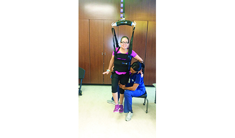
One highly recognized piece of equipment is the LiteGait (Mobility Research, Tempe, Ariz) with body weight support treadmill training, to enable nonambulatory or limited ambulatory hemiparetic stroke patients to practice coordinated stepping:
• during progressively increased demands for postural control;
• with control over gait speed;
• with potential to stimulate normal walking pattern; and
• with reduced oxygen demand.
The use of body weight support (BWS) allows the walking process to be comfortable for the patient. Such support provides the ability to focus on stepping with the paretic leg with assistance from the therapist. It also reduces the need for intensive support from therapists during gait training, which often leads to fatigue and risk of injury. It in fact results in lower intensity of therapy for the patient. Use of a treadmill permits a greater number of steps to be performed within a training session, thereby increasing the amount of task-specific practice completed. Hesse, 2003, reported that people after stroke can perform up to 1,000 steps in a 20-minute treadmill training session, compared with only 50 to 100 steps during a 30-minute session of conventional physical therapy.5
Cochrane systematic review concluded that patients with stroke, who received electro-mechanics assisted gait training in combination with physical therapy, were more likely to achieve independent walking than those who do not.2 The review also found that the individuals most likely to benefit from this therapy appear to be those who are within the first 3 months after stroke as well as those who are unable to walk. Treadmill training (with or without BWS) at higher speeds (2.0 mph) is more effective at improving walking after stroke than training at slower speeds.6 In addition, the task-specific nature of gait training on the treadmill requires the patient to engage in walking practice with high repetition. Such training results in long-term changes in functional ability. During BWS treadmill training, prescription of specific parameters such as percent of body weight support, speed of the treadmill, support stiffness, and handrail hold can affect treatment outcomes in stroke patients.7
Ankle dorsiflexor weakness affects 20% to 30% of patients undergoing rehabilitation and has been identified as a predictor of decreased walking speed and endurance.8 Electrical stimulation orthotic substitute devices, such as the Bioness L300 Foot Drop System (Bioness, Valencia, Calif) and WalkAide (Innovative Neurotronics, Reno, Nev), have been shown to improve gait speed in patients who require dorsiflexion assist during acute, subacute, and chronic phases of stroke recovery.9 Increased patient satisfaction, confidence in walking, and compliance have also been reported with the use of electrical stimulation devices compared to AFOs in poststroke patients.8 These devices have the advantage of monitoring a patient’s compliance with a walking program at home and facilitating correct kinematics without manual assistance both on a treadmill with or without BWS and overground walking.

Spatiotemporal Symmetry

Gait asymmetry is a particularly important problem for stroke survivors due to
(i) reduced walking speed;
(ii) deficits in propulsive force production;
(iii) reductions in dynamic standing balance; and
(iv) musculoskeletal imbalances, which can lead to
• pain;
• reductions in general activity levels; and
• reductions in loading, which can affect bone density and reduced efficiency of walking.
Rehabilitation targeting gait symmetry is an important consideration for treatment, during both the acute and the chronic stages of stroke rehabilitation. Studies have shown that cortical and subcortical strokes causing a range of motor and sensory deficits did not impair a person’s ability to make immediate reactions or slower adaptations during split-belt locomotion.10 Instead, it could temporarily store a new interlimb relationship, producing a more normal gait pattern.10
One method of improving gait symmetry in stroke patients is through the use of split-belt treadmill training. It is an adaptation paradigm where one leg is driven to move faster than the other. In this method, the patients learn a new spatial and temporal coordination pattern between limbs when the belts are driven at different speeds. A critical component of split-belt treadmill training is the error augmentation that provides the nervous system with a cue to correct the asymmetry. Studies have shown that 4 weeks of error-augmenting patient specific split-belt training can lead to more symmetric over-ground walking patterns.11 Therapists should consider each individual patient’s baseline asymmetry before commencing use of split-belt treadmill, since the same split-belt perturbation can result in different after-effects.12 The recommended speed for split belt treadmill training is at a rate of 2:1.12 The patient’s baseline asymmetry decides which leg will be placed on the slow moving belt. Table 1 explains the recommended paradigms for correcting temporospatial gait asymmetry.12
Technologies such as the GAITRite (CIR Systems Inc, Franklin, NJ) and Zeno Walkway (ProtoKinetics, Havertown, Pa) provide quantitative measures of spatio-temporal parameters of gait such as cadence, step length, double support as a percentage of gait cycle, and velocity. It enables the therapist to devise appropriate treatment plans to correct specific gait asymmetries, monitor effectiveness of intervention over time, and select appropriate assistive devices. The GAITRite system and the Zeno Walkway have been used with balance and gait outcome measures such as TUG, 6MWT, and 10MWT to monitor the changes in spatio-temporal gait variables with different functional tasks and its significance on activities of daily living.
Body weight support allows the walking process to be comfortable for the patient. It also reduces the need for intensive support from therapists during gait training, which often leads to fatigue and risk of injury.

Proactive and Reactive Balance

Falls are one of the most common medical complications after stroke, and are major safety concern during rehabilitation. The incidence ranges from 7% in the first week after stroke to 25%-36% from the first month to sixth months poststroke.2 Between 6 and 12 months, the incidence probability increases to 70%.2 This has a significant impact on a stroke survivor’s quality of life and cost of healthcare. The reduced confidence in one’s ability to balance and move around in an upright position leads to fear of falls with limited independence, functional mobility, and participation in ADLs.
Retraining of dynamic standing balance is an integrative component of stroke rehabilitation. Dynamic BWS systems such as the SafeGait 360° Balance and Mobility Trainer (Gorbel Inc-Medical Division, Fishers, NY), Vector (Bioness, Valencia, Calif) and ZeroG (Aretech, Ashburn, Va) allows therapists to work on proactive and reactive balance in standing without risk of falls. It simultaneously creates confidence in patients to practice functional activities. In addition, the different functional tasks incorporated in the SafeGait system enable the patient and therapist to keep track of the number of repetitions and efficiency of tasks during each session. SafeGait also enables the patient and therapist to work on pre-gait activities, overground walking, managing steps, curbs, quick turns and reaching in multiple directions simulating normal environment.
Biodex Balance System SD (Biodex Medical Systems Inc, Shirley, NY) is another technological advance available in the rehabilitation of balance in poststroke patients. It offers postural stability training, weight shift training in multiple directions, fine and random movement training, and fall risk screening. It allows the clinician to study the postural control recovery of stroke patients and provides audio-visual and proprioceptive feedback to patients to improve weight bearing asymmetry and strategies to work on improving postural control and balance.

Toward Increased Intensity, Efficiency, and Quality in Gait Rehabilitation

Gait training incorporating electro-mechanical devices assists therapists and patients to work on improving the quality and intensity of gait training without risking safety and fatigue from early on. In the current healthcare environment, patients are faced with reduced inpatient hospital days and outpatient therapy visits. There is a high expectation from insurance companies to improve functional mobility. The variety of electromechanical devices available, such as SafeGait, Split Belt Treadmill, LiteGait, Vector, ZeroG, Bioness L300 Foot Drop System, WalkAide, etc, enables therapists to address specific impairments in gait and balance, thereby improving quality of life, reducing risk of falls and additional healthcare costs. PTP
Mithu Lijo, Msc PT, NCS, CBIS, MSCS, is a physical therapist at TIRR Memorial Hermann Hospital, The Woodlands, Texas. She has more than a decade of international experience across Asia, Europe, and the United States. She has a postgraduate degree in Neurological Physical Therapy from Sheffield Hallam University, United Kingdom (UK). She also holds Basic Bobath certification from the UK. She has specialized experience in the rehabilitation of stroke, brain injury, spinal cord injury, and various neurodegenerative disorders.
Jamie Bolt, PT, DPT, NCS, has practiced in multiple specialty areas, including acute care, adult inpatient rehab, women’s health, cancer rehabilitation, lymphedema therapy, and vestibular rehabilitation. Jamie received a certification in vestibular rehabilitation from Emory University School of Medicine in 2013. Jamie has worked for TIRR Memorial Hermann in The Woodlands since opening in 2013 and is now the clinical coordinator of Physical Therapy services. Jamie also is a LSVT-certified clinician and enjoys working with patients with degenerative diseases. For more information, contact PTPEditor@allied360.com.
References
1. Mehrholz J, Elsner B, Werner C, Kugler J, Pohl M. Electromechanical-assisted training for walking after stroke. Cochrane Database Syst Rev. 2013 Jul 25;(7):CD006185.
2. Winstein CJ, Stein J, Arena R, et al. Guidelines for adult stroke rehabilitation and recovery: a guideline for healthcare professionals from the American Heart Association/American Stroke Association. Stroke. 2016;47(6):e98-e169.
3. Duncan PW, Sullivan KW, Behrman AL, et al. Body-weight-supported treadmill rehabilitation after stroke. N Engl J Med. 2011;364(21):2026-36.
4. Mehrholz J, Pohl M, Elsner B. Treadmill training and body weight support for walking after stroke. Cochrane Database Syst Rev. 2014 Jan 23;(1):CD002840.
5. Hesse S. Treadmill training with partial body weight support after stroke: A review. NeuroRehabilitation. 2008;23(1):55-65.
6. Sullivan KJ, Brown DA, Klassen T, et al. Effects of task-specific locomotor and strength training in adults who were ambulatory after stroke: results of the STEPS randomized clinical trial. Phys Ther. 2007;87(12):1580–1602.
7. Sheffler LR, Chae J. Technological advances in interventions to enhance post-stroke gait. Phys Med Rehabil Clin N Am. 2013 May;24(2):305-323.
8. Bosch PR, Harris JE, Wing K, American Congress of Rehabilitation Medicine (ACRM) Stroke Movement Interventions Subcommittee. Review of therapeutic electrical stimulation for dorsiflexion assist and orthotic substitution. From the American Congress of Rehabilitation Medicine Stroke Movement Interventions Subcommittee. Arch Phys Med Rehabil. 2014;95(2):390-396.
9. Robbins SM, Houghton PE, Woodbury MG, Brown JL. The therapeutic effect of functional and transcutaneous electric stimulation on improving gait speed in stroke patients: a meta-analysis. Arch Phys Med Rehabil. 2006;87(6):853-859.
10. Reisman DS, Wityk R, Silver K, Bastian AJ. Locomotor adaptation on a split-belt treadmill can improve walking symmetry post-stroke. Brain. 2007 July;130(Pt 7):1861-1872.
11. Reisman DS, Wityk R, Silver K, Bastian AJ. Split-belt treadmill adaptation transfers to overground walking in persons poststroke. Neurorehabil Neural Repair. 2009 Sep;23(7):735-744.
12. Malone LA, Bastian AJ. Spatial and temporal asymmetries in gait predict split-belt adaptation behavior in stroke. Neurorehabil Neural Repair. 2014 Mar-Apr;28(3):230-240.

Low physical capacity second only to smoking as highest death risk

Well I guess I proved this out. I was in incredible physical shape when I survived my stroke.  3 years post-stroke I still had a resting heart rate of an athlete.
http://www.alphagalileo.org/ViewItem.aspx?ItemId=166201&CultureCode=e
A 45 year study in middle-aged men has shown that the impact of low physical capacity on risk of death is second only to smoking. The research is published today in the European Journal of Preventive Cardiology.1
“The benefits of being physically active over a lifetime are clear,” said lead author Dr Per Ladenvall, a researcher in the Department of Molecular and Clinical Medicine, Sahlgrenska Academy at University of Gothenburg, Sweden. “Low physical capacity is a greater risk for death than high blood pressure or high cholesterol.”
The study included 792 men from the “Study of Men Born in 1913”, a representative sample of 50 year old men in Gothenburg recruited in 1963. The study was designed to investigate risk factors for cardiovascular disease and mortality.
In 1967, at 54 years of age, the 792 men did an exercise test. Of those, 656 men also did a maximum exercise test in which they pushed themselves to the limit. The remaining men were excluded from the maximum exercise test because they had a health condition that could make it unsafe. Maximal oxygen uptake, called VO2 max, was measured in a subpopulation of the 656 men using ergospirometry.
Dr Ladenvall said: “VO2 max is a measure of aerobic capacity and the higher the figure, the more physically fit a person is. In 1967 it was difficult to do ergospirometry in large populations, so the researchers derived a formula using the measurements in the subpopulation, and then calculated predicted VO2 max for the remaining 656 men who had done the maximum exercise test.”
After the initial examination in 1967, the men were followed up until 2012, at the age of 100 years. Several physical examinations were performed, about one every 10 years. Data on all-cause death was obtained from the National Cause of Death Registry.
To analyse the association between predicted VO2 max and mortality the men were divided into three groups (tertiles) ranging from low to high: 2.00 l/min, 2.26 l/min, and 2.56 l/min.
The researchers found that each tertile increase in predicted VO2 max was associated with a 21% lower risk of death over 45 years of follow up, and after adjusting for other risk factors (smoking, blood pressure and serum cholesterol).
Dr Ladenvall said: “We found that low aerobic capacity was associated with increased rates of death. The association between exercise capacity and all-cause death was graded, with the strongest risk in the tertile with the lowest maximum aerobic capacity. The effect of aerobic capacity on risk of death was second only to smoking.”
“The length of follow up in our study is unique,” continued Dr Ladenvall. “When this study began, most data was derived from hospital cohorts and there was very limited data on exercise testing in a large general population. Our sample is representative of the male population in Gothenburg at that time. The risk associated with low aerobic capacity was evident throughout more than four decades and suggests that being physically active can have a big impact over a lifetime.”
He concluded: “We have come a long way in reducing smoking. The next major challenge is to keep us physically active and also to reduce physical inactivity, such as prolonged sitting.
1Ladenvall P, Persson CU, Mandalenakis Z, Wilhelmsen L, Grimby G, Svärdsudd K, Hansson PO. Low aerobic capacity in middle-aged men associated with increased mortality rates during 45 years of follow-up. European Journal of Preventive Cardiology. DOI: 10.1177/2047487316655466

From vision to hand action - Neuroscientists at the German Primate Center decipher how our brain controls grasping movements

What has your doctor have you doing to recover those three grasping areas?
http://www.alphagalileo.org/ViewItem.aspx?ItemId=166424&CultureCode=en
Our hands are highly developed grasping organs that are in continuous use. Long before we stir our first cup of coffee in the morning, our hands have executed a multitude of grasps. Directing a pen between our thumb and index finger over a piece of paper with absolute precision appears as easy as catching a ball or operating a doorknob. The neuroscientists Stefan Schaffelhofer and Hansjörg Scherberger of the German Primate Center (DPZ) have studied how the brain controls the different grasping movements. In their research with rhesus macaques, it was found that the three brain areas AIP, F5 and M1 that are responsible for planning and executing hand movements, perform different tasks within their neural network. The AIP area is mainly responsible for processing visual features of objects, such as their size and shape. This optical information is translated into motor commands in the F5 area. The M1 area is ultimately responsible for turning this motor commands into actions. The results of the study contribute to the development of neuroprosthetics that should help paralyzed patients to regain their hand functions (eLife, 2016).
The three brain areas AIP, F5 and M1 lay in the cerebral cortex and form a neural network responsible for translating visual properties of an object into a corresponding hand movement. Until now, the details of how this “visuomotor transformation” are performed have been unclear. During the course of his PhD thesis at the German Primate Center, neuroscientist Stefan Schaffelhofer intensively studied the neural mechanisms that control grasping movements. "We wanted to find out how and where visual information about grasped objects, for example their shape or size, and motor characteristics of the hand, like the strength and type of a grip, are processed in the different grasp-related areas of the brain", says Schaffelhofer.
For this, two rhesus macaques were trained to repeatedly grasp 50 different objects. At the same time, the activity of hundreds of nerve cells was measured with so-called microelectrode arrays. In order to compare the applied grip types with the neural signals, the monkeys wore an electromagnetic data glove that recorded all the finger and hand movements. The experimental setup was designed to individually observe the phases of the visuomotor transformation in the brain, namely the processing of visual object properties, the motion planning and execution. For this, the scientists developed a delayed grasping task. In order for the monkey to see the object, it was briefly lit before the start of the grasping movement. The subsequent movement took place in the dark with a short delay. In this way, visual and motor signals of neurons could be examined separately.
The results show that the AIP area is primarily responsible for the processing of visual object features. “The neurons mainly respond to the three-dimensional shape of different objects”, says Stefan Schaffelhofer. “Due to the different activity of the neurons, we could precisely distinguish as to whether the monkeys had seen a sphere, cube or cylinder. Even abstract object shapes could be differentiated based on the observed cell activity.”
In contrast to AIP, area F5 and M1 did not represent object geometries, but the corresponding hand configurations used to grasp the objects. The information of F5 and M1 neurons indicated a strong resemblance to the hand movements recorded with the data glove. “In our study we were able to show where and how visual properties of objects are converted into corresponding movement commands”, says Stefan Schaffelhofer. “In this process, the F5 area plays a central role in visuomotor transformation. Its neurons receive direct visual object information from AIP and can translate the signals into motor plans that are then executed in M1. Thus, area F5 has contact to both, the visual and motor part of the brain.”
Knowledge of how to control grasp movements is essential for the development of neuronal hand prosthetics. “In paraplegic patients, the connection between the brain and limbs is no longer functional. Neural interfaces can replace this functionality”, says Hansjörg Scherberger, head of the Neurobiology Laboratory at the DPZ. “They can read the motor signals in the brain and use them for prosthetic control. In order to program these interfaces properly, it is crucial to know how and where our brain controls the grasping movements”. The findings of this study will facilitate to new neuroprosthetic applications that can selectively process the areas’ individual information in order to improve their usability and accuracy.


http://www.dpz.eu

Attached files

  • Dr. Stefan Schaffelhofer prepares the setup for grasp tests with a data glove and an object carrousel. Photo: Thomas Steuer

  • All finger and hand movements of the monkeys were recorded with an electromagnetic data glove. Picture: Benjamin Lamplmair

EEG scans could help diagnose levels of awareness in patients with a disorder of consciousness

Sounds like something useful to identify locked-in-syndrome. Are you positive your emergency department has this available and knows how to use it? What is your ER protocol to identify such patients?
http://www.alphagalileo.org/ViewItem.aspx?ItemId=166432&CultureCode=en
New research suggests that an electroencephalogram (EEG) could be a strong indicator of the level of awareness of patients in a vegetative state after a severe brain injury.
Functional magnetic resonance imaging (fMRI) has repeatedly shown that a significant minority of patients diagnosed as in the vegetative state are actually aware, but unable to show it reliably with their behaviour.
The new research findings, published in Annals of Neurology, suggest a correspondence between a patient’s ability to generate an EEG marker of attention to tactile stimulation, and their ability to produce the critical clinical marker of awareness by following verbal commands.
Crucially, this relationship existed for patients who could only follow commands with the more expensive methods of fMRI.
The mental demands of the EEG task are lower than the demands of the fMRI tasks. Furthermore, EEG is entirely portable, inexpensive, and available in the majority of hospitals.
The researchers state that this more simple EEG assessment may be capable of diagnosing a patient’s level of awareness without the need for expensive and challenging fMRI scans, thereby increasing the number of patients who may benefit from a more accurate diagnosis.
14 patients were selected for the study, across levels of awareness and behavioural ability; seven in a vegetative state, four in a minimally conscious state, two emerging from a minimally conscious state, and one with locked in syndrome.
Each patient’s surrogate decision maker provided informed, written consent for the patient’s participation in the study. As a scientific control, a sample of fifteen healthy volunteers also participated in the tasks.
The patients completed two sets of brain imaging tasks:
·         Vibrating stimulators affixed to each wrist and the upper back administered non-painful pulses five times per second while the patients’ EEGs were recorded. 80% of these vibrations occurred on the upper back. The relatively more infrequent vibrations on the wrists (20% of the time) produce changes in a healthy individual’s EEG that reflect attention being drawn toward the new location of stimulation.
·         During separate fMRI scans, patients were asked to engage in three established measures of a covert ability to follow commands – imagining playing tennis, imagining walking around the house, and counting target words in a stream of distractors.
All patients whose EEGs showed evidence of attention being directed toward the infrequent tactile stimuli were also able to display evidence of following commands in the fMRI tasks.
Similarly, most patients (five of six) who did not generate a response to the EEG task did not generate evidence of command following.
Dr Damian Cruse, from the University of Birmingham, explained, “A bedside EEG may work as a cost-efficient and portable way of improving the accuracy of diagnosis in disorders of consciousness. While current clinical diagnoses are accurate for many patients, recent reports estimate that as many as 15% of patients considered to be in a vegetative state could retain awareness that cannot be detected reliably from their behaviour alone.”
“The ultimate aim is to provide more accurate diagnoses for all patients, thus directing appropriate rehabilitation and therapy to those most likely to benefit.”

What can be done when mild cognitive impairment occurs?

I got nothing useful out of this, so you will have to ask your personal stroke researcher how to use this information. It should be done once by a great stroke association but instead we have fucking failures of stroke associations. And will need to be done for 10 million yearly stroke survivors.

What can be done when mild cognitive impairment occurs? 


A randomized controlled study has evaluated the effects of two treatments for mild cognitive impairment. Authors examined the efficacy of group-based cognitive intervention (GCI) and home-based cognitive intervention (HCI) in amnestic mild cognitive impairment (aMCI) and intervention effects on serum brain-derived neurotrophic factor (BDNF). Methods: In this randomized and rater-blinded trial, 293 patients with aMCI from 18 nationwide hospitals were randomized as follows: 96 to the GCI group, 98 to the HCI group and 99 to the control group.
For 12 weeks, participants receiving GCI participated twice per week in group sessions led by trained instructors, and those receiving HCI completed homework materials 5 days per week. They were assessed at baseline, post intervention (PI) and at the 6-month follow-up. The primary endpoint was the change from baseline to PI in the modified Alzheimer's Disease Assessment Scale-cognitive subscale (ADAS-Cog). Results showed that compared to controls (a 0.8-point decrease), subjects receiving GCI (a 2.3-point decrease, p = 0.01) or HCI (a 2.5-point decrease, p = 0.02) reported significant improvements in the modified ADAS-Cog at PI, respectively. These findings were confirmed at 6-month follow-up, where those receiving GCI or HCI still had better scores in the modified ADAS-Cog than controls.
In addition, changes in BDNF levels significantly correlated with the changes in the modified ADAS-Cog in the GCI (r = -0.29, p = 0.02 at PI) and HCI (r = -0.27, p = 0.03 at 6-month follow-up) groups. Authors concluded that an enhanced brain plasticity may be a crucial component of the mechanism underpinning cognitive improvements associated with cognitive interventions.

Sonification of Arm Movements in Stroke Rehabilitation – A Novel Approach in Neurologic Music Therapy

This has been out there for years. When will the presidents of our fucking failures of stroke associations read the riot act to researchers that duplicate previous research and don't write up stroke protocols?

Interactive Sonification of Human Movements for Stroke Rehabilitation  May, 2012 

Approaching a new stroke rehabilitation therapy with a SonicPainter  Nov. 2013

A mobile sonification system for stroke rehabilitation  July, 2014 

Sonification as a possible stroke rehabilitation strategy  Oct. 2014 

Moving with music for stroke rehabilitation: a sonification feasibility study  March 2015 


The latest here:

Sonification of Arm Movements in Stroke Rehabilitation – A Novel Approach in Neurologic Music Therapy

imageDaniel S. Scholz1, imageSönke Rohde1, imageNikou Nikmaram1, imageHans-Peter Brückner1, imageMichael Großbach1, imageJens D. Rollnik2 and imageEckart O. Altenmüller1*
  • 1Institute of Music Physiology and Musicians’ Medicine, University of Music, Drama and Media, Hannover, Germany
  • 2Institute for Neurorehabilitational Research (InFo), BDH-Clinic Hessisch Oldendorf, Teaching Hospital of Hannover Medical School (MHH), Hessisch Oldendorf, Germany
Gross motor impairments are common after stroke, but efficient and motivating therapies for these impairments are scarce. We present an innovative musical sonification therapy, especially designed to retrain patients’ gross motor functions. Sonification should motivate patients and provide additional sensory input informing about relative limb position. Twenty-five stroke patients were included in a clinical pre–post study and took part in the sonification training. The patients’ upper extremity functions, their psychological states, and their arm movement smoothness were assessed pre and post training. Patients were randomly assigned to either of two groups. Both groups received an average of 10 days (M = 9.88; SD = 2.03; 30 min/day) of musical sonification therapy [music group (MG)] or a sham sonification movement training [control group (CG)], respectively. The only difference between the two protocols was that in the CG no sound was played back during training. In the beginning, patients explored the acoustic effects of their arm movements in space. At the end of the training, the patients played simple melodies by coordinated arm movements. The 15 patients in the MG showed significantly reduced joint pain (F = 19.96, p < 0.001) in the Fugl–Meyer assessment after training. They also reported a trend to have improved hand function in the stroke impact scale as compared to the CG. Movement smoothness at day 1, day 5, and the last day of the intervention was compared in MG patients and found to be significantly better after the therapy. Taken together, musical sonification may be a promising therapy for motor impairments after stroke, but further research is required since estimated effect sizes point to moderate treatment outcomes.

Introduction

Stroke is a major cause of mortality and morbidity in both the developed and developing world (1). In Germany, stroke is one of the most common disorders with an estimated 200,000 first events and 66,000 recurrent events in 2008 (2). The World Health Organization stresses the need to collect high quality longitudinal data on rehabilitation and to improve the comparability between studies (3).
The rehabilitation of stroke patients remains a challenge, although there are currently several new training programs under development that aim at improved efficiency and sustainability of stroke rehabilitation (4). Some of the traditional rehabilitation programs lack general acceptance by patients, due to the required endurance and high demands on the patients’ cooperation, which sometimes is perceived as a frustrating experience (5). Yet, even the well-established standard physiotherapies do not unambiguously provide evidence of efficacy when it comes to improvement of skilled motor behavior (6–8). Therefore, there is an urgent need for innovative, motivating, and goal-directed training protocols in stroke rehabilitation.
In this article, we present an innovative approach to rehabilitation by retraining the gross motor functions of the affected upper limbs using musical sonification. In an earlier clinical feasibility study (9), we showed how a musical sonification therapy could be applied. The data presented herein were obtained with this method from a larger number of patients. Sonification stands for the usage of non-speech sound representing otherwise not audible information (10). One of the first sonification devices was the Geiger–Müller counter, which detects electromagnetic radiation and communicates a decay by a click sound. In the present study, arm movements were translated into sound. In two earlier studies, we demonstrated the efficacy of a music-supported stroke rehabilitation training utilizing a MIDI drumset and a MIDI piano (11, 12). Stroke patients with some residual abilities to move the arm and the fingers were instructed to play simple tunes (nursery rhymes or folk songs) on either instrument. We could show that auditory sensorimotor circuits established via this form of music-supported therapy (MST) promotes beneficial neuroplasticity in stroke patients (13, 14). One of the few constraints of MST was that it was mainly designed to retrain fine-motor skills on MIDI instruments. And it did not provide continuous real-time feedback for the gross motor functions of the arm, which are more frequently impaired in early rehabilitation stages. A real-time movement feedback may be beneficial since it informs the patients about the way they move, not only whether they hit the target or not. With the musical sonification therapy presented here, patients repeatedly train movements with their affected arm in a predefined space. They form associations of their relative armposition in space and the corresponding sound at this specific position. At the end, they play familiar melodies by moving their arm. This musical sonification therapy, therefore, broadens the scope to train stroke patients from an earlier stage on, when still suffering from gross motor dysfunction. Musical sonification will not only contribute to the motivation of the patients due to its playful and positive emotional character, but may also improve motor control, since auditory real-time feedback of the patient’s arm movements can be substituted for potentially lost proprioception. There are several preliminary studies with healthy participants that apply non-musical sonification in motor control and the perception of movements (15–17). Schmitz et al. found that sonifying breast stroke movements led to more precise perceptual judgments of movement velocity. They showed that sonification of movements amplifies the human action observation system as indicated by more pronounced fMRI connectivity patterns between the activation peaks of the left superior and medial posterior temporal regions with the basal ganglia, the thalamus, and frontal regions for movement congruent sonification stimuli. Thus, sonification may be an important method to enhance training and therapy effects in neurological rehabilitation. Chen et al. developed a real-time, multimodal feedback system for stroke rehabilitation (18). This sonification system was tested with stroke patients and showed promising results (19). However, in their design, music was only a passive byproduct of arm movements. That means participants did not play with the sonification sound intentionally. They moved their arms and harmonic music progressions were played back to them. In contrast to that, we developed a musical sonification therapy to train stroke patients to explicitly and consciously play music through intended movements of their affected upper extremity. Thus, we hoped to be able to use the beneficial effects of music on neuroplasticity to facilitate the recovery after stroke (13). Since in other studies repetitive exercise has been shown to be effective (8, 20), our training is of a repetitive nature too. We hypothesize that the auditory cues provided by the sonification may make multimodal associative learning possible where otherwise mere visual and motor learning would have taken place. We assume that patients will benefit in their rehabilitation process from guided attention, necessary concentration, and long-term motivation to play music. Rohrer et al. (21) (see also references therein) describe an increase of several movement smoothness indices in both acute and chronic stroke patients during movement therapy. Hence, the present study additionally investigated changes in movement smoothness over the course of the therapy. After having evaluated an optimal two-dimensional sonification mapping (22), we now present a more detailed analysis of our three-dimensional musical sonification therapy with a larger sample (9).

New study sounds the alarm on dietary supplements

Be careful out there.
http://www.cbsnews.com/news/dietary-supplements-health-risks-consumer-reports-15-ingredients-to-avoid/
A new investigation may have you rethinking some of your vitamins.
Consumer Reports finds certain ingredients in dietary supplements sold around the country can carry major health risks, including heart palpitations, allergic reactions and pain, reports CBS Sports' Dana Jacobson.
Eighteen-year-old Logan Stiner died after overdosing on a caffeine powder supplement he bought online.
A new study by Consumer Reports outlined health risks associated with dietary supplements -- including vitamins, probiotics and weight-loss aids. Unlike drug products that must be proven safe and effective, dietary supplements do not have to go through FDA approval.
"If it could kill someone like Logan, it has no borders - it will kill again," said his mother, Katie Stiner.
"It's what you don't know - I think that's the thing that we're most concerned about," said Lisa Gill, deputy content editor at Consumer Reports. "Just because it's not prescription, you say, 'oh, it's safe,' but that's not necessarily true."
"What is the biggest misconception about supplements?" Jacobson asked.
"Oh, that they're safe. A manufacturer doesn't have to prove to the FDA before it gets put on the shelves -- that what's in those tablets, is what they say is there," Gill said.
A new study by Consumer Reports outlined health risks associated with dietary supplements -- including vitamins, probiotics and weight-loss aids. Unlike drug products that must be proven safe and effective, dietary supplements do not have to go through FDA approval.
Gill said this leaves the consumer at risk.
"It could be adulterated, it could be counterfeit, it could be hiding prescription drugs," Gill said.
Consumer Reports worked with independent doctors and dietary experts to identify 15 ingredients they say consumers should always avoid. They include caffeine powder found in some weight-loss supplements - like Kava, which claims to reduce anxiety and red yeast rice in supplements, which claims to reduce cholesterol.
Gill urges consumers to always avoid these 15 ingredients.
"They are known to have very specific harms. In some cases they can cause seizures or they can cause liver or kidney damage, there have been deaths associated with each of these," Gill said.
But they found all 15 ingredients are available in supplements online or in major retailers. The Council for Responsible Nutrition -- which represents the supplement industry -- responded in a statement:
"More than 150 million Americans take dietary supplements each year... Overwhelmingly, dietary supplements are safe and play a valuable role in helping Americans live healthy lifestyles."
But Dr. Pieter Cohen, an assistant professor at Harvard Medical School, disagrees.
"Consumers need to know that they cannot trust that anything sold as a supplement is what's actually listed on the label," Cohen said. "Nor that it works. Or that it's safe."
The FDA acknowledged its limited role in regulating the industry, saying "it's important to remind consumers, that just because you can buy supplements in stores doesn't mean the FDA has reviewed them for safety or efficacy."
Gill recommends consumers look for the United States Pharmacopoeia or USP label and consult a medical expert.
"Tell your doctor and your pharmacist what you're taking. Treat it like a medication. It's that important -- it's really about your health," Gill said.