Use the labels in the right column to find what you want. Or you can go thru them one by one, there are only 34,370 posts. Searching is done in the search box in upper left corner. I blog on anything to do with stroke. DO NOT DO ANYTHING SUGGESTED HERE AS I AM NOT MEDICALLY TRAINED, YOUR DOCTOR IS, LISTEN TO THEM. BUT I BET THEY DON'T KNOW HOW TO GET YOU 100% RECOVERED. I DON'T EITHER BUT HAVE PLENTY OF QUESTIONS FOR YOUR DOCTOR TO ANSWER.
Changing stroke rehab and research worldwide now.Time is Brain!trillions and trillions of neuronsthatDIEeach day because there areNOeffective hyperacute therapies besides tPA(only 12% effective). I have 523 posts on hyperacute therapy, enough for researchers to spend decades proving them out. These are my personal ideas and blog on stroke rehabilitation and stroke research. Do not attempt any of these without checking with your medical provider. Unless you join me in agitating, when you need these therapies they won't be there.
What this blog is for:
My blog is not to help survivors recover, it is to have the 10 million yearly stroke survivors light fires underneath their doctors, stroke hospitals and stroke researchers to get stroke solved. 100% recovery. The stroke medical world is completely failing at that goal, they don't even have it as a goal. Shortly after getting out of the hospital and getting NO information on the process or protocols of stroke rehabilitation and recovery I started searching on the internet and found that no other survivor received useful information. This is an attempt to cover all stroke rehabilitation information that should be readily available to survivors so they can talk with informed knowledge to their medical staff. It lays out what needs to be done to get stroke survivors closer to 100% recovery. It's quite disgusting that this information is not available from every stroke association and doctors group.
Explore the
multifaceted journey of stroke recovery, from the golden hour of timely
intervention to the latest advancements in therapy. Discover how
personalized approaches are reshaping the landscape of post-stroke care.(This word alone tells me you don't understand what survivors want.)
Bridging the Gap: Innovative Rehabilitation for Stroke Survivors
Imagine waking up one
day, and the world as you know it has changed. Simple tasks such as
speaking or moving your arm become Herculean efforts. This is the
reality for many stroke survivors. In the bustling heart of healthcare
innovation, scientists and doctors are tirelessly working to bridge the
gap between a life paused by stroke and a future of possibilities.
Through the lens of personal stories, like that of Nithin Kamath, and
cutting-edge research, we delve into the transformative world of stroke
rehabilitation.
The Road to Recovery: More Than Just Physical Healing
Stroke
recovery is a multifaceted journey that encompasses much more than the
physical aspects of healing. The condition, resulting from a blockage or
burst of blood vessels in the brain, can leave survivors facing a range
of physical and cognitive impairments. Rehabilitation is crucial,
involving therapies such as speech, physical, and occupational therapy. However, the introduction of innovative approaches like robotics-assisted equipment, virtual reality, and non-invasive brain stimulation is revolutionizing the recovery process. Stroke rehabilitation is not a one-size-fits-all; it's a tailored journey that considers the unique needs of each survivor.
Timely Intervention: The Golden Hour
The
concept of the 'golden hour' is well-known in emergency medicine,
referring to the critical window following traumatic health events when
timely medical intervention can significantly affect outcomes. This
principle is paramount in stroke treatment. The importance of early
intervention cannot be overstated, as it plays a pivotal role in
minimizing brain damage and enhancing the recovery trajectory. The role
of family support in this journey is equally significant, providing not
just emotional but practical support that underpins the rehabilitation
process.
Exploring New Frontiers in Stroke Therapy
Research
into stroke rehabilitation methods is continually evolving, pushing the
boundaries of what is possible in post-stroke recovery. Studies like
the VIGoROUS multisite trial offer promising insights into game-based
motor practice and its effectiveness in improving motor capacity in
stroke survivors. Furthermore, the use of repetitive Transcranial Magnetic Stimulation (rTMS)
has shown considerable promise in managing post-stroke dysphagia, as
highlighted in a comprehensive umbrella review. These innovative
therapies not only offer hope for improved outcomes but also underscore
the importance of personalized treatment plans in addressing the diverse
needs of stroke survivors.
The
journey of stroke recovery is complex, marked by challenges and
triumphs. Behind every statistic is a person fighting to reclaim their
life, supported by a dedicated team of healthcare professionals and
loved ones. As science advances, so too does our understanding and
ability to support those affected by stroke, offering a beacon of hope
for a brighter, more independent future.
I can't see anything here that even remotely looks like a protocol on how to handle these dissections. So completely useless! If we had survivors in charge we would get protocols out of stroke research that gets survivors recovered!
Cervical
artery dissection is an important cause of stroke, particularly in
young adults. Data conflict on the diagnostic evaluation and treatment
of patients with suspected cervical artery dissection, leading to
variability in practice. We aim to provide an overview of cervical
artery dissection in the setting of minor or no reported mechanical
trigger with a focus on summarizing the available evidence and providing
suggestions on the diagnostic evaluation, treatment approaches, and
outcomes. Writing group members drafted their sections using a
literature search focused on publications between January 1, 1990, and
December 31, 2022, and included randomized controlled trials,
prospective and retrospective observational studies, meta-analyses,
opinion papers, case series, and case reports. The writing group chair
and vice chair compiled the manuscript and obtained writing group
members’ approval. Cervical artery dissection occurs as a result of the
interplay among risk factors, minor trauma, anatomic and congenital
abnormalities, and genetic predisposition. The diagnosis can be
challenging both clinically and radiologically. In patients with acute
ischemic stroke attributable to cervical artery dissection, acute
treatment strategies such as thrombolysis and mechanical thrombectomy
are reasonable in otherwise eligible patients. We suggest that the
antithrombotic therapy choice be individualized and continued for at
least 3 to 6 months. The risk of recurrent dissection is low, and
preventive measures may be considered early after the diagnosis and
continued in high-risk patients. Ongoing longitudinal and
population-based observational studies are needed to close the present
gaps on preferred antithrombotic regimens considering clinical and
radiographic prognosticators of cervical artery dissection.
Cervical artery (internal carotid or vertebral artery) dissection can occur in the absence of major trauma.1
It is usually attributable to an intimal tear or rupture of the vasa
vasorum. This can lead to an intraluminal thrombus, vascular stenosis,
occlusion, or dissecting aneurysm formation. Cervical artery dissections
can present with local signs and symptoms (eg, pain or cranial nerve
compression) or cerebral or spinal cord ischemia.1
Cervical artery dissection is an important cause of stroke and stroke-related disability in young adults.2
Despite high-quality studies, data conflict on the diagnostic
evaluation, treatment, and outcomes of patients with suspected non–major
trauma–associated cervical artery dissection with or without cerebral
ischemia.2 Conflicting evidence has led to clinical equipoise and variability in practice across clinicians.3
In
this scientific statement, we aim to provide an overview of non–major
trauma–associated cervical artery dissection, hereafter referred to as
cervical artery dissection, with a focus on summarizing the available
evidence on the diagnostic evaluation, treatment approaches, and
outcomes.
METHODS
A
multidisciplinary team of 11 writing group members from neurology,
neuroradiology, neurosurgery, emergency medicine, and interventional
neurology were identified to cover the sections. Each writing group
member wrote and reviewed at least 1 section. In this scoping review,
authors performed a literature search using key words relevant to each
section, and the search results were reviewed for relevant publications.
The literature search focused on publications between January 1, 1990,
and December 31, 2022, and included randomized controlled trials,
prospective and retrospective observational studies, meta-analyses,
opinion papers, case series, and case reports. Apart from meeting
presentations for clinical trials, meeting abstracts were not included.
Using
the information from relevant publications, the writing group members
drafted their sections. Once the sections were complete, the writing
group chair and vice chair compiled the manuscript and circulated it to
the writing group members for review and feedback. Multiple drafts were
circulated among writing group members until a consensus was achieved. A
summary of suggestions to clinicians pertinent to each section is
provided in the Table.
Section Summary of suggestions
Table. Summary of Conclusions From Each Section
Epidemiology
Cervical
artery dissection is a common cause of stroke in younger adults and
caused by the interplay between comorbidities, genetic or congenital
factors, anatomic factors, and environmental triggers.
Clinical diagnosis
It
is reasonable to perform urgent cervical vascular imaging with CTA or
MRI with MRA in patients with headache or neck pain associated with
symptoms of ischemia or a partial Horner syndrome and considered in
those with new or worsening headache or neck pain, especially when there
is a history of minor cervical trauma or other risk factors for
cervical artery dissection.
Imaging tools for diagnosis
In
patients with suspected cervical artery dissection, an MRA or CTA is a
reasonable test to consider. In patients with negative CTA and continued
clinical concern for cervical artery dissection, MRA with
fat-suppressed images may be considered, given the high sensitivity to
visualize a mural hematoma. DSA should be avoided as a first-line
diagnostic tool but may be considered in patients with clinical concern
and negative MRA and CTA. Ultrasound might be useful for follow-up
assessments of arterial remodeling.
Imaging diagnostic challenges
Imaging
signs of dissection include a tapering stenosis or occlusion,
intramural hematoma, dissecting aneurysm, double lumen, and dissection
flap. It is crucial to carefully review the cervical vascular imaging
studies of patients with ≥1 signs of dissection present and to recognize
dissection mimics such as carotid artery web, fenestrated artery,
pseudo-occlusion, and imaging artifacts.
Role of genetic testing
It
is reasonable to screen patients with cervical artery dissection for
clinical signs of monogenic connective tissue disorders (eg, recurrent
dissection, family history, physical examination) and to perform
appropriate genetic counseling and testing based on the previously
discussed screening tools. Routine genetic testing in the absence of
signs or symptoms of monogenic connective tissue disorder is not
suggested because of its increased cost and low yield but may be
considered in patients with recurrent dissection.
Diagnostic testing after dissection diagnosis
In
patients with cervical artery dissection, it is reasonable to screen
for cerebral aneurysm and aortic root dilation. In those with
hypertension or evidence of fibromuscular dysplasia, screening for renal
artery stenosis with a renovascular Doppler is reasonable.
Timing and predictors of ischemic stroke
The
early ischemic stroke risk supports the timely recognition, diagnosis,
and initiation of optimal antithrombotic treatment for cervical artery
dissection. Clinicians can use demographics, clinical characteristics,
and imaging findings to predict which patients are at higher risk of
developing an ischemic stroke after cervical artery dissection.
IVT
In
the absence of data suggesting safety concerns and given the proven
efficacy of IVT in otherwise eligible patients with acute ischemic
stroke, it is reasonable to consider IVT for patients with acute
ischemic stroke with cervical artery dissection if they meet other
standard criteria, as recommended by current guidelines. For patients
with intracranial extension of the dissection, the risks and benefits of
IVT are not well established.
Mechanical thrombectomy
It
is reasonable to perform mechanical thrombectomy in otherwise eligible
patients with a large-vessel occlusion in the setting of cervical artery
dissection.
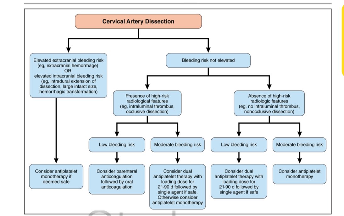
Antithrombotic treatment
Because
most ischemic events occur within the first several days after
diagnosis, optimizing antithrombotic treatment (when safe) early is of
paramount importance. In addition, it is reasonable that clinicians
start immediate antithrombotic treatment if deemed safe. Parenteral
followed by oral anticoagulation may be considered in particular in
patients at low risk of intracranial hemorrhage (small infarct size, no
intradural extension, and no intracranial hemorrhage) but at high risk
for ischemic stroke (eg, intraluminal thrombus, occlusive dissection).
Although the evidence for use in cervical artery dissection is weak, a
short course of dual antiplatelet therapy with a loading dose (followed
by single antiplatelet agent) might be preferred over monotherapy when
deemed safe, particularly in patients who would have qualified for the
dual antiplatelet trials for early prevention after minor stroke/TIA.
Otherwise, antiplatelet monotherapy could be used. In the absence of a
general superiority of any antithrombotic regimen, it is reasonable that
clinicians individualize treatment on the basis of the risk of ischemic
stroke and ICH, as well as a shared decision-making process with their
patients.
Antithrombotic treatment duration
It
is reasonable that the duration of antithrombotic therapy in patients
with cervical artery dissection be 3–6 mo. Decisions to extend
antithrombotic therapy past the 6-mo mark may be considered in the
context of an individual’s overall vascular risk factor profile and in
the context of neuroimaging features as remodeling occurs.
Subacute stenting
Patients
with cervical artery dissection with significant stenosis causing
distal hemodynamic compromise AND recurrent ischemic stroke despite
optimal medical treatment AND who can withstand surgery may be
considered for stenting as a measure for secondary stroke prevention.
Risk and predictors of recurrent dissection
The
risk of recurrent cervical artery dissection is 1%–2% per y but is
particularly increased in the first few months after the initial
cervical artery dissection. Younger age and fibromuscular dysplasia are
risk factors for dissection recurrence.
Precautions to reduce the risk of dissection recurrence
Because
the risk of recurrent or worsening dissection is highest in the first
few months after the initial dissection, it is reasonable that all
patients with cervical artery dissection avoid activities that increase
the risk of cervical injury for 1–6 mo from diagnosis and until healing
of the index dissection. Furthermore, although there are no proven
precautions to reduce the long-term risk of recurrent dissection, it is
reasonable for health care clinicians to suggest that patients with
cervical artery dissection who are at high risk for recurrent cervical
dissection (eg, known connective disorder, recurrent dissection) avoid
such activities lifelong.
Radiological recanalization of the dissection
Recanalization
of cervical artery dissections occurs mostly in the first 12 mo after
diagnosis. Cervical artery–dissecting aneurysms infrequently increase in
size, become symptomatic, and require treatment. Recanalization and
development or resolution of cervical artery–dissecting aneurysms do not
seem to be affected by antithrombotic treatment regimens.
CTA
indicates computed tomography angiography; DSA, digital subtraction
angiography; ICH, intracranial hemorrhage; IVT, intravenous
thrombolysis; MRA, magnetic resonance angiography; MRI, magnetic
resonance imaging; and TIA, transient ischemic stroke.
EPIDEMIOLOGY/RISK FACTORS
Cervical artery dissection contributes to 2% of all ischemic strokes4 but up to 25% of ischemic stroke in adults <50 years of age.5,6 Prior studies reported an incidence rate of 2.6 to 3.0 per 100 000 people4,7,8 but the true incidence is likely higher,9 partly because some patients with dissection may not seek medical attention because of self-limited or minor symptoms.
The mean age at diagnosis is 45 years.7
Although the proportion of dissection-related stroke is higher in
younger patients, the absolute prevalence of dissection-related ischemic
strokes increases with age.10 The incidence is slightly higher in men,11
but the age at onset and the peak age prevalence of dissection-related
stroke are lower in women (30–49 years) compared with men (50–89 years).10,11
The
pathogenesis of cervical artery dissection is multifactorial and
involves the interplay of comorbidities, environmental triggers, genetic
or congenital factors, including connective tissue disorders, and
anatomic factors such as elongated styloid process (>30 mm; Eagle
syndrome) or increased vascular tortuosity (Figure S1).1,12–14
Furthermore, risk factors may differ according to dissection location;
recent systemic infection is more common in carotid artery dissection,
and history of minor trauma is more common in vertebral artery
dissection.15
CHALLENGES IN DIAGNOSIS
Clinical Diagnostic Challenges
Because
of the often nonspecific nature of symptoms (eg, headache, neck pain,
dizziness, and tinnitus), diagnosis of cervical artery dissection is
challenging, especially in the absence of ischemic or localizing
symptoms. In a statewide study of claims data, 3.1% of patients with
cervical artery dissection were previously seen in the emergency
department for possible related symptoms in the 14 days preceding
diagnosis16; younger and female patients were more likely to have a probable missed diagnosis.16
Evaluation
for suspected cervical artery dissection should include a detailed
symptom history, questions about minor trauma, and a detailed
neurological examination. Common initial symptoms of cervical artery
dissection include facial pain, headache (≈65%), and neck pain (≈50%),15 which tend to precede cerebrovascular ischemic symptoms17,18;
thus, early diagnosis and treatment could reduce the risk of
cerebrovascular ischemia. Isolated pain (without cerebral ischemia)
occurs in 8% to 12% of diagnosed cases.19,20
For internal carotid dissections, pain is often facial, frontal, or
temporal, whereas vertebral artery dissections generally cause pain in
the cervical or occipital area.17,21 In carotid dissection, pain may also be associated with a (partial) Horner syndrome (≈25%7) and cranial nerve palsies (≈12%),22 and both carotid and vertebral dissection can be associated with pulsatile tinnitus (≈8%).17,23,24
In fact, a partial Horner syndrome in a patient with new or worsening
headache may suggest a carotid dissection diagnosis. Headache
characteristics are not specific but can rarely be acute and of
thunderclap nature.
Diagnostic Imaging Tools and Their Yield
The
diagnostic modalities for cervical artery dissection are magnetic
resonance imaging (MRI)/magnetic resonance angiography (MRA), computed
tomography angiography (CTA), ultrasound, and conventional digital
subtraction angiography (DSA). Historically, DSA has been considered the
reference imaging technique to demonstrate the lumen content and can
delineate the presence of an intimal flap, double lumen, and dissecting
aneurysm.25
DSA, however, provides limited information on the arterial wall (ie,
intramural hematoma). Moreover, DSA carries periprocedural risks (≈0.5%
iatrogenic dissection26 and 0.15% stroke).27
CTA
is an effective noninvasive alternative to DSA that can display luminal
contour alterations with rapid acquisition time, ready availability,
and high spatial resolution; disadvantages include the exposure to
radiation and iodinated contrast, and for intramural hematoma, CTA may
show an eccentric or crescent-shaped vessel wall thickening or the rind
sign in vertebral dissection28 (Figure 1),
but these findings are neither sensitive nor specific to cervical
artery dissection. A false-positive CTA can be due to streak artifact
mimicking a double lumen and pulsation artifact mimicking an intramural
hematoma. A false-negative CTA could be seen in the setting of a
non–flow-limiting nondominant vertebral artery dissection but most
commonly is due to failure of attributing an abnormality to dissection.29
Figure 1.Imaging signs suggestive of dissection diagnosis. A, Double lumen. B, Tapered or flame-shaped occlusion at typical dissection site. C, Intimal flap. D, Pseudoaneurysm. E, Intramural hematoma on fat-suppressed T1. F,
Carotid imaging duplex mural hematoma (white arrows) as hypoechogenic
structure compromising the arterial blood flow in the vertebral artery
(V3 segment). G, Axial source image from computed
tomography angiography (CTA) neck in a patient with complete occlusion
of the left internal carotid artery (ICA) related to a dissection,
showing the ring sign, with enhancement of the carotid wall and fresh
nonenhancing thrombus within the carotid lumen. This sign is nonspecific
for ICA dissection but points to a recent rather than a chronic
occlusion. H, Axial source image from CTA neck in a
patient with left vertebral artery dissection, with thrombus surrounding
the hypoplastic but apparently normal lumen vertebral artery, known as
the suboccipital rind sign, involving the atlanto-occipital (V3)
segment.
MRA may be superior to CTA
for the identification of the intramural hematoma when the appropriate
protocol including axial fat-suppressed T1-weighted images is used.
Limitations of MRA include the cost, availability, patient-related
restrictions (eg, pacemakers and claustrophobia), and diagnostic
limitations of time-of-flight MRA of the neck (eg, signal loss due to
turbulence or vessel turn, susceptibility to artifact, and flow reversal
artifact). False-positive MRA can be due to a hyperintense signal in
the setting of turbulent flow and adjacent structures simulating a
periarterial double-lumen sign. False-negative MRA can be related to the
absence of hyperintense signal of the intramural hematoma in the
hyperacute setting.29
Overall,
both MRA and CTA have good sensitivity and specificity for diagnosis of
cervical arterial dissection. MRA has high sensitivity in diagnosing
carotid dissection (95%), but its sensitivity is lower for vertebral
artery dissection (60%) compared with DSA.30
This is not the case with CTA, which has been shown to have similar
sensitivity and specificity compared with DSA in diagnosing vertebral
artery dissection.25
Ultrasound with color Doppler is noninvasive but is operator dependent and is of poor diagnostic utility,31
especially when the dissection is high cervical. Ultrasound may be
helpful in rare cases with hyperacute dissection where the intramural
hematoma can be visualized on ultrasound but not MRA (Figure 1).
Ultrasound generally requires confirmation by CTA or MRA. Ultrasound
has been shown to be useful for follow-up assessments within the first 4
weeks, when arterial remodeling is most prevalent.32
The findings could help in developing public policies to combat the risks of climate change.
High hourly heat exposure was associated with elevated risk for acute ischemic stroke, researchers reported in JAMA Network Open.
“A nationwide case-crossover investigation based on individual acute ischemic stroke onset time is imperative to quantify the association between acute ischemic stroke onset and hourly ambient heat exposure,” Xinlei Zhu, MS,
from the School of Public Health, Key Lab of Public Health Safety of
the Ministry of Education and National Health Commission Key Lab of
Health Technology Assessment, Fudan University, Shanghai, and colleagues
wrote. “As one of the world’s largest middle-income countries, China
has the highest stroke burden and is among the most vulnerable to global
climate change. Accordingly, in the present time-stratified
case-crossover analysis, we aimed to evaluate the association between
hourly high ambient temperature and the onset of acute ischemic stroke
using a nationwide registry of acute ischemic stroke. Additionally, we
assessed possible association modifiers, such as geographic region,
smoking, alcohol drinking, sex, age and history of other disease.”
The analysis involved 82,455 patients with acute ischemic stroke
(mean age, 66 years; 63% men) included in a national registry of more
than 200 stroke centers in China who were hospitalized in the warm
seasons from 2019 to 2021.
The researchers found that a monotonically increasing risk for acute
ischemic stroke onset was associated with higher temperatures, and that
the excess acute ischemic stroke risk occurred immediately at lag 0
hours and persisted for 10 hours.
Compared with the reference temperature of 12.1°C, the cumulative OR
over lag of 0 to 10 hours of acute ischemic stroke onset for extremely
high temperatures (33.3°C) was 1.88 (95% CI, 1.65-2.13), Zhu and
colleagues wrote.
The exposure-response curve was steeper in Northern institutions (OR =
1.8; 95% CI, 1.53-2.11) compared with Southern institutions (OR = 1.57;
95% CI, 1.31-1.87; P for z score = .27), according to the researchers.
The ORs were greater among men than women and for those with a
history of dyslipidemia or atrial fibrillation compared with those
without a history, but the differences were not significant, Zhu and
colleagues wrote.
The study “presents robust evidence of high temperature as an
important factor in increased risk of acute ischemic stroke onset,” the
researchers wrote. “The results add valuable insights to the adverse
cardiovascular outcome of climate warming and may benefit the
formulation of public health strategies to reduce cerebrovascular risk
associated with high ambient temperature under global warming.
Additionally, the findings underscore the ongoing need for public health
agencies to advocate for interventions that mitigate heat exposure and
bolster cooling measures, particularly among populations at high risk
for acute ischemic stroke.”
1School of Electrical and Information Engineering, Zhengzhou University, China
2Zhengzhou University, China
3The Rehabilitation Department, Fifth Affiliated Hospital of Zhengzhou University, China
The final, formatted version of the article will be published soon.
Stroke is the second leading cause of death globally and a
primary factor contributing to disability. Unilateral limb motor
impairment caused by stroke is the most common scenario. The bilateral
movement pattern plays a crucial role in assisting stroke survivors on
the affected side to relearn lost skills. However, motion compensation
often lead to decreased coordination between the limbs on both sides.
Furthermore, muscle fatigue resulting from imbalanced force exertion on
both sides of the limbs can also impact the rehabilitation outcomes.In
this study, an assessment method based on muscle synergy indicators was
proposed to objectively quantify the impact of motion compensation
issues on rehabilitation outcomes.Muscle synergy describes the body's
neuromuscular control mechanism, representing the coordinated activation
of multiple muscles during movement. 8 post-stroke hemiplegia patients
and 8 healthy subjects participated in this study. During hand-cycling
tasks with different resistance levels, surface electromyography signals
were synchronously collected from these participants before and after
fatigue. Additionally, a simulated compensation experiment was set up
for healthy participants to mimic various hemiparetic states observed in
patients.: Synergy symmetry and synergy fusion were chosen as potential
indicators for assessing motion compensation. The experimental results
indicate significant differences in synergy symmetry and fusion levels
between the healthy control group and the patient group (p ≤ 0.05), as
well as between the healthy control group and the compensation group.
Moreover, the analysis across different resistance levels showed no
significant variations in the assessed indicators (p > 0.05),
suggesting the utility of synergy symmetry and fusion indicators for the
quantitative evaluation of compensation behaviors. Although muscle
fatigue did not significantly alter the symmetry and fusion levels of
bilateral synergies (p > 0.05), it did reduce the synergy
repeatability across adjacent movement cycles, compromising movement
stability and hindering patient recovery. Based on synergy symmetry and
fusion indicators, the degree of bilateral motion compensation in
patients can be quantitatively assessed, providing personalized
recommendations for rehabilitation training and enhancing its
effectiveness.
You don't understand ONE GODDAMN THING ABOUT SURVIVOR MOTIVATION, DO YOU?
You create 100% recovery protocols and your survivor will be motivated
to do the millions of reps needed because they are looking forward to
100% recovery. GET THERE!
The problem is stroke researchers are not motivated to solve stroke. What the fuck is your solution to that failure? We still don't know how to motivate stroke medical 'professionals' to solve stroke to 100% recovery!
To cite this article: Claire Stewart, Emma Power, Annie McCluskey, Suzanne Kuys & Meryl Lovarini (22 Feb 2024): Implementing ward-based practice books to increase the amount of practice completed during inpatient stroke rehabilitation: a mixed-methods process evaluation, Disability and Rehabilitation, DOI: 10.1080/09638288.2024.2315502 To link to this article: https://doi.org/10.1080/09638288.2024.2315502
Claire Stewarta, Emma Powerb,c, Annie McCluskeyc,d, Suzanne Kuyse and Meryl Lovarinic aaustralasian Rehabilitation outcomes Centre, University of Wollongong, Wollongong, australia; bthe University of technology sydney, Graduate school of health, sydney, australia; csydney school of health sciences, the University of sydney, sydney, australia; dthe strokeed Collaboration, sydney, australia; eschool of allied health, australian Catholic University, banyo, australia
ABSTRACT
Purpose: Stroke survivors must complete large amounts of practice to achieve functional improvements but spend many hours inactive during their rehabilitation. We conducted a mixed methods process evaluation exploring factors affecting the success of a 6-month behaviour change intervention to increase use of ward-based practice books. Methods: Audits of the presence, quality and use of ward based-practice books were conducted, alongside focus groups with staff (n = 19), and interviews with stroke survivors (n = 3) and family members (n = 4). Quantitative data were analysed descriptively. Focus group and interview transcripts were analysed using qualitative analysis. Results: Personal (patient-related) factors (including severe weakness, cognitive and communication deficits of stroke survivors), staff coaching skills, understanding and beliefs about their role, affected practice book use. Staff turnover, nursing shift work and a lack of action planning reduced success of the behaviour change intervention. Conclusions: Staff with the necessary skills and understanding of their role in implementing ward practice overcame personal (patient-related) factors and assisted stroke survivors to successfully practice on the ward. To improve success of the intervention, repeated training of new staff is required. In addition to audit and feedback, team action planning is needed around the presence, quality, and use of ward practice books. h IMPLICATIONS FOR REHABILITATION • Ward-based practice books are one evidence-based strategy that can be used by rehabilitation teams to increase the amount of practice completed by stroke survivors during inpatient rehabilitation. • Stroke survivors’ personal factors (including severe weakness, cognitive and communication deficits), staff beliefs about their role and coaching skills, affected stroke survivors ability to practice on the ward using practice books. • Staff with the necessary skills, understanding and belief about their role in implementing ward practice can overcome personal (patient related) factors (such as severe weakness) and assist stroke survivors to successfully practice on the ward. • To increase the success of ward practice, repeated booster training of staff is required along with audit and feedback and team action planning on the presence, quality, and use of ward practice books
Since there is a 23% chance of stroke survivors getting PTSD what is your doctor doing to reduce that further risk of stroke due to PTSD? How EXACTLY is your doctor preventing PTSD from stroke? And then treating it?
Posttraumatic
stress disorder (PTSD) has been associated with ischemic heart disease
in women veterans, but evidence for associations with other
cardiovascular disorders remains limited in this population. This
retrospective longitudinal cohort study evaluated the association of
PTSD with incident stroke/transient ischemic attack (TIA) in women
veterans.
Methods and Results
Veterans
Health Administration electronic health records were used to identify
women veterans aged ≥18 years engaged with Veterans Health
Administration health care from January 1, 2000 to December 31, 2019. We
identified women veterans with and without PTSD without a history of
stroke or TIA at start of follow‐up. Propensity score matching was used
to match groups on age, race or ethnicity, traditional cardiovascular
risk factors, female‐specific risk factors, a range of mental and
physical health conditions, and number of prior health care visits.
PTSD, stroke, TIA, and risk factors used in propensity score matching
were based on diagnostic codes. Cox proportional hazards models were
used to estimate hazard ratios (HRs) and 95% CIs for associations of
PTSD with an incident stroke/TIA composite. Subanalyses considered
stroke and TIA separately, plus age‐ and race‐ or ethnicity‐stratified
analyses were carried out.
The analytic sample
included 208 092 women veterans (104 046 with and 104 046 without PTSD).
PTSD was associated with a greater rate of developing stroke/TIA (HR,
1.33 [95% CI, 1.25–1.42], P<0.001). This elevated risk was especially pronounced in women <50 years old and in Hispanic/Latina women.
Conclusions
Findings
indicate a strong association of PTSD with incident stroke/TIA in women
veterans. Research is needed to determine whether addressing PTSD and
its downstream consequences can offset this risk.
Predicting post stroke epilepsy rather than preventing it is the HEIGHT OF STUPIDITY! And you're still employed in stroke? Look how long we've known of the problem.
Recurrence in Post-Stroke seizure (PSS) is associated with significant functional decline in stroke patients.
•
Choice of anti-seizure medications (ASMs) may influence the morbidity and mortality.
•
Future research is needed to explore the effects of ASMs on PSS pathophysiology.
Abstract
Background
The
occurrence of seizures following a stroke is a well-recognized
complication associated with a significant increase in morbidity and
mortality. Despite the numerous studies examining outcomes and risk
factors related to post-stroke seizures (PSS), there remains a lack of
clarity(And you incompetently didn't create a protocol on how to prevent this problem?) regarding the clinical characteristics, treatment, and PSS
recurrence (PSSR) rates in patients experiencing their initial episode
of PSS.
Purpose
This
study aimed to determine the risk factors for developing recurrent
seizures after first PSS and their effects on functional outcomes and
mortality.
Methods
All
patients underwent an electroencephalography (EEG) and were monitored
for a minimum of 24 months following the first PSS. The primary endpoint
was the recurrence of seizures. Predictive factors for PSSR were
determined by using the Cox-proportional hazards model, and the
cumulative latency of recurrence at 90, 180, 360, and 720 days was
estimated using Kaplan-Meier analysis.
Results
Seizure
recurred in 36.8% (39/106). Significant association of PSSR was noted
with female gender, use of older anti-seizure medications (ASMs)
(p<0.001), EEG findings as focal slow wave activity (p<0.001),
Ictal epileptiform abnormalities (p=0.015), status epilepticus
(p=0.015), and with severe disability (p=0.008). However, multivariate
cox-proportional hazards model showed significant association of female
gender (HR=3.28; 95% CI: 1.42-7.58; p=0.006). Hazard ratio (HR) was
increased with older ASMs use, focal aware seizure types, Ictal EAs, and
periodic discharges on EEG; though, statistically significant.
Conclusion
Factors
such as the type of ASMs, EEG findings, and seizure type were
significantly linked to PSSR. Female gender was the only independent
predictor established. Additionally, significant functional decline was
reported with recurrence.
1Department and Research Institute of Rehabilitation Medicine, Yonsei University College of Medicine, Seoul, Republic of Korea
2Department of Neurosurgery and Brain Research Institute, Yonsei University College of Medicine, Seoul, Republic of Korea
3Graduate Program of Biomedical Engineering, Yonsei University College of Medicine, Seoul, Republic of Korea
4Brain Korea 21 FOUR Project for Medical Science, Yonsei University College of Medicine, Seoul, Republic of Korea
5Rehabilitation Institute of Neuromuscular Disease, Yonsei University College of Medicine, Seoul, Republic of Korea
Background: Intrathecal baclofen (ITB) therapy, a
viable alternative for unsuitable candidates of conventional spasticity
medications, is a preferred method of administration over the oral
route. Owing to its enhanced bioavailability, ITB ensures a more
effective delivery at the target site.
Objective: There is a lack of conclusive evidence
regarding the use of ITB treatment in managing ambulatory patients with
spastic dystonia. Before ITB pump implantation, patients commonly
undergo an ITB bolus injection trial to rule out potential adverse
reactions and verify the therapeutic effects on hypertonic issues. In
this report, we highlight a case of spastic dystonia, particularly
focusing on an ambulatory patient who demonstrated significant
improvement in both the modified Ashworth scale (MAS) score and gait
pattern following the ITB injection trial.
Case report: This case report outlines the
medical history of a 67-year-old male diagnosed with left-side
hemiplegia and spastic dystonia, resulting from his second episode of
intracranial hemorrhage in the right thalamus. An ITB injection trial
was initiated because the patient was not suitable for continued
botulinum toxin injections and oral medications. This was due to the
persistent occurrence of spastic dystonia in both the upper and lower
extremities. The patient underwent a four-day ITB injection trial with
progressively increasing doses, resulting in improved MAS scores and
gait parameters, including cadence, step length, step time, stride
length, and stride time were increased. Particularly, kinematic gait
analysis demonstrates a substantial improvement of increased knee
flexion in the swing phase in stiff knee gait pattern. These findings
indicated a gradual reduction in spasticity-related symptoms, signifying
the positive effect of the ITB injection trial. The patient eventually
received an ITB pump implantation.
Conclusion: In this post-stroke patient with
spastic dystonia, ITB therapy has demonstrated effective and substantial
management of spasticity, along with improvement in gait patterns.
Introduction
Spasticity is defined as velocity-dependent hypertonia
and tendon jerk hyperreflexia, resulting from stretch reflex
excitability (1, 2). In contrast, dystonia involves involuntary muscle contractions, often leading to abnormal posturing and twisting movements (3).
There are different types of dystonia, including focal dystonia such as
blepharospasm, oromandibular dystonia, spasmodic dysphonia,
torticollis, and writer’s cramp (4, 5). Generalized dystonia involves the leg, trunk, and at least one other body part (6).
Spastic dystonia, which is a specific aspect of the upper motor neuron
syndrome, is characterized by involuntary tonic contractions caused by
the inability to relax the muscles (1).
Spastic overactivity, a prevalent subtype of muscle hypertonia is
frequently observed in patients with spinal or cerebral dysfunction. The
term “spastic overactivity,” frequently employed to describe
stretch-sensitive muscle overactivity, poses difficulties in precisely
defining this intricate condition, as it includes spasms, involuntary
motions, and undesired muscle activity (7).
The primary treatment for muscle overactivity and
generalized dystonia typically involves oral medications, including
baclofen, benzodiazepines, anticholinergics, antispasmodics, and
levodopa (8, 9).
Baclofen, an agonist for gamma-aminobutyric acid (GABA) B receptors,
inhibits mono- and polysynaptic reflexes in the central and peripheral
nervous systems, contributing to its therapeutic effect in reducing
spasticity (10).
Previous research explored the effectiveness of oral baclofen in
managing poststroke spasticity, focusing on ankle stiffness and clonus
during passive stretch, and identified factors influencing
responsiveness (2).
Nevertheless, oral baclofen may not always be effective for all
patients due to the low concentration of baclofen in the cerebrospinal
fluid (2).
These non-responders exhibited a positive effect at higher
cerebrospinal fluid levels achievable through intrathecal baclofen
injection (2). Benzodiazepines demonstrate limited effectiveness, with approximately 16 to 23% showing a good clinical response (11).
Trihexyphenidyl, a widely used agent, is a muscarinic acetylcholine
receptor antagonist with a notably variable therapeutic dosage. While
children may effectively respond to doses as low as 4 mg/day, adults are
less likely to tolerate increasing dosages when necessary as they tend
to be more sensitive to side effects such as memory loss, confusion,
restlessness, insomnia, and nightmares (11). Intramuscular botulinum toxin injections can be beneficial in reducing focal spasticity in specific muscle groups (12, 13).
Adverse effects, including dry mouth, fatigue, and flu-like symptoms,
may occur when the drug spreads to unintended muscles and organs during
botulinum toxin therapy (13).
In some cases, these manifestations can escalate to systemic botulism
signs. Although recent studies support the safety of botulinum toxin
high-dose therapy, official documents recommend maximum botulinum toxin
doses of onabotulinumtoxinA (Botox®, Allergan, Irvine, CA, USA)
400 units for upper limb or lower limb spasticity (14, 15).
In the 1980s, intrathecal baclofen (ITB) therapy emerged as potentially
effective next-in-line management for severe spasticity unresponsive to
oral medications (9, 16–18), delivering concentrated drug doses directly into the intrathecal space to minimize systemic toxicity (19).
Given the delicate nature of baclofen therapy and its potential
complications, ITB bolus injections are administered to rule out adverse
reactions and ensure the drug’s therapeutic effect on hypertonia before
ITB pump implantation (9).
ITB therapy offers reversible treatment of spasticity and effective
modulation of muscle tone to reduce severe spasticity, for it was
approved for the management of spasticity (20, 21).
However, current literature highlights the necessity for additional
research on the effectiveness of ITB treatment for mixed hypertonia,
including spastic dystonia (21). In particular, additional research dedicated to ambulatory patient is essential.
Evaluating the effectiveness of treatment is crucial in
managing spastic overactivity, and commonly employed methods include
high-speed passive muscle stretching assessments like the modified
Ashworth scale (MAS) and the modified Tardieu scale. However, these
evaluations solely gauge resistance to passive movement and lack insight
into muscle dynamics during dynamic tasks like gait (22).
These scales also rely on subjective interpretation by clinicians,
making it challenging to differentiate specific types of spastic
hyperactivity, such as spasticity, spastic dystonia, and spastic
co-contraction. Therefore, surface electromyography or gait analysis
reflecting muscle activity during dynamic tasks has recently been used
to classify spastic overactivity in detail (23, 24).
Gait analysis, in particular, assesses muscle hyperactivity during
functional tasks, allowing for a comprehensive understanding of specific
movement patterns and continuous monitoring of various gait factors (23).
Moreover, a transient decrease in spastic dystonia following ITB
therapy can induce changes in gait pattern that are not easily
discernible through casual observation. In such cases, gait analysis
offers precise and objective measurements, detecting even subtle
modifications in ambulatory performance (25).
Herein, we report an ambulatory patient with spastic
dystonia demonstrating significant improvement in both the MAS score and
gait pattern following the ITB bolus injection trial. The objective of
this case study is to enhance comprehension of the response of adult
spastic dystonia patients to ITB bolus injections and identify
prospective recipients who may experience improved gait after ITB pump
implantation. This study also represents the first stage in assessing
whether the walking performance observed during the ITB bolus injection
trial, evaluated through computerized gait analysis, is associated with
changes following ITB pump implantation.

 Follow Us




 Yanhong Liu
 Yaowei Li
 Zan Zhang
 Benyan Huo
 Anqin Dong
 Kyung Min Kim
 Tae Kwon Lee
 Su Min Lee
 Won Seok Chang
 Su Ji Lee1
 Jihye Hwang
 Sung-Rae Cho
