I thought I'd analyze myself. I know I shouldn't be doing a doctors' work but being a stroke survivor you just automatically assume that your doctor knows nothing and will do nothing useful.
http://www.alphagalileo.org/ViewItem.aspx?ItemId=143428&CultureCode=en
How often do you have six or more drinks on one occasion?
At our monthly wine potlucks I probably have six drinks over the course of 6 hours.
As a result of your drinking, did anything happen in the last year that you wish didn’t happen?
Nope.
More at link if you fail these questions.
Monday, July 7, 2014


Saturday, July 5, 2014
INCOG Guidelines for Cognitive Rehabilitation Following Traumatic Brain Injury: Methods and Overview
You will need to ask your doctor if any of this is applicable to your stroke rehabilitation. If there is anything like this for stroke it obviously is not public. That is what our damnable stroke associations should be doing for us but we get useless prevention tips and F.A.S.T.
Good luck on getting anything from your doctor.
http://www.ncbi.nlm.nih.gov/pubmed/24984093
Good luck on getting anything from your doctor.
http://www.ncbi.nlm.nih.gov/pubmed/24984093
Abstract
INTRODUCTION:
Traumatic brain injury results in complex cognitive sequelae. However, clinicians have difficulty implementing the available evidence. An international group of researchers and clinicians (known as INCOG) convened to develop clinical practice guidelines for cognitive rehabilitation posttraumatic brain injury.METHODS:
The Guidelines Adaptation and Development cycle was used to derive the recommendations. Previously published cognitive rehabilitation recommendations were identified and tabulated. An expert panel met to select appropriate recommendations. Afterward, the team enhanced the recommendations by reviewing available literature. To address shortfalls of previous guidelines, the team developed decision algorithms incorporating the recommendations based on inclusion and exclusion criteria of published trials and expert opinion. The team then prioritized the recommendations for implementation and developed audit criteria to evaluate adherence to best practice.RESULTS:
The team recommends that individuals have detailed assessments of cognition after resolution of posttraumatic amnesia. Cognitive assessment and rehabilitation should be tailored to the patient's neuropsychological profile, premorbid cognitive characteristics, and goals for life activities and participation. Clinical algorithms and audit tools to evaluate current practice are provided.CONCLUSION:
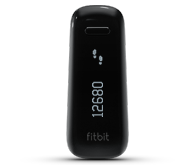
Cognitive rehabilitation should be offered to select??? individuals with traumatic brain injury. These guidelines provide assistance to clinicians who want to provide evidence-based care.Fitbit
Just got one. My first wearing of it was to clear off some more waist high grasses in the farthest loop in the natural area using this;

The two inner loops are mostly done. The 2 16-18 inch trees lying across those trails can by bypassed.
I probably have 200 yards of waist high grass to go yet. Put in 3.12 miles, 2017 calories, 6516 steps, 0 stairs. It's good to know how far I'm walking since I need to lose a fair amount of weight. My social activities have caused me to gain weight. They were worth it.
The two inner loops are mostly done. The 2 16-18 inch trees lying across those trails can by bypassed.
First evidence for painless Atrial Fibrillation treatment
Remember only your doctor can tell you about your medical options. That however does assume your doctor knows about the available options. As a former project manager once told her group, never assume, 'That just makes an ass out of u and me' e.g. ass-u-me. Good luck treating your doctors lack of knowledge.
http://www.alphagalileo.org/ViewItem.aspx?ItemId=143268&CultureCode=en
Cardiac optogenetics achieve defibrillation without the pain of electric shocks
The first evidence for a shockless treatment for atrial fibrillation (AF) will be presented today at Frontiers in CardioVascular Biology (FCVB) 2014 in Barcelona, Spain. The meeting is organised by the Council on Basic Cardiovascular Science of the European Society of Cardiology (ESC) in collaboration with 13 European cardiovascular science societies. http://spo.escardio.org/SessionDetails.aspx?eevtid=65&sessId=13104
Dr Brian O. Bingen, first author, said: “AF is the most common cardiac arrhythmia. Symptoms range from the feeling of fish flapping in the chest, to tiredness and exercise intolerance. AF can lead to tachycardia induced cardiomyopathy and thromboembolic events which increase the risk of morbidity and death.”
He added: “Preventing these symptoms and complications requires bringing the patient out of AF and back to the normal sinus rhythm. The quickest way to do that is to deliver an electric shock. The shock depolarises and synchronises the heart muscle and allows the sinus node to re-establish a normal rhythm.”
Dr Bingen continued: “Shocks are currently the most effective way to get patients directly back into sinus rhythm but they are very painful. To deliver a shock you have to give anaesthesia which comes with its own possible adverse effects.”
AF usually progresses from a paroxysmal form, in which episodes of AF last from several minutes up to 7 days, to a persistent and eventually a chronic form. People with the latter are in AF 24 hours a day, 7 days a week, and shock treatment no longer works. Dr Bingen said: “AF causes structural changes to the atrium which make patients more prone to subsequent induction of AF. That’s another reason to get patients back into sinus rhythm as soon as possible.”
For the current study, the researchers devised a method of shockless defibrillation. They used optogenetics to genetically insert depolarising ion channels into the heart that can be activated by light. Dr Bingen said: “The theory was that we could just turn a light switch on and depolarise the entire myocardium without needing a shock. In theory, the patient could be given an implantable device with a mesh of light emitting diodes (LEDs) and when AF occurs you turn the light on and the AF stops.”
During arrhythmias there is activity subepicardially but the heart is a complex 3D structure and it is only possible to directly observe the epicardium (outside layer). To see how their method worked subepicardially, the researchers developed 2D hearts. They isolated cardiac muscle cells from the rat atrium, replated them in a culture dish and allowed the cells to form intercellular connections, creating a 2D heart.
AF was induced in 31 of these 2D hearts. The researchers used a lentivirus to insert a gene into the 2D hearts called calcium-translocating channelrhodopsin (CatCh), which is a light sensitive depolarising channel.
Dr Bingen said: “Then it was just a matter of switching on the light and seeing what happened. We found that in all 31 of these 2D hearts we were able to achieve the 2D equivalent of cardioversion into sinus rhythm. The mechanism we saw was a bit different than the normal defibrillation but was equally effective.”
He continued: “We now have to test our method in the 3D setting. In that scenario we won’t be able to see the defibrillating mechanism in as much detail, but we hope that it will be possible to terminate AF in the complete heart. We will also test other types of light or energy sources that penetrate the body more deeply and could be applied externally, avoiding the need for an implanted device.”
Dr Bingen continued: “This is the first evidence of a shockless defibrillation. Our method of using optogenetics to defibrillate by light is completely painless and looks promising but more research is needed before it can be applied in patients.”
Please note Spanish version is available for this Press Release
http://www.alphagalileo.org/ViewItem.aspx?ItemId=143268&CultureCode=en
Cardiac optogenetics achieve defibrillation without the pain of electric shocks
The first evidence for a shockless treatment for atrial fibrillation (AF) will be presented today at Frontiers in CardioVascular Biology (FCVB) 2014 in Barcelona, Spain. The meeting is organised by the Council on Basic Cardiovascular Science of the European Society of Cardiology (ESC) in collaboration with 13 European cardiovascular science societies. http://spo.escardio.org/SessionDetails.aspx?eevtid=65&sessId=13104
Dr Brian O. Bingen, first author, said: “AF is the most common cardiac arrhythmia. Symptoms range from the feeling of fish flapping in the chest, to tiredness and exercise intolerance. AF can lead to tachycardia induced cardiomyopathy and thromboembolic events which increase the risk of morbidity and death.”
He added: “Preventing these symptoms and complications requires bringing the patient out of AF and back to the normal sinus rhythm. The quickest way to do that is to deliver an electric shock. The shock depolarises and synchronises the heart muscle and allows the sinus node to re-establish a normal rhythm.”
Dr Bingen continued: “Shocks are currently the most effective way to get patients directly back into sinus rhythm but they are very painful. To deliver a shock you have to give anaesthesia which comes with its own possible adverse effects.”
AF usually progresses from a paroxysmal form, in which episodes of AF last from several minutes up to 7 days, to a persistent and eventually a chronic form. People with the latter are in AF 24 hours a day, 7 days a week, and shock treatment no longer works. Dr Bingen said: “AF causes structural changes to the atrium which make patients more prone to subsequent induction of AF. That’s another reason to get patients back into sinus rhythm as soon as possible.”
For the current study, the researchers devised a method of shockless defibrillation. They used optogenetics to genetically insert depolarising ion channels into the heart that can be activated by light. Dr Bingen said: “The theory was that we could just turn a light switch on and depolarise the entire myocardium without needing a shock. In theory, the patient could be given an implantable device with a mesh of light emitting diodes (LEDs) and when AF occurs you turn the light on and the AF stops.”
During arrhythmias there is activity subepicardially but the heart is a complex 3D structure and it is only possible to directly observe the epicardium (outside layer). To see how their method worked subepicardially, the researchers developed 2D hearts. They isolated cardiac muscle cells from the rat atrium, replated them in a culture dish and allowed the cells to form intercellular connections, creating a 2D heart.
AF was induced in 31 of these 2D hearts. The researchers used a lentivirus to insert a gene into the 2D hearts called calcium-translocating channelrhodopsin (CatCh), which is a light sensitive depolarising channel.
Dr Bingen said: “Then it was just a matter of switching on the light and seeing what happened. We found that in all 31 of these 2D hearts we were able to achieve the 2D equivalent of cardioversion into sinus rhythm. The mechanism we saw was a bit different than the normal defibrillation but was equally effective.”
He continued: “We now have to test our method in the 3D setting. In that scenario we won’t be able to see the defibrillating mechanism in as much detail, but we hope that it will be possible to terminate AF in the complete heart. We will also test other types of light or energy sources that penetrate the body more deeply and could be applied externally, avoiding the need for an implanted device.”
Dr Bingen continued: “This is the first evidence of a shockless defibrillation. Our method of using optogenetics to defibrillate by light is completely painless and looks promising but more research is needed before it can be applied in patients.”
Please note Spanish version is available for this Press Release
Reliability of the Balance Evaluation Systems Test (BESTest) and BESTest sections for adults with hemiparesis
This is still a subjective test. I never really passed the Berg Balance Test but the only time I fell was on snow or ice.
http://www.scielo.br/scielo.php?pid=S1413-35552014005040033&script=sci_arttext
http://www.scielo.br/scielo.php?pid=S1413-35552014005040033&script=sci_arttext
1Graduate Program in Human Movement Sciences, Health
Sciences and Sports Center, Universidade do Estado de Santa Catarina (UDESC),
Florianópolis, SC, Brazil
2Physical Therapy Department, Health Sciences and
Sports Center, UDESC, Florianópolis, SC, Brazil
3Master's Program in Physical Therapy, UDESC,
Florianópolis, SC, Brazil
BACKGROUND:
The Balance Evaluation Systems Test (BESTest) was recently created to allow the
development of treatments according to the specific balance system affected in
each patient. The Brazilian version of the BESTest has not been specifically
tested after stroke.
OBJECTIVE:
To evaluate the intra- and inter-rater reliability and concurrent and convergent
validity of the total score of the BESTest and BESTest sections for adults with
hemiparesis after stroke.
METHOD:
The study included 16 subjects (61.1±7.5 years) with chronic hemiparesis
(54.5±43.5 months after stroke). The BESTest was administered by two raters in the
same week and one of the raters repeated the test after a one-week interval.
Intraclass correlation coefficient (ICC) was calculated to assess intra- and
interrater reliability. Concurrent validity with the Berg Balance Scale (BBS) and
convergent validity with the Activities-specific Balance Confidence scale
(ABC-Brazil) were assessed using Pearson's correlation coefficient.
RESULTS:
Both the BESTest total score (ICC=0.98) and the BESTest sections (ICC between
0.85 and 0.96) have excellent intrarater reliability. Interrater reliability for
the total score was excellent (ICC=0.93) and, for the sections, it ranged between
0.71 and 0.94. The correlation coefficient between the BESTest and the BBS and
ABC-Brazil were 0.78 and 0.59, respectively.
CONCLUSIONS:
The Brazilian version of the BESTest demonstrated adequate reliability when
measured by sections and could identify what balance system was affected in
patients after stroke. Concurrent validity was excellent with the BBS total score
and good to excellent with the sections. The total scores but not the sections
present adequate convergent validity with the ABC-Brazil. However, other
psychometric properties should be further investigated.Friday, July 4, 2014
Decision Support for Stroke Rehabilitation Therapy via Describable Attribute-based Decision Trees
Does your therapist have an objective decision tree for how they approach your therapy? Or are they enamored of 'All strokes are different, all stroke recoveries are different'?
http://www.public.asu.edu/~pturaga/papers/StrokeDecisionTree.pdf
http://www.public.asu.edu/~pturaga/papers/StrokeDecisionTree.pdf
Vinay Venkataraman, Pavan Turaga, Nicole Lehrer, Michael Baran, Thanassis Rikakis, and Steven L. Wolf
Abstract
—This paper proposes a computational framework
for movement quality assessment using a decision tree model
that can potentially assist a physical therapist in a telereha-
bilitation context. Using a dataset of key kinematic attributes
collected from eight stroke survivors, we demonstrate that the
framework can be reliably used for movement quality assess-
ment of a reach-to-grasp cone task, an activity commonly used
in upper extremity stroke rehabilitation therapy. The proposed
framework is capable of providing movement quality scores that
are highly correlated to the ratings provided by therapists, who
used a custom rating rubric created by rehabilitation experts.
Our hypothesis is that a decision tree model could be easily
utilized by therapists as a potential assistive tool, especially in
evaluating movement quality on a large-scale dataset collected
during unsupervised rehabilitation (e.g., training at the home),
thereby reducing the time and cost of rehabilitation treatment.
6 more pages and some great math equations.
Adult neurogenesis and its promise as a hope for brain repair
It has been 52 years since adult neurogenesis was discovered so I'm sure we won't get a breakthrough to help us survivors in our lifetime. Read it all at the link.
http://journal.frontiersin.org/Journal/10.3389/fnins.2014.00165/full?
http://journal.frontiersin.org/Journal/10.3389/fnins.2014.00165/full?
Do not disturb! How the brain filters out distractions
This seems incredibly important for our doctors to be able to fix in stroke survivors. Everyone I know complains about not being able to concentrate on tasks due to distractions.
http://www.alphagalileo.org/ViewItem.aspx?ItemId=143385&CultureCode=en
Tübingen neuroscientists: brain uses varying strategies to prevent the notepad of working memory from being wiped clean
You know the feeling? You are trying to dial a phone number from memory… you have to concentrate…. then someone starts shouting out other numbers nearby. In a situation like that, your brain must ignore the distraction as best it can so as not to lose vital information from its working memory. A new paper published in Neuron by a team of neurobiologists led by Professor Andreas Nieder at the University of Tübingen gives insight into just how the brain manages this problem.
The researchers put rhesus monkey in a similar situation. The monkeys had to remember the number of dots in an image and reproduce the knowledge a moment later. While they were taking in the information, a distraction was introduced, showing a different number of dots. And even though the monkeys were mostly able to ignore the distraction, their concentration was disturbed and their memory performance suffered.
Measurements of the electrical activity of nerve cells in two key areas of the brain showed a surprising result: nerve cells in the prefrontal cortex signaled the distraction while it was being presented, but immediately restored the remembered information (the number of dots) once the distraction was switched off. In contrast, nerve cells in the parietal cortex were unimpressed by the distraction and reliably transmitted the information about the correct number of dots.
These findings provide important clues about the strategies and division of labor among different parts of the brain when it comes to using the working memory. “Different parts of the brain appear to use different strategies to filter out distractions,” says Dr. Simon Jacob, who carried out research in Tübingen before switching to the Psychiatric Clinic at the Charité hospitals in Berlin. “Nerve cells in the parietal cortex simply suppress the distraction, while nerve cells in the prefrontal cortex allow themselves to be momentarily distracted – only to return immediately to the truly important memory content.”
The researchers were surprised by the two brain areas’ difference in sensitivity to distraction. “We had assumed that the prefrontal cortex is able to filter out all kinds of distractions, while the parietal cortex was considered more vulnerable to disturbances,” says Professor Nieder. “We will have to rethink that. The memory-storage tasks and the strategies of each brain area are distributed differently from what we expected.”
http://www.alphagalileo.org/ViewItem.aspx?ItemId=143385&CultureCode=en
Tübingen neuroscientists: brain uses varying strategies to prevent the notepad of working memory from being wiped clean
You know the feeling? You are trying to dial a phone number from memory… you have to concentrate…. then someone starts shouting out other numbers nearby. In a situation like that, your brain must ignore the distraction as best it can so as not to lose vital information from its working memory. A new paper published in Neuron by a team of neurobiologists led by Professor Andreas Nieder at the University of Tübingen gives insight into just how the brain manages this problem.
The researchers put rhesus monkey in a similar situation. The monkeys had to remember the number of dots in an image and reproduce the knowledge a moment later. While they were taking in the information, a distraction was introduced, showing a different number of dots. And even though the monkeys were mostly able to ignore the distraction, their concentration was disturbed and their memory performance suffered.
Measurements of the electrical activity of nerve cells in two key areas of the brain showed a surprising result: nerve cells in the prefrontal cortex signaled the distraction while it was being presented, but immediately restored the remembered information (the number of dots) once the distraction was switched off. In contrast, nerve cells in the parietal cortex were unimpressed by the distraction and reliably transmitted the information about the correct number of dots.
These findings provide important clues about the strategies and division of labor among different parts of the brain when it comes to using the working memory. “Different parts of the brain appear to use different strategies to filter out distractions,” says Dr. Simon Jacob, who carried out research in Tübingen before switching to the Psychiatric Clinic at the Charité hospitals in Berlin. “Nerve cells in the parietal cortex simply suppress the distraction, while nerve cells in the prefrontal cortex allow themselves to be momentarily distracted – only to return immediately to the truly important memory content.”
The researchers were surprised by the two brain areas’ difference in sensitivity to distraction. “We had assumed that the prefrontal cortex is able to filter out all kinds of distractions, while the parietal cortex was considered more vulnerable to disturbances,” says Professor Nieder. “We will have to rethink that. The memory-storage tasks and the strategies of each brain area are distributed differently from what we expected.”
Test your stress level with this free brain teaser
I only found one difference so I'm good to go. Your doctor immediately upon you entering the hospital should be testing your stress and giving you protocols to ameliorate that to make sure it doesn't turn into PTSD.
http://sharpbrains.com/blog/2014/07/04/test-your-stress-level-with-this-free-brain-teaser/
Does your doctor and hospital even know about these problems and how to correct them?
http://sharpbrains.com/blog/2014/07/04/test-your-stress-level-with-this-free-brain-teaser/
Does your doctor and hospital even know about these problems and how to correct them?
When a Stressful Hospital Stay Makes You Sick
Self-Tuning Neurons Promote Resilience to Stress, Depression
Study: Stress Makes Women Nicer and Men Meaner
A Whole New Way To Think About Stress That Changes Everything We've Been Taught
How to make stress your friend
Mindfulness meditation may ease anxiety, mental stress
Nociceptin: Nature’s Balm for the Stressed Brain
Epigenetics and the regulation of stress vulnerability and resilience
Being a grump only makes things worse: a transactional account of acute stress on mind wandering
636,120 Ways to Have Posttraumatic Stress Disorder
Understanding stress resilience
The role of orphanin FQ/nociceptin in neuroplasticity: relationship to stress, anxiety and neuroinflammation
Arousal and Physiological Toughness: Implications for Mental and Physical Health
Sexual activity counteracts the suppressive effects of chronic stress on adult hippocampal neurogenesis and recognition memory
Does psychosocial distress elevate your risk of stroke?
This heart rate sensor is worn around the ear, connects with an app aimed at reducing stress
Deric Bownds - Neural mechanisms of stress vulnerability and resilience
Psychosocial Distress Associated With Increased Stroke Risk
How to Prevent Stress from Shrinking Your Brain
Chewing during chronic stress ameliorates stress-induced suppression of neurogenesis in the hippocampal dentate gyrus in aged SAMP8 mice
How stress eats holes in your brain
Religious Experiences Shrink Part of the Brain
I'd be willing to bet your doctor has not read a single one of the underlying research articles. So they are relying on outdated knowledge from their medical training. Ask them what the last research article they read having to do with stroke.
Thursday, July 3, 2014
Does your stroke doctor believe in magic?
I bet they will say no.
But have they said, 'All strokes are different, all stroke recoveries are different'? Total magical expectation there.
'You will fully recover.' With only 10% getting to full recovery, that is a whopping lot of magical thinking.
'If you work on movements and exercise, neuroplasticity will help you recover' They have no explanation for how it works so it has to be magic.
And to think we are actually paying these people for their magical wishes.
But have they said, 'All strokes are different, all stroke recoveries are different'? Total magical expectation there.
'You will fully recover.' With only 10% getting to full recovery, that is a whopping lot of magical thinking.
'If you work on movements and exercise, neuroplasticity will help you recover' They have no explanation for how it works so it has to be magic.
And to think we are actually paying these people for their magical wishes.
Why aren't we doing eStim to relax spastic muscles?
I'm obviously stupid because electrical signals are needed to contract and relax muscles. Why can't we use eStim to relax spastic muscles? I'm sure all the therapists out there know the answer and are laughing at me. Amy, stop laughing.
The application of precisely controlled functional electrical stimulation to the shoulder, elbow and wrist for upper limb stroke rehabilitation: a feasibility study
This is great but we need someone to write up an exact stroke protocol from this. Otherwise we'll end up with hundreds of thousands of therapists creating something different.
http://www.jneuroengrehab.com/content/11/1/105/abstract
http://www.jneuroengrehab.com/content/11/1/105/abstract
Journal of NeuroEngineering and Rehabilitation 2014, 11:105
doi:10.1186/1743-0003-11-105
Published: 30 June 2014
Published: 30 June 2014
Abstract (provisional)
Background
Functional electrical stimulation (FES) during repetitive practice of everyday tasks
can facilitate recovery of upper limb function following stroke. Reduction in impairment
is strongly associated with how closely FES assists performance, with advanced iterative
learning control (ILC) technology providing precise upper-limb assistance. The aim
of this study is to investigate the feasibility of extending ILC technology to control
FES of three muscle groups in the upper limb to facilitate functional motor recovery
post-stroke.
Methods
Five stroke participants with established hemiplegia undertook eighteen intervention
sessions, each of one hour duration. During each session FES was applied to the anterior
deltoid, shoulder, and wrist/finger extensors to assist performance of functional
tasks with real-objects, including closing a drawer and pressing a light switch. Advanced
model-based ILC controllers used kinematic data from previous attempts at each task
to update the FES applied to each muscle on the subsequent trial. This produced stimulation
profiles that facilitated accurate completion of each task while encouraging voluntary
effort by the participant. Kinematic data were collected using a Microsoft Kinect,
and mechanical arm support was provided by a SaeboMAS. Participants completed Fugl-Meyer
and Action Research Arm Test clinical assessments pre- and post-intervention, as well
as FES-unassisted tasks during each intervention session.
Results
Fugl-Meyer and Action Research Arm Test scores both significantly improved from pre-
to post-intervention by 4.4 points. Improvements were also found in FES-unassisted
performance, and the amount of arm support required to successfully perform the tasks
was reduced.
Conclusions
This feasibility study indicates that technology comprising low-cost hardware fused
with advanced FES controllers accurately assists upper limb movement and may reduce
upper limb impairments following stroke.
The complete article is available as a provisional PDF. The fully formatted PDF and HTML versions are in production.
Turn your wheelchair into a motorcycle
Batec E-Handbike - Electric powered front wheel that attaches to your wheelchair
You will have to run it thru Google translate here;
https://translate.google.com/translate?sl=auto&tl=en&js=y&prev=_t&hl=en&ie=UTF-8&u=http%3A%2F%2Fbatec-mobility.com&edit-text=


http://batec-mobility.com
Other mobility possibilities here:
Or maybe you'd prefer one of these other ones
http://www.wimp.com/newdevice/
http://inhabitat.com/equal-a-compact-electric-vehicle-specially-designed-for-people-with-disabilities/


You will have to run it thru Google translate here;
https://translate.google.com/translate?sl=auto&tl=en&js=y&prev=_t&hl=en&ie=UTF-8&u=http%3A%2F%2Fbatec-mobility.com&edit-text=
The Spanish version here if you want to practice your Spanish.
http://batec-mobility.com
Other mobility possibilities here:
Or maybe you'd prefer one of these other ones
http://www.wimp.com/newdevice/

or Hondas;

Or this;
Or this cool one;
Or this;

Or a:
Extreme Offroad Tracked Wheelchair the Original Ripchair 2.0
Sue Austin: Deep sea diving … in a wheelchair
Smartest Neurologist Quiz: Dementia
From looking at the questions you can see why neurologists don't have any clue about dementia prevention. The smartest neurologists obviously need to know nothing about that. We are totally f*cking screwed until we overthrow the complete stroke medical system.Who gives a shit about your 33% dementia chance post-stroke from an Australian study?
I however will prevent my descent into dementia like this:
Do not follow my ideas, they are not medically approved.
Dear Smartest Neurologist Player,
We invite you to take the
"Dementia" Quiz
>>Click Here to Play! |
When the Joint Commission is at the hospital, leave
This might not be possible for us as stroke patients. A great blog posting on MedPageTodays' KevinMD.com
I personally believe the Joint Commissions stuff on stroke is worthless.
You can check out Joint Commission standards here:
http://www.jointcommission.org/assets
I saw absolutely nothing about what should be done the first week or anything about measuring 30-day deaths and 100% recovery.
I personally believe the Joint Commissions stuff on stroke is worthless.
You can check out Joint Commission standards here:
http://www.jointcommission.org/assets
I saw absolutely nothing about what should be done the first week or anything about measuring 30-day deaths and 100% recovery.
The Comforting Trap of Blaming the Victim
An excellent writeup By Heidi Moawad, MD
http://stroke.about.com/od/rehab/fl/The-Comforting-Trap-of-Blaming-the-Victim.htm?nl=1
Our stroke associations have perfected this by only focusing on prevention messages and F.A.S.T.
F.A.S.T. is a wonderful way to blame the victim. You didn't recognize you had a stroke and get to the hospital fast enough to avail yourself of the medical interventions of stroke. Nevermind tPA hiding behind the curtain with only a 12% efficacy. This may be the best they can do right now but it leads directly to blaming the victim.
----------------------------------------------------------------------------------------------------
ASA statement;
Use FAST to remember the warning signs:
F. FACE:
Ask the person to smile.
Does one side of the face droop?
A. ARMS:
Ask the person to raise both arms.
Does one arm drift downward?
S. SPEECH:
Ask the person to repeat a simple phrase.
Is their speech slurred or strange?
T. TIME:
If you observe any of these signs,
call 9-1-1 immediately.
----------------------------------------------------------------------------------------------------------
WSO statement;
These are the warning signs that someone is having a stroke:
Call your emergency medical services and get to a hospital right away!
http://stroke.about.com/od/rehab/fl/The-Comforting-Trap-of-Blaming-the-Victim.htm?nl=1
Our stroke associations have perfected this by only focusing on prevention messages and F.A.S.T.
F.A.S.T. is a wonderful way to blame the victim. You didn't recognize you had a stroke and get to the hospital fast enough to avail yourself of the medical interventions of stroke. Nevermind tPA hiding behind the curtain with only a 12% efficacy. This may be the best they can do right now but it leads directly to blaming the victim.
----------------------------------------------------------------------------------------------------
ASA statement;
THINK YOU ARE HAVING A STROKE? CALL 9-1-1 IMMEDIATELY!
F.A.S.T. is an easy way to remember the sudden signs of stroke. When you can spot the signs, you'll know that you need to call 9-1-1 for help right away. F.A.S.T. is:
F. Face Drooping – Does one side of the face droop or is it numb? Ask the person to smile. Is the person's smile uneven?
A. Arm Weakness – Is one arm weak or numb? Ask the person to raise both arms. Does one arm drift downward?
S. Speech Difficulty
– Is speech slurred? Is the person unable to speak or hard to
understand? Ask the person to repeat a simple sentence, like "The sky is
blue." Is the sentence repeated correctly?
T. Time to call 9-1-1
– If someone shows any of these symptoms, even if the symptoms go away,
call 9-1-1 and get the person to the hospital immediately. Check the
time so you'll know when the first symptoms appeared.
-----------------------------------------------------------------------------------------------------------
NSA statement:
Learn the many warning signs of a stroke. Act FAST and
CALL 9-1-1 IMMEDIATELY at any sign of a stroke.Use FAST to remember the warning signs:
F. FACE:
Ask the person to smile.
Does one side of the face droop?
A. ARMS:
Ask the person to raise both arms.
Does one arm drift downward?
S. SPEECH:
Ask the person to repeat a simple phrase.
Is their speech slurred or strange?
T. TIME:
If you observe any of these signs,
call 9-1-1 immediately.
----------------------------------------------------------------------------------------------------------
WSO statement;
Stroke Warning Signs
- Sudden numbness or weakness of the face, arm or leg, especially on one side of the body
- Sudden confusion, trouble speaking or understanding
- Sudden trouble seeing in one or both eyes
- Sudden trouble walking, dizziness, loss of balance or coordination
- Sudden, severe headache with no known cause
Call your emergency medical services and get to a hospital right away!
Dark Chocolate Enhances Artery Dilation
An explanation as to why it is good for you. Is this in your hospital diet? Wouldn't you want your brain arteries dilated to get that life-saving oxygen to your damaged neurons? But since I bet nothing is being done your doctor and hospital don't care enough to even understand the problem and fix it.
Dark Chocolate Enhances Artery Dilation
A couple of interesting paragraphs from here:
Dark Chocolate Enhances Artery Dilation
IC involves impaired blood flow to the limbs, especially during exercise.
"Endothelial dysfunction, reduced glucose oxidation, accumulation of toxic metabolites, impaired nitric oxide (NO) generation, and oxidative stress seem to play a role among the factors contributing to reduce blood flow in PAD," the researchers wrote.
In previous studies, these researchers and others have found that dark chocolate enhanced artery dilation by lowering NOX2, which is the catalytic subunit of nicotinamide-adenine dinucleotide phosphate (NADPH) oxidates, which exert vasoconstrictor activity in human and animal models.
The goal of the Loffredo group's latest work was to determine whether eating dark chocolate could improve walking autonomy in PAD patients with IC.
The single, blind, crossover study included 20 patients (14 males, 6 females) who routinely exhibited IC symptoms after walking less than 200 meters (Fontaine stage IIb) and who were in stable condition without abrupt changes in walking distance and ankle brachial index (ABI) in the month prior to entry.
The patients were randomized to "treatment" with 40 grams of dark chocolate (>85% cocoa) or milk chocolate (<35% cocoa) in a crossover, single-blind design, with at least 1 week of washout between the study's two phases.
Flow-mediated dilation (FMD), oxidative stress, serum levels of NOx and epicatechin (EC) were assessed at baseline, after 24 hours' abstinence from food rich in polyphenols, and 2 hours after ingestion of chocolate.
Fasting blood samples were drawn and analyzed early in the morning (8 a.m.) and ABI and FMD were also performed. At 9 a.m. a first treadmill test was performed and 2 minutes after maximal walking distance (MWD) and maximal walking time (MWT) were measured, ABI was measured again.
At 9:25 a.m. the participants were given the chocolate (40 grams), and they had 15 minutes to eat it. At 11:25 a.m. blood samples were again drawn to analyze oxidative stress and epicatechin levels. At 11:30 a.m. a second ABI at rest and FMD were performed, and at 11:50 a.m. the participants underwent a second treadmill test. Once again, ABI was performed 2 minutes after MWD and MWT were measured.
Wednesday, July 2, 2014
Harrington wins National Stroke Care Award - SOUTHBRIDGE, MA
It's not exceptional care if you don't tell us the results that prove your point.
Big f*cking whoopee. Meeting specific quality achievement measures say nothing about 30-day deaths or 100% recovery so as such are useless for survivors.
http://www.telegram.com/article/20140701/TOWNNEWS/307019650&TEMPLATE=TOWNPORTAL
Excellence would be reducing 30-day deaths every month and increasing the number getting to 100% recovery. No one gives a shit about procedures, You measure results. Hasn't anyone taken any Business 101 courses?
Guidelines here: You can see how this is nothing to be impressed about. This is all indirect action, not results.
http://www.heart.org/HEARTORG/HealthcareResearch/GetWithTheGuidelinesHFStrokeResus/GetWithTheGuidelinesStrokeHomePage/Get-With-The-Guidelines-Stroke-Overview_UCM_308021_Article.jsp
Big f*cking whoopee. Meeting specific quality achievement measures say nothing about 30-day deaths or 100% recovery so as such are useless for survivors.
http://www.telegram.com/article/20140701/TOWNNEWS/307019650&TEMPLATE=TOWNPORTAL
Excellence would be reducing 30-day deaths every month and increasing the number getting to 100% recovery. No one gives a shit about procedures, You measure results. Hasn't anyone taken any Business 101 courses?
Guidelines here: You can see how this is nothing to be impressed about. This is all indirect action, not results.
http://www.heart.org/HEARTORG/HealthcareResearch/GetWithTheGuidelinesHFStrokeResus/GetWithTheGuidelinesStrokeHomePage/Get-With-The-Guidelines-Stroke-Overview_UCM_308021_Article.jsp
Subscribe to:
Posts (Atom)