Changing stroke rehab and research worldwide now.Time is Brain! trillions and trillions of neurons that DIE each day because there are NO effective hyperacute therapies besides tPA(only 12% effective). I have 523 posts on hyperacute therapy, enough for researchers to spend decades proving them out. These are my personal ideas and blog on stroke rehabilitation and stroke research. Do not attempt any of these without checking with your medical provider. Unless you join me in agitating, when you need these therapies they won't be there.

What this blog is for:

My blog is not to help survivors recover, it is to have the 10 million yearly stroke survivors light fires underneath their doctors, stroke hospitals and stroke researchers to get stroke solved. 100% recovery. The stroke medical world is completely failing at that goal, they don't even have it as a goal. Shortly after getting out of the hospital and getting NO information on the process or protocols of stroke rehabilitation and recovery I started searching on the internet and found that no other survivor received useful information. This is an attempt to cover all stroke rehabilitation information that should be readily available to survivors so they can talk with informed knowledge to their medical staff. It lays out what needs to be done to get stroke survivors closer to 100% recovery. It's quite disgusting that this information is not available from every stroke association and doctors group.

Saturday, March 16, 2019

Targeted Vagus Nerve Stimulation for Rehabilitation After Stroke

Just who the fuck is incompetent enough to not know of all the previous vagus nerve research that they approved this new one? Names please, we need to embarrass the hell out of these people and get them out of the stroke world.
It is so easy, here are 38 posts on vagus nerve. If I, a stroke addled survivor can pull them up in 10 seconds, your stroke leaders should be able to do it in one quarter that time.  Isn't it the responsibility of researchers to be up to date in their field of study? Especially their mentors and senior researchers? And still nothing that resembles a protocol for use of this.


Targeted Vagus Nerve Stimulation for Rehabilitation After Stroke

  • 1MicroTransponder (United States), United States
  • 2School of Health and Rehabilitation Sciences, MGH Institute of Health Professions, United States
  • 3Institute of Cardiovascular and Medical Sciences, University of Glasgow, United Kingdom
  • 4Texas Biomedical Device Center, Erik Jonsson School of Engineering and Computer Science, The University of Texas at Dallas, United States
  • 5Department of Bioengineering, Erik Jonsson School of Engineering and Computer Science, The University of Texas at Dallas, United States
Stroke is a leading cause of disability worldwide. In approximately 60% of individuals, upper limb deficits persist six months after stroke. These deficits adversely affect functional use of the upper limb and restrict participation in day to day activities. An important goal of stroke rehabilitation is to improve the quality of life by restoring functional independence and participation in activities. Since upper limb deficits are one of the best predictors of quality of life after stroke, effective interventions targeting upper limb deficits may represent a means to improve quality of life.
An increased understanding of the neurobiological processes underlying stroke recovery has led to the development of targeted approaches to improve motor deficits. One such targeted strategy uses brief bursts of Vagus Nerve Stimulation (VNS) paired with rehabilitation to enhance plasticity and support recovery of upper limb function after chronic stroke. Stimulation of the vagus nerve triggers release of plasticity promoting neuromodulators, such as acetylcholine and norepinephrine, throughout the cortex. Timed engagement of neuromodulators concurrent with motor training drives task-specific plasticity in the motor cortex to improve function and provides the basis for paired VNS therapy.
A number of studies in preclinical models of ischemic stroke demonstrated that VNS paired with rehabilitative training significantly improved the recovery of forelimb motor function compared to equivalent rehabilitative training without VNS. The improvements were associated with synaptic reorganization of cortical motor networks and recruitment of residual motor neurons controlling the impaired forelimb, demonstrating the putative neurobiological mechanisms underlying recovery of motor function. These preclinical studies provided the basis for conducting two multisite, randomized controlled pilot trials in individuals with moderate to severe upper limb weakness after chronic ischemic stroke. VNS paired with rehabilitation improved motor deficits compared to rehabilitation alone. The trials provided support for a 120-patient pivotal study designed to evaluate the efficacy of VNS paired with rehabilitation in individuals with chronic ischemic stroke. This review will discuss the neurobiological rationale for VNS therapy, examine the preclinical and clinical evidence of VNS therapy in the context of stroke, and outline the challenges and opportunities for the future use of VNS therapy.
Keywords: Stroke, Vagus nerve (VN) stimulation, plasticity, Rehabiliation, Neuromodulation
Received: 31 Oct 2018; Accepted: 08 Mar 2019.
Edited by:
Gottfried Schlaug, Beth Israel Deaconess Medical Center, Harvard Medical School, United States
Reviewed by:
Kevin J. Otto, University of Florida, United States
Karim Oweiss, University of Florida, United States  

Implementing a function-based cognitive strategy intervention within inter-professional stroke rehabilitation teams: Changes in provider knowledge, self-efficacy and practice

Once again the stroke survivor is responsible for every piece of their recovery, their doctor is doing nothing. 

Implementing a function-based cognitive strategy intervention within inter-professional stroke rehabilitation teams: Changes in provider knowledge, self-efficacy and practice


Abstract

Background

The Cognitive Orientation to daily Occupational Performance (CO-OP) approach is a complex rehabilitation intervention in which clients are taught to use problem-solving cognitive strategies to acquire personally-meaningful functional skills, and health care providers are required to shift control regarding treatment goals and intervention strategies to their clients. A multi-faceted, supported, knowledge translation (KT) initiative was targeted at the implementation of CO-OP in inpatient stroke rehabilitation teams at five freestanding rehabilitation hospitals. The study objective was to estimate changes in rehabilitation clinicians’ knowledge, self-efficacy, and practice related to implementing CO-OP.

Methods

A single arm pre-post and 6-month follow up study was conducted. CO-OP KT consisted of a 2-day workshop, 4 months of implementation support, a consolidation session, and infrastructure support. In addition, a sustainability plan was implemented. Consistent with CO-OP principles, teams were given control over specific implementation goals and strategies. Multiple choice questions (MCQ) were used to assess knowledge. A self-efficacy questionnaire with 3 subscales (Promoting Cognitive Strategy Use, PCSU; Client-Focused Therapy, CFT; Top-Down Assessment and Treatment, TDAT) was developed for the study. Medical record audits were used to investigate practice change. Data analysis for knowledge and self-efficacy utilized mixed effects models. Medical record audits were analyzed with frequency counts and chi-squares.

Results

Sixty-five health care providers consisting mainly of occupational and physical therapists entered the study. Mixed effects models revealed intervention effects for MCQs, CFT, and PCSU at post intervention and follow-up, but no effect on TDAT. No charts showed any evidence of CO-OP use at baseline, compared to 8/40 (20%) post intervention. Post intervention there was a trend towards reduction in impairment goals and significantly more component goals were set (z = 2.7, p = .007).

Concepts for brain aging: resistance, resilience, reserve, and compensation

You probably lost all your brain reserve just surviving your stroke. Your doctor needs to provide an EXACT PROTOCOL on how to rebuild that brain reserve. 

Concepts for brain aging: resistance, resilience, reserve, and compensation

  • ,
  • ,
  • ,
  • Email authorView ORCID ID profile,
  • ,
  • ,
  • and
Alzheimer's Research & Therapy201911:22
  • Published:

Abstract

A primary goal of research in cognitive impairment and dementia is to understand how some individuals retain sufficient cognitive function for a fulfilling life while many others are robbed of their independence, sometimes their essence, in the last years and decades of life. In this commentary, we propose operational definitions of the types of factors that may help individuals retain cognitive function with aging. We propose operational definitions of resistance, resilience, reserve, with an eye toward how these may be measured and interpreted, and how they may enable research aimed at prevention. With operational definitions and quantification of resistance, resilience, and reserve, a focused analytic search for their determinants and correlates can be undertaken. This approach, essentially a search to identify protective risk factors and their mechanisms, represents a relatively unexplored pathway toward the identification of candidate preventive interventions.

Keywords

  • Reserve capacity
  • Resistance
  • Resilience
  • Alzheimer’s disease

Commentary

A primary goal of research in cognitive impairment and dementia is to understand how some individuals retain sufficient cognitive function for a fulfilling life while many others are robbed of their independence, sometimes their essence, in the last years and decades of life. Here, we propose to define key concepts for which there is not yet a consensus. At the outset, we recognize that our focus is biological (molecules, cells, systems, organism), appreciate the major impact of environmental and social determinants of health and admit our prejudice that environmental and social factors ultimately impact cognition through biological processes.
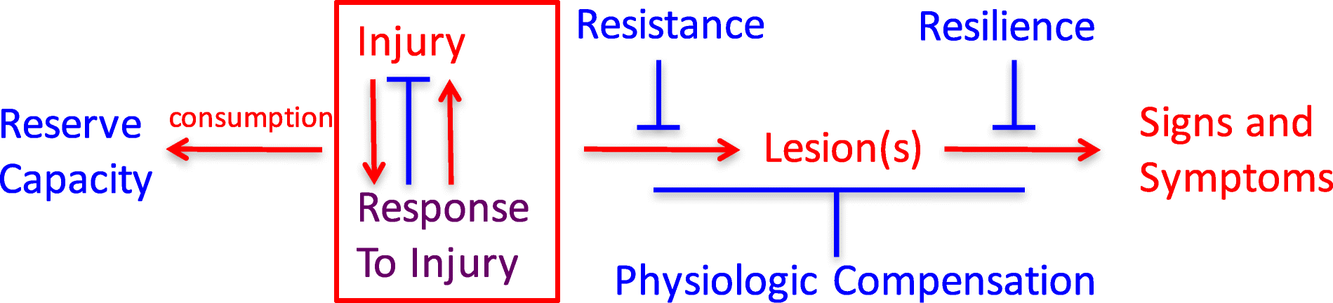
It seems likely that a host of diverse factors active during fetal development, childhood, and throughout adult life may initiate, aggravate, or protect against relevant pathophysiologic processes that underlie neurodegeneration and its clinical expression. These factors—some adverse and some protective—may operate independently, synergistically, antagonistically, sequentially, or even differentially (Fig. 1). While some may be examined individually and in exquisite molecular detail in animal or in vitro models, most will require careful, longitudinal validation in humans. From this perspective, it is not surprising that so far we have had only limited success in identifying risk factors and their underlying mechanisms to guide effective primary and secondary preventive interventions.
Fig. 1
Fig. 1
Relationships among adverse (red), protective (blue), and mixed (purple) processes that culminate in signs and symptoms of neurodegenerative diseases
Until quite recently, “late onset Alzheimer’s disease” was widely viewed as a specific disease entity responsible for the vast majority of late-life dementia. However, longitudinal epidemiologic studies of brain aging and cognitive decline with brain autopsy have consistently demonstrated a central role for multiple co-morbidities as the dominant determinants of late-life dementia. It is important to recognize that current intra vitam measures of these several common diseases of the aging brain are limited, and consequently, despite limitations, brain histopathologic evaluation remains the only means to assess comprehensively the impact of co-morbid diseases on cognitive performance during life.
In combination with functional assessments obtained during life, histopathologic features (lesions) determined with brain autopsy define the presence of specific clinico-pathologic entities, which may or may not reliably correspond to specific mechanism(s) of disease. As a result of the highly consistent findings from longitudinal epidemiologic studies with brain autopsy from across the globe, the view of cognitive decline and dementia in older adults is shifting from being the result of a single disease to a conspiracy of multiple, common age-related disease processes that combine idiosyncratically in each individual. The most common is Alzheimer’s disease, defined by amyloid beta accumulation and neurofibrillary degeneration in certain regions of the brain. Four other commonly recognized pathophysiologic processes that can contribute to cognitive decline and dementia in late life include Lewy body disease, vascular brain injury (especially from small vessel disease), hippocampal sclerosis, and generalized atrophy beyond what can be explained by these other diseases. While the brain lesions of AD are more prevalent at autopsy than any of the other lesions, the combined frequencies of the non-AD abnormalities are usually greater. Indeed, in both the Nun Study and the Honolulu Asia Aging Study, > 90% of participants with severe cognitive impairment can be fully attributed to the collective or individual influences of these five abnormalities [1]. It is critically important, but infrequently appreciated, that the exponential influence of co-morbid disease is reflected in the multiplication of individual relative risks (or odds ratios) for each disease related to cognitive impairment or dementia (Table 1).
Table 1
Point estimates of odds ratios (OR) from ordinal logistic regression of the impact of the coprevalence of five brain lesions on cognitive performance within 2 years of death
Lesion co-morbidity index
OR for the Nun Study (n = 334)
OR for the Honolulu Asia Aging Study (n = 774)
0
1.0 (reference)
1.0 (reference)
0.4–0.8
2.8
2.4
1.0–1.8
5.0
4.6
2.0–2.4
23.1
16.3
2.6–4.4
99.1
37.6
Severity of each of the five brain lesions (Braak stage for neurofibrillary degeneration, cerebral cortical Lewy body disease, cerebral cortical microinfarcts, hippocampal sclerosis, low brain weight) was scored as none/mild (0), moderate (0.4), or severe (1.0) by established criteria, and the lesion co-morbidity index was calculated as the sum of scores for each of the five lesions [1]
To frame a discussion of resistance, resilience, reserve, and compensation, we conventionally consider the diseases that cause late-life cognitive impairment and dementia to derive from injury and response to injury that begin before there are signs or symptoms, but that the resulting damage, distortion, disruption, and/or degeneration ultimately becomes overwhelmingly evident as impairments of cognitive and behavioral function.
The recognition of risk factors linked to measures of different types and amount of brain lesions may illuminate fundamental mechanisms and primary instigating exposures. A systematic search to identify specific protective factors and the mechanisms that underlie them has been conducted relatively infrequently. We propose the following operational definitions as a step toward systematically investigating each of these processes in individuals:
Resistance is inferred from an observed absence or lower level of dementia-associated brain injury, relative to an expected greater frequency or severity based on age, genetic factors, or other characteristics of the individual. This state of unexpectedly low or absent brain injury theoretically may be intrinsic, meaning in someone with greater defenses to forces that usually lead to brain lesions, or environmental, meaning in someone with usual defenses but who avoided exposure to these forces. While resistance now can be assessed comprehensively only with neuropathologic evaluation, specific facets (e.g., beta amyloid, pathologic tau burden, neuron damage) can be estimated during life with biomarkers and imaging.
Resilience is inferred from an observed level of cognitive functioning higher than expected in the face of demonstrated brain injury. Resilience only can be recognized or measured when injury exists and can be related to (near) coincident assessment of function. We prefer to consider two forms of resilience: apparent and essential. Apparent resilience refers to a specific lesion type without consideration of common co-morbidities. Consider two individuals who both are positive by PET imaging for fibrillar amyloid and pathologic tau; one is cognitively normal and the other has dementia. The first person has apparent resilience to AD neuropathologic change. Imagine further a future state when there also is a PET ligand for pathologic alpha-synuclein. Now, we learn that the first person lacks Lewy body disease and the second has co-morbid neocortical Lewy body disease. Is the difference between these two individuals explained by resilience to AD neuropathologic change or by resistance to Lewy body in the first person? Once comprehensive assessment of brain lesions associated with dementia is achieved, then essential resilience can be evaluated. Currently, this is accomplished best with neuropathologic assessment, but even this approach is limited. Our brain autopsy data suggest that much, and perhaps most, of what is referred to currently as (apparent) resilience actually is resistance to co-morbid disease.
Consumption or retention of reserve can be measured or inferred either as brain structural and/or physiological pre-morbid capacity. Examples might be greater than usual synaptic density (analogous to computational “hardware”) or enhanced cognitive effectiveness or redundancy because of learned language, educational richness, or occupational complexity (analogous to computational “software”) prior to the onset of disease. The salutary influence of such resources may be apparent in cognitive test performance well before the onset of cognitive decline. This definition requires that measures of reserve capacity must have been estimated or inferred prior to the development of brain injury. Mechanisms underlying physiologic compensation also may be changes in “hardware” or “software,” but in distinction to pre-existing reserve capacity, physiologic compensation occurs following injury rather than developing prior to injury/response to injury. An example of physiologic compensation might be recruitment of additional regions of the brain to subserve memory function following damage to the hippocampus or in recovery of language functioning after an infarction or brain injury.

Conclusions

With operational definitions of resistance, resilience, and reserve, a focused analytic search for their predictors and correlates can be undertaken. This will require distinguishing and measuring each independently, and then employing those measures as distinct endpoints to identify their individual determinants. This approach, essentially a search to identify protective risk factors and their mechanisms, represents a relatively unexplored pathway toward the identification of candidate preventive interventions.

Re-imagining Stroke Environments with Virtual Reality (RiSE-VR) – research - Australian request for research guinea pigs

Join if you can. 

Re-imagining Stroke Environments with Virtual Reality (RiSE-VR) – research

Florey Institute of Neuroscience and Mental Health, 30 Royal Parade, Parkfille, VIC, 3052
0 comments

This project is examining how responses and behaviours of stroke survivors can be influenced by hospital ward physical environment. There is limited scientific knowledge informing the design of hospital environments for people who have had a stroke. You are eligible for this study if you have had a stroke and are discharged from hospital at least 1 month.
Researchers from The Florey Institute and Swinburne University are exploring the responses of stroke survivors to a novel patient room design immersive virtual reality experience (VR).
Participation involves completing 2 VR sessions lasting 60 minutes each, and answering some questionnaires. This will be followed by a single interview with you to gather further details from you about the VR experience.
Researchers will measure your responses using preference and emotional ratings while using the VR experience. Your physical responses will also be tracked using a pulse and blood pressure probe, and a wearable motion sensor. This study will run for 2 years at The Florey Institute, Melbourne.
Contact:
Michelle Shannon
mshannon1@student.unimelb.edu.au

Synergistic Benefits of Combined Aerobic and Cognitive Training on Fluid Intelligence and the Role of IGF-1 in Chronic Stroke

Useless for survivors, no protocols. No objective starting point for use of these guidelines. No protocols. No understandable objective endpoint.  

Synergistic Benefits of Combined Aerobic and Cognitive Training on Fluid Intelligence and the Role of IGF-1 in Chronic Stroke 

First Published February 28, 2019 Research Article
Background. Paired exercise and cognitive training have the potential to enhance cognition by “priming” the brain and upregulating neurotrophins.  
Methods. Two-site randomized controlled trial. Fifty-two patients >6 months poststroke with concerns about cognitive impairment trained 50 to 70 minutes, 3× week for 10 weeks with 12-week follow-up. Participants were randomized to 1 of 2 physical interventions: Aerobic (>60% VO2peak using <10% body weight–supported treadmill) or Activity (range of movement and functional tasks). Exercise was paired with 1 of 2 cognitive interventions (computerized dual working memory training [COG] or control computer games [Games]). The primary outcome for the 4 groups (Aerobic + COG, Aerobic + Games, Activity + COG, and Activity + Games) was fluid intelligence measured using Raven’s Progressive Matrices Test administered at baseline, posttraining, and 3-month follow-up. Serum neurotrophins collected at one site (N = 30) included brain-derived neurotrophic factor (BDNF) at rest (BDNFresting) and after a graded exercise test (BDNFresponse) and insulin-like growth factor–1 at the same timepoints (IGF-1rest, IGF-1response).  
Results. At follow-up, fluid intelligence scores significantly improved compared to baseline in the Aerobic + COG and Activity + COG groups; however, only the Aerobic + COG group was significantly different (+47.8%) from control (Activity + Games −8.5%). Greater IGF-1response at baseline predicted 40% of the variance in cognitive improvement. There was no effect of the interventions on BDNFresting or BDNFresponse; nor was BDNF predictive of the outcome. Conclusions. Aerobic exercise combined with cognitive training improved fluid intelligence by almost 50% in patients >6 months poststroke. Participants with more robust improvements in cognition were able to upregulate higher levels of serum IGF-1 suggesting that this neurotrophin may be involved in behaviorally induced plasticity.

Is Unilateral Spatial Neglect Associated With Motor Recovery of the Affected Upper Extremity Poststroke? A Systematic Review

You lazy fuckers. We need protocols that fix your described problem, not just the description of the problem. 

Is Unilateral Spatial Neglect Associated With Motor Recovery of the Affected Upper Extremity Poststroke? A Systematic Review 

First Published February 20, 2019 Review Article
Background and Purpose. Individuals with stroke often present symptoms of multiple domains, such as weakness of the affected upper extremity (UE) and unilateral spatial neglect (USN), which are both associated with poor functional outcome. The aims of this systematic review were to search and review studies that investigated (1) the relationship between USN and affected UE sensorimotor recovery poststroke and (2) the effectiveness of sensorimotor interventions to improve the affected UE in patients with USN.
Methods. An electronic search of databases (MEDLINE, EMBASE, CINAHL and Cochrane CENTRAL) was conducted using a combination of the following terms: stroke, USN, and affected UE. Studies meeting the inclusion criteria were rated using a modified version of the Quality Index, and relevant data were extracted.  
Results. A total of 850 studies were identified, and 14 were included; 13 studies assessed correlations between USN and the affected UE capacity/recovery, and 1 study assessed an intervention to improve the UE of individuals with USN. An association between presence of USN and UE capacity/recovery was found in most studies and USN did not interfere with recovery of the affected UE in the single experimental study.  
Conclusions. USN is associated with poor UE motor capacity and less UE recovery poststroke. Therefore, these impairments should be considered when planning rehabilitation and discharge. Because USN is a well-researched phenomenon, the lack of studies and insufficient evidence related to UE interventions in individuals with USN was unexpected. These interventions should be developed and researched to improve UE and overall functional outcome poststroke.(So you punted the actual work to some unspecified future person? I call that lazy and a fireable cause.)

Article available in:

Related Articles

Citing Articles: 0

Language as a Predictor of Motor Recovery: The Case for a More Global Approach to Stroke Rehabilitation

Will you stop trying to predict recovery and just deliver recovery protocols you blithering idiots. There is not a survivor in the world who cares about predictions, they just want the facts about the efficacy of stroke rehab protocols so they know exactly what they have to do to recover. Have you no brains at all? 

Language as a Predictor of Motor Recovery: The Case for a More Global Approach to Stroke Rehabilitation

First Published February 13, 2019 Research Article
Stroke is the third leading cause of death in the developed world and the primary cause of adult disability. The most common site of stroke is the middle cerebral artery (MCA), an artery that supplies a range of areas involved in both language and motor function. As a consequence, many stroke patients experience a combination of language and motor deficits. Indeed, those suffering from Broca’s aphasia have an 80% chance of also suffering hemiplegia. Despite the prevalence of multifaceted disability in patients, the current trend in both clinical trials and clinical practice is toward compartmentalization of dysfunction. In this article, we review evidence that aphasia and hemiplegia do not just coexist, but that they interact. We review a number of clinical reports describing how therapies for one type of deficit can improve recovery in the other and vice versa. We go on to describe how language deficits should be seen as a warning to clinicians that the patient is likely to experience motor impairment and slower motor recovery, aiding clinicians to optimize their choice of therapy. We explore these findings and offer a tentative link between language and arm function through their shared need for sequential action, which we term fluency. We propose that area BA44 (part of Broca’s area) acts as a hub for fluency in both movement and language, both in terms of production and comprehension.

Article available in:

Related Articles

Citing Articles: 0

Sociodemographic disparities in long-term mortality among stroke survivors in the United States: The REGARDS Study

Well fuck, we don't need better long term care, we need better RESULTS. That will come from setting up and using stroke rehab protocols. Anyone who uses the term 'care' in stroke needs to be fired.  That is just being fucking lazy. 

Sociodemographic disparities in long-term mortality among stroke survivors in the United States: The REGARDS Study

StrokeElfassy T, et al. | March 14, 2019

In this REGARDS study, which included 1,329 black and white participants, researchers described long-term (> 30 days) mortality rates among stroke survivors, and determined whether there exist socioeconomic disparities. They found that long-term mortality was high in older individuals as well as among men vs women. The age-adjusted mortality rate among adults who survived ≥ 30 days poststroke was 82.3 per 1,000 person-years. Investigators found that long-term mortality rates among stroke survivors were higher among people with lower socioeconomic status (SES) and those living in lower SES neighborhood. No differences, however, were found in age-adjusted rates of long-term post-stroke mortality by race, rurality, or US region. These results highlighted the need for improvements in post-stroke long-term care, particularly among lower SES individuals.
Read the full article on Stroke

Intensive blood pressure reduction with intravenous thrombolysis therapy for acute ischaemic stroke (ENCHANTED): An international, randomised, open-label, blinded-endpoint, phase 3 trial

Why would you even expect blood pressure lowering to improve functional outcomes?  You are reducing the amount and quantity of oxygen carrying blood to the brain. Does not sound like a good idea. 

Intensive blood pressure reduction with intravenous thrombolysis therapy for acute ischaemic stroke (ENCHANTED): An international, randomised, open-label, blinded-endpoint, phase 3 trial


The LancetAnderson CS, et al. | March 11, 2019

In this trial that included a sample of nearly 2,200 alteplase-eligible adults with acute ischemic stroke and systolic blood pressure of ≥ 150 mmHg, researchers assessed intensive blood pressure lowering vs guideline-recommended blood pressure lowering in these patients. A total of 1,081 participants were allocated to the intensive group and 1,115 were allocated to the guideline group. Among the 2,175 given intravenous alteplase, 1,466 received a standard dose. Intensive blood pressure lowering was identified as safe, but led to no improved clinical outcome vs guideline treatment—despite the observed reduction in intracranial hemorrhage. Results, thus, did not support a major shift towards this treatment for those receiving alteplase for mild-to-moderate acute ischemic stroke.
Read the full article on The Lancet

Heterozygous Deletion of EphrinA5 Does Not Improve Functional Recovery After Experimental Stroke

Completely and totally useless for survivors. Not even an abstract to get a clue of what it is about. A great stroke association would make all stroke research freely available to all survivors. 

Heterozygous Deletion of EphrinA5 Does Not Improve Functional Recovery After Experimental Stroke

Friday, March 15, 2019

Stroke Treatment Academic Industry Roundtable X

Oh God, what stupidity. Changing wording from neuroprotection to Brain Cytoprotection. It still doesn't sound important enough to immediately fix.  Neuronal cascade of death implies immediate interventions are needed. 

Stroke Treatment Academic Industry Roundtable X

Brain Cytoprotection Therapies in the Reperfusion Era

First page at link.