Changing stroke rehab and research worldwide now.Time is Brain! trillions and trillions of neurons that DIE each day because there are NO effective hyperacute therapies besides tPA(only 12% effective). I have 523 posts on hyperacute therapy, enough for researchers to spend decades proving them out. These are my personal ideas and blog on stroke rehabilitation and stroke research. Do not attempt any of these without checking with your medical provider. Unless you join me in agitating, when you need these therapies they won't be there.

What this blog is for:

My blog is not to help survivors recover, it is to have the 10 million yearly stroke survivors light fires underneath their doctors, stroke hospitals and stroke researchers to get stroke solved. 100% recovery. The stroke medical world is completely failing at that goal, they don't even have it as a goal. Shortly after getting out of the hospital and getting NO information on the process or protocols of stroke rehabilitation and recovery I started searching on the internet and found that no other survivor received useful information. This is an attempt to cover all stroke rehabilitation information that should be readily available to survivors so they can talk with informed knowledge to their medical staff. It lays out what needs to be done to get stroke survivors closer to 100% recovery. It's quite disgusting that this information is not available from every stroke association and doctors group.

Thursday, June 6, 2024

Evaluation of walking activity and gait to identify physical and mental fatigue in neurodegenerative and immune disorders: preliminary insights from the IDEA-FAST feasibility study

 With the high occurrence rate of stroke survivor fatigue this research should also be done on stroke survivors. But with NO leadership and NO strategy nothing will be done because stroke medical 'professionals' don't have two functioning neurons to rub together!

At least half of all stroke survivors experience fatigue Known since March 2017

Or is it 70%? Known since March 2015.

Or is it 40%? Known since September 2017.

Evaluation of walking activity and gait to identify physical and mental fatigue in neurodegenerative and immune disorders: preliminary insights from the IDEA-FAST feasibility study

Abstract

Background

Many individuals with neurodegenerative (NDD) and immune-mediated inflammatory disorders (IMID) experience debilitating fatigue. Currently, assessments of fatigue rely on patient reported outcomes (PROs), which are subjective and prone to recall biases. Wearable devices, however, provide objective and reliable estimates of gait, an essential component of health, and may present objective evidence of fatigue. This study explored the relationships between gait characteristics derived from an inertial measurement unit (IMU) and patient-reported fatigue in the IDEA-FAST feasibility study.

Methods

Participants with IMIDs and NDDs (Parkinson's disease (PD), Huntington's disease (HD), rheumatoid arthritis (RA), systemic lupus erythematosus (SLE), primary Sjogren’s syndrome (PSS), and inflammatory bowel disease (IBD)) wore a lower-back IMU continuously for up to 10 days at home. Concurrently, participants completed PROs (physical fatigue (PF) and mental fatigue (MF)) up to four times a day. Macro (volume, variability, pattern, and acceleration vector magnitude) and micro (pace, rhythm, variability, asymmetry, and postural control) gait characteristics were extracted from the accelerometer data. The associations of these measures with the PROs were evaluated using a generalised linear mixed-effects model (GLMM) and binary classification with machine learning.

Results

Data were recorded from 72 participants: PD = 13, HD = 9, RA = 12, SLE = 9, PSS = 14, IBD = 15. For the GLMM, the variability of the non-walking bouts length (in seconds) with PF returned the highest conditional R2, 0.165, and with MF the highest marginal R2, 0.0018. For the machine learning classifiers, the highest accuracy of the current analysis was returned by the micro gait characteristics with an intrasubject cross validation method and MF as 56.90% (precision = 43.9%, recall = 51.4%). Overall, the acceleration vector magnitude, bout length variation, postural control, and gait rhythm were the most interesting characteristics for future analysis.

Conclusions

Counterintuitively, the outcomes indicate that there is a weak relationship between typical gait measures and abnormal fatigue. However, factors such as the COVID-19 pandemic may have impacted gait behaviours. Therefore, further investigations with a larger cohort are required to fully understand the relationship between gait and abnormal fatigue.

Background

Many people with neurodegenerative disorders (NDD) and immune-mediated inflammatory diseases (IMID) experience abnormal fatigue. For instance, abnormal fatigue has been reported in over 85% of those with systemic lupus erythematosus (SLE) [1, 2], 33% to 58% of those with Parkinson’s disease (PD) [3], 67% of people with primary Sjogren’s syndrome (PSS) [4], and over 41% of patients with rheumatoid arthritis (RA) showed clinically important levels of fatigue [5]. These symptoms can be debilitating for those who experience them and are key contributors to poor quality of life. Accordingly, a key goal of the IDEA-FAST consortium [6] is to explore and identify digital endpoints that provide reliable, objective, and sensitive evaluations of abnormal fatigue, which in turn will facilitate therapeutic development to alleviate these symptoms.

A common method for assessing fatigue is patient reported outcomes (PRO)s, where the patients answer questionnaires and diaries designed to record how the patient is feeling. This approach gives a snapshot view of fatigue, either during a one-off visit at the clinic or with regular at-home questionnaires. PROs, however, are subjective, susceptible to recall bias [7], and the measurement of granular changes over time requires high patient burden through repeated assessments. Furthermore, studies have shown that individuals who are sleep-deprived or are out of their circadian phase are prone to underestimating their fatigue-related impairments [8,9,10,11]. These issues are therefore worsened in cases where neurological functionality or sleep quality is impacted by an illness. Wearable devices may circumvent the pitfalls of PRO-based assessments since they could objectively and continuously monitor physiological changes related to physical and mental fatigue. Inertial measurement units (IMUs)—e.g., wearable devices comprising triaxial accelerometers and gyroscopes—are becoming an increasingly popular option for continuous remote monitoring, due to their affordability and ease of use in users’ natural environment. These recordings can be wirelessly transmitted to a device, such as a smartphone, where the signals can be processed, assessed, and reported to the user or clinicians.

Mobility (e.g., walking, gait) is considered as the 6th vital sign and represents an essential component of health and quality of life, being key to physical, mental, and social well-being [12]. Loss in mobility has been associated with morbidity, falls, dementia, cognitive decline, hospitalisations, mortality, and symptoms of chronic disorders [13,14,15,16]. As such, the current study will explore the relationship between gait characteristics and abnormal fatigue in people with NDD and IMID.

The current knowledge in the relationships between walking and abnormal fatigue in NDDs and IMIDs is very limited. Many studies exploring fatigue focus on muscle or exercise-induced fatigue in healthy participants with parameters such as accelerometer spectra [17], measures of acceleration, jerk, and posture [18,19,20,21], and temporal measures of gait (“micro” gait, e.g., gait speed, step time) [22, 23]. Most notably, the majority of existing literature has been conducted within in laboratory-based environment, where the participants are monitored whilst doing an instructed task. This includes the six-minute walking distance test (6-MWDT) to assess the impacts of fatigue on gait (exercise) capacity [24,25,26,27,28]; gait speed i.e., the ten-meter walking test (10-MWT) [24, 29,30,31,32]; an electronic walkway [33, 34]; and a predefined path for unaided walking [35]. We found three studies that explored fatigue in NDD or IMID specifically with free-living gait assessment. The first analysed physical activity over a seven-day period from a hip-worn tri-axial accelerometer in 123 participants with SLE [36]. Light and moderate/vigorous activity and moderate/vigorous activity periods > 10 min were identified from the accelerometer’s vector magnitude and compared to the participants’ Fatigue Severity Score (FSS). The second study investigated the impact of physical activity on non-motor symptoms with the Movement Disorder Society-Unified Parkinson’s Disease Rating Scale (MDS-UPDRS) in 45 PD participants with a hip-worn accelerometer for at least three days to record daily step count and assess sedentary behaviour, light physical activity, and moderate-to-vigorous activity [37]. In the third, participants with multiple sclerosis (MS)—134 fatigued and 76 non-fatigued—wore an accelerometer above the dominant hip for seven days to record light and moderate-to-vigorous activity counts, whilst reporting on their fatigue severity with FSS [38].

In many cases, the exploration of gait measures is limited to characteristics such as gait speed [31, 39], and ambulatory activity (step count) [30]. Other studies were more focussed on gait, but only explored different activity levels based on step count [37], vector magnitude [36], and activity counts [38]. Another explored 6-MWT, dynamic activity, number of postural transitions, and walking bouts longer than ten seconds [28]. Three studies included a more comprehensive analysis of macro (reflecting activity) and micro (reflecting discrete gait outcomes) gait characteristics within a laboratory: one study assessed the associations of Multidimensional Fatigue Inventory (MFI) with measures of pace, rhythm, variability, asymmetry, and postural control [33]; one analysed the cycle time, stride length, swing time, and double support time and their variability (coefficient of variation (CoV)) [34]; and one compared gait duration (10-MWT), gait speed (m/s), cadence (steps/min), and stride length (m) to the Parkinson FSS [32]. Furthermore, the participants explored by the studies in the literature typically include only healthy subjects [33] or only one disease cohort such as PD [32, 37, 39], MS [35, 38], IBD [28], SLE [36], symptomatic knee osteoarthritis [31], fibromyalgia [34], and stroke survivors [30].

Therefore, in-depth analyses of the associations between real-world gait and abnormal fatigue in NND and IMID are currently lacking. The current study will analyse free-living data from several differing disease cohorts and will conduct an extensive exploration of various measures of gait and walking activity as a preliminary assessment of data collected by IDEA-FAST [6].

This study aims to:

  1. (i)

    comprehensively explore the feasibility of using macro and micro gait characteristics from an IMU attached to the lower back to objectively identify PRO scores of physical and mental fatigue in NDD and IMID participants;

  2. (ii)

    assess the associations of the macro and micro characteristics with selected PROs using a generalised linear mixed effect model and low vs. high fatigue binary classification performances of popular machine learning models;

  3. (iii)

    explore the usefulness of the gait-model’s physiological feature groups using the associations from the linear mixed effect model.

Brain-Protected TAVR Results in Fewer Real-World Disabling Strokes

 If you need this demand your doctor guarantee no stroke at all.

Brain-Protected TAVR Results in Fewer Real-World Disabling Strokes

Also, patients with prior stroke ID'd as likely responders in TVT registry analysis

NEW YORK CITY -- Researchers eked out evidence of a small clinical benefit with the Sentinel cerebral embolic protection (CEP) device in transcatheter aortic valve replacement (TAVR) -- and pointed to the patients more likely to benefit -- on the basis of disabling strokes.

The incidence rate of in-hospital disabling stroke, defined as stroke associated with either in-hospital death or discharge to a non-home location, was 0.79% with the CEP device versus 0.91% without the device (RR 0.87, 95% CI 0.73-1.00) in nationwide STS/ACC TVT registry data from 2018 to 2023, the largest dataset to date in this field.

This reduction in disabling stroke in the real world was supported by a secondary analysis using propensity score weighting (OR 0.79, 95% CI 0.70-0.90), reported Neel Butala, MD, of Rocky Mountain Regional VA Medical Center and the University of Colorado School of Medicine, both in Aurora.

"Our prespecified primary analysis using an instrumental variable approach demonstrated a small, borderline significant reduction in in-hospital disabling stroke that is likely to be causal in nature," Butala said here at the New York Valvesopens in a new tab or window meeting, the successor to the annual TVT conferenceopens in a new tab or window previously hosted by the Cardiovascular Research Foundation. The study was simultaneously published in Circulation: Cardiovascular Interventionsopens in a new tab or window.

Panelist Samir Kapadia, MD, of the Cleveland Clinic, called the study an "important contribution" that signals that the CEP field is still "active," with newer, better devices on the horizon.

Despite FDA approval of the Sentinel deviceopens in a new tab or window, CEP remains controversial following multiple trial setbacks. The technology never entered mainstream practice because of what many consider to be an unproven benefit of capturing debris dislodged during TAVR.

For example, the Sentinel CEP device failed to reduce periprocedural strokes in the randomized PROTECTED-TAVR trialopens in a new tab or window, even though it prevented embolic material from reaching the brain. In that study, the researchers tried and failed to find subgroup responders who would likely have strokes prevented by the CEP device.

Based on the TVT registry, however, Butala and colleagues did identify TAVR patients with prior stroke as a subgroup that disproportionately benefited from fewer disabling strokes with CEP.

"We've been trying to find this, a cohort of patients that would benefit more from EPD [embolic protection devices] and I think you kind of nailed it," said session discussant Alexandra Lansky, MD, of Yale School of Medicine in New Haven, Connecticut. "The sample size is large enough that patients with prior stroke are probably the ones we want to target."

There was no reduction in nondisabling strokes in the cohort overall or in any subgroup. This may be explained by the Sentinel device allowing smaller particles to get to the head and causing smaller strokes, Butala told the audience.

"These findings are congruent with those from the PROTECTED-TAVR trial and suggest that the reduction in disabling stroke from EPD use is real, but the effect size may be relatively small in all-comer patients," he concluded.

The investigators had conducted a retrospective study of first-time TAVR recipients in the STS/ACC TVT registry. They excluded emergent procedures, alternative access, and people who received a non-Sentinel CEP device.

In the end, there were 414,649 TAVR recipients included in the study (mean age 78-79, 39-44% women), of whom nearly 13% received a Sentinel device.

The Sentinel group had more bicuspid valves (8.9% vs 5.5%) and more prior aortic valve procedures (11.6% vs 8.1%) compared with controls. Otherwise, baseline characteristics were relatively well-matched, according to Butala's group.

Use of CEP devices had plateaued at 15% until the 3,000-patient PROTECTED-TAVR results were released in 2022 -- dropping down to 12% by mid-2023, Butala said. He highlighted the variation in CEP use across the study period, with 40% of sites reporting no CEP use and 6% using the devices for over half their cases.

The investigators acknowledged that their analysis was not randomized, but maintained that causal inference was supported by their quasi-experimental approach of an instrumental variable analysis with site-level preference for CEP use as the instrument.

Lansky added that the potential underestimating of event rates and lack of long-term mortality data were limitations to the study.

In addition, Butala and colleagues had relied on discharge location as a proxy for stroke severity, a variable that is not explicitly identified in the TVT registry. The disabling stroke endpoint was separately validated in a population pooling data from PARTNER 2A, PARTNER 3, the Sapien S3i Registry, SURTAVI, Evolut Low Risk, and PROTECTED TAVR -- with sensitivity of 80%, specificity of 66.7%, and a positive predictive value (PPV) of 58.2%.

"While we can be fairly certain that a patient discharged home after TAVR did not have a disabling stroke, the lower sensitivity and PPV temper our ability to verify if discharge to non-home location or death was truly due to a disabling stroke post-TAVR or due to another postprocedure complication(s) or for other reasons such as frailty," cautioned Dharam Kumbhani, MD, SM, of the University of Texas Southwestern Medical Center in Dallas, and Frederick Welt, MD, of the University of Utah Health Sciences Center in Salt Lake City, in an accompanying editorialopens in a new tab or window. "In addition, baseline residential status is not factored into this definition."

"The current endeavor by Butala and colleagues is a creative and interesting approach to try and shed light on a question that has yet to be definitively answered by randomized data," they added. "It potentially sets the stage for future studies and action items on this topic."

The duo pointed to the BHF PROTECT TAVIopens in a new tab or window study as an ongoing randomized trial in the CEP space, but said it is unlikely to be powered for a disabling stroke endpoint.

For now, "we don't find evidence of harm. It's not like we're harming people using this device," Butala said, adding that the only downsides with the CEP device are longer procedure time and higher cost.

  • author['full_name']

    Nicole Lou is a reporter for MedPage Today, where she covers cardiology news and other developments in medicine. Follow

Disclosures

The study was funded by a grant from Boston Scientific.

Butala disclosed receiving consulting fees/honoraria from Shockwave Medical and Boston Scientific.

Kapadia had no disclosures.

Lansky reported personal ties to Boston Scientific, Cordis/MedAlliance, and Abiomed; and grant support from Abbott, Sinomed, and Abiomed.

Kumbhani and Welt had no disclosures.

Primary Source

Circulation: Cardiovascular Interventions

Source Reference: opens in a new tab or windowButala NM, et al "Impact of cerebral embolic protection devices on disabling stroke after transcatheter aortic valve replacement: updated results from the STS/ACC TVT registry" Circ Cardiovasc Interv 2024; DOI: 10.1161/CIRCINTERVENTIONS.123.013697.

Secondary Source

Circulation: Cardiovascular Interventions

Source Reference: opens in a new tab or windowKumbhani DJ, Welt FG "Supplementing randomized trial data to answer a real world question: discharge to home status as a heuristic for stroke severity after transcatheter aortic valve replacement" Circ Cardiovasc Interv 2024; DOI: 10.1161/CIRCINTERVENTIONS.124.014374.

Better Cognitive Outcomes Tied to Vigorous Exercise in Hypertensive Adults

 Your competent? doctor is responsible for getting you 100% recovered with EXACT 100% recovery protocols so you can do this vigorous exercise. There is NO valid excuse for not having those protocols, your doctor has known since medical school that only 10% get rehab full recovery!

So if s/he didn't immediately start advocating and working on getting 100% recovery protocols, that is a sign of COMPLETE INCOMPETENCE! Why are you seeing incompetent doctors?

Send me hate mail on this: oc1dean@gmail.com. I'll print your complete statement with your name and my response in my blog. Or are you afraid to engage with my stroke-addled mind?  You'll want 100% recovery when you are the 1 in 4 per WHO that has a stroke! My definition of competence is 100% recovery, what is your definition?

Better Cognitive Outcomes Tied to Vigorous Exercise in Hypertensive Adults

SPRINT MIND analysis looks at 7,600 older adults at high risk of cognitive decline

A photo of senior man digging in the garden on the side of his house.

Key Takeaways

  • Vigorous physical activity appeared to preserve cognitive function in older adults with hypertension, a post hoc analysis showed.
  • Hypertension is associated with an increased risk of cognitive decline.
  • Among older adults with hypertension, at least one weekly vigorous exercise session was associated with a lower risk of future cognitive impairment.

Vigorous exercise appeared to preserve cognitive function in older adults with hypertension, a post hoc analysis of SPRINT MIND trial data showed.

At least one vigorous physical activity session per week reduced the risk of future mild cognitive impairment and probable dementia for people with hypertension over 4.5 years of follow-up, reported Richard Kazibwe, MD, of Wake Forest University School of Medicine in Winston-Salem, North Carolina, and co-authors.

Participants who had one or more vigorous physical exercise sessions weekly had a significantly lower risk of mild cognitive impairment (HR 0.80, 95% CI 0.67-0.95) and a composite of mild cognitive impairment/probable dementia (HR 0.82, 95% CI 0.70-0.94) compared with those who had less than one vigorous session a week, Kazibwe and colleagues wrote in Alzheimer's and Dementiaopens in a new tab or window.

Higher vigorous physical activity trended toward a lower risk of probable dementia, but the relationship didn't reach statistical significance (HR 0.80, 95% CI 0.63-1.02).

"We know that physical exercise offers many benefits, including lowering blood pressure, improving heart health, and potentially delaying cognitive decline," Kazibwe said in a statement. "However, the amount and the intensity of exercise needed to preserve cognition is unknown."

Many epidemiological studies support the role of lifestyle in dementia risk. In 2020, the Lancet Commissionopens in a new tab or window reported that up to 40% of dementia cases may be prevented or delayed by modifying 12 risk factors, including physical inactivity. Studies of U.K. Biobank participants have linked moderate and vigorous exerciseopens in a new tab or window with midlife cognition, and sedentary lifestylesopens in a new tab or window in older adults with incident dementia.

Hypertension is associated with an increased risk of cognitive impairment. In their analysis, Kazibwe and co-authors evaluated data from 7,670 people who participated in the SPRINT MINDopens in a new tab or window substudy of the SPRINTopens in a new tab or window randomized clinical trial. The SPRINT trial was stopped early when its primary outcome showed a significant benefit of intensive systolic blood pressure treatment compared with standard treatment.

The SPRINT MIND substudy showed that treating hypertensive older adults to a systolic blood pressure goal of less than 120 mm Hg, compared with treating them to a goal of less than 140 mm Hg, reduced the risk of probable dementia by 17%, a statistically non-significant difference. Intensive blood pressure control showed statistically significant benefits in secondary outcomes, including a 19% lower rate of mild cognitive impairment.

At enrollment, SPRINT participants were asked about their frequency of vigorous-intensity physical activity over the past 12 months. The researchers categorized vigorous physical activity into two groups: less than one session per week (low activity group), and one or more sessions per week (high activity group). Participants with significant limitations in physical function or missing data were excluded from the post hoc analysis.

Mean participant age was 68 years, 34.5% were women, and 59.6% were white. Kazibwe noted that nearly 60% of study participants reported vigorous activity at least once a week, even those ages 75 and older. This "suggests that older adults who recognize the importance of exercise may be more inclined to exercise at higher intensity," he observed.

Over a maximum follow-up of 7.4 years (median 4.5 years), 570 mild cognitive impairment events, 273 probable dementia events, and 759 mild cognitive impairment/probable dementia events were reported.

Among participants in the high activity group compared with the low activity group, the incidence of mild cognitive impairment events was 6.5% versus 8.8%, 3.1% versus 4.3% for probable dementia events, and 8.7% versus 11.7% for mild cognitive impairment/probable dementia events.

Compared with participants in the low activity group, those in the high activity group experienced lower event rates per 1,000 person-years of mild cognitive impairment (13.9 vs 19.7), probable dementia (6.3 vs 9.0), and mild cognitive impairment/probable dementia (18.5 vs 25.8). Overall, the protective relationship between vigorous exercise and cognitive outcomes was more pronounced for people under age 75.

In the post hoc analysis, participants in the SPRINT intensive and standard blood pressure treatment groups had comparable results for the relationship between vigorous physical activity and risk of cognitive outcomes.

The study had several limitations. The relatively fewer cases of probable dementia might mean the study lacked statistical power to detect whether vigorous activity influenced its risk, the researchers noted. In addition, all physical activity was self-reported at baseline.

"While this study provides evidence that vigorous exercise may preserve cognitive function in high-risk patients with hypertension, more research is needed to include device-based physical activity measurements and more diverse participant populations," Kazibwe said.

  • Judy George covers neurology and neuroscience news for MedPage Today, writing about brain aging, Alzheimer’s, dementia, MS, rare diseases, epilepsy, autism, headache, stroke, Parkinson’s, ALS, concussion, CTE, sleep, pain, and more. Follow

Disclosures

The SPRINT trial is funded by the NIH.

Kazibwe and co-authors reported no conflicts of interest.

Primary Source

Alzheimer's & Dementia

Source Reference: opens in a new tab or windowKazibwe R, et al "Effect of vigorous-intensity physical activity on incident cognitive impairment in high-risk hypertension" Alzheimer's Dement 2024; DOI: 10.1002/alz.13887.

Brain-Protected TAVR Results in Fewer Real-World Disabling Strokes

 Why are there disabling strokes at all? Because we have fucking failures of stroke associations DOING NOTHING to get survivors to 100% recovery!

Brain-Protected TAVR Results in Fewer Real-World Disabling Strokes

Also, patients with prior stroke ID'd as likely responders in TVT registry analysis

NEW YORK CITY -- Researchers eked out evidence of a small clinical benefit with the Sentinel cerebral embolic protection (CEP) device in transcatheter aortic valve replacement (TAVR) -- and pointed to the patients more likely to benefit -- on the basis of disabling strokes.

The incidence rate of in-hospital disabling stroke, defined as stroke associated with either in-hospital death or discharge to a non-home location, was 0.79% with the CEP device versus 0.91% without the device (RR 0.87, 95% CI 0.73-1.00) in nationwide STS/ACC TVT registry data from 2018 to 2023, the largest dataset to date in this field.

This reduction in disabling stroke in the real world was supported by a secondary analysis using propensity score weighting (OR 0.79, 95% CI 0.70-0.90), reported Neel Butala, MD, of Rocky Mountain Regional VA Medical Center and the University of Colorado School of Medicine, both in Aurora.

"Our prespecified primary analysis using an instrumental variable approach demonstrated a small, borderline significant reduction in in-hospital disabling stroke that is likely to be causal in nature," Butala said here at the New York Valvesopens in a new tab or window meeting, the successor to the annual TVT conferenceopens in a new tab or window previously hosted by the Cardiovascular Research Foundation. The study was simultaneously published in Circulation: Cardiovascular Interventionsopens in a new tab or window.

Panelist Samir Kapadia, MD, of the Cleveland Clinic, called the study an "important contribution" that signals that the CEP field is still "active," with newer, better devices on the horizon.

Despite FDA approval of the Sentinel deviceopens in a new tab or window, CEP remains controversial following multiple trial setbacks. The technology never entered mainstream practice because of what many consider to be an unproven benefit of capturing debris dislodged during TAVR.

For example, the Sentinel CEP device failed to reduce periprocedural strokes in the randomized PROTECTED-TAVR trialopens in a new tab or window, even though it prevented embolic material from reaching the brain. In that study, the researchers tried and failed to find subgroup responders who would likely have strokes prevented by the CEP device.

Based on the TVT registry, however, Butala and colleagues did identify TAVR patients with prior stroke as a subgroup that disproportionately benefited from fewer disabling strokes with CEP.

"We've been trying to find this, a cohort of patients that would benefit more from EPD [embolic protection devices] and I think you kind of nailed it," said session discussant Alexandra Lansky, MD, of Yale School of Medicine in New Haven, Connecticut. "The sample size is large enough that patients with prior stroke are probably the ones we want to target."

There was no reduction in nondisabling strokes in the cohort overall or in any subgroup. This may be explained by the Sentinel device allowing smaller particles to get to the head and causing smaller strokes, Butala told the audience.

"These findings are congruent with those from the PROTECTED-TAVR trial and suggest that the reduction in disabling stroke from EPD use is real, but the effect size may be relatively small in all-comer patients," he concluded.

The investigators had conducted a retrospective study of first-time TAVR recipients in the STS/ACC TVT registry. They excluded emergent procedures, alternative access, and people who received a non-Sentinel CEP device.

In the end, there were 414,649 TAVR recipients included in the study (mean age 78-79, 39-44% women), of whom nearly 13% received a Sentinel device.

The Sentinel group had more bicuspid valves (8.9% vs 5.5%) and more prior aortic valve procedures (11.6% vs 8.1%) compared with controls. Otherwise, baseline characteristics were relatively well-matched, according to Butala's group.

Use of CEP devices had plateaued at 15% until the 3,000-patient PROTECTED-TAVR results were released in 2022 -- dropping down to 12% by mid-2023, Butala said. He highlighted the variation in CEP use across the study period, with 40% of sites reporting no CEP use and 6% using the devices for over half their cases.

The investigators acknowledged that their analysis was not randomized, but maintained that causal inference was supported by their quasi-experimental approach of an instrumental variable analysis with site-level preference for CEP use as the instrument.

Lansky added that the potential underestimating of event rates and lack of long-term mortality data were limitations to the study.

In addition, Butala and colleagues had relied on discharge location as a proxy for stroke severity, a variable that is not explicitly identified in the TVT registry. The disabling stroke endpoint was separately validated in a population pooling data from PARTNER 2A, PARTNER 3, the Sapien S3i Registry, SURTAVI, Evolut Low Risk, and PROTECTED TAVR -- with sensitivity of 80%, specificity of 66.7%, and a positive predictive value (PPV) of 58.2%.

"While we can be fairly certain that a patient discharged home after TAVR did not have a disabling stroke, the lower sensitivity and PPV temper our ability to verify if discharge to non-home location or death was truly due to a disabling stroke post-TAVR or due to another postprocedure complication(s) or for other reasons such as frailty," cautioned Dharam Kumbhani, MD, SM, of the University of Texas Southwestern Medical Center in Dallas, and Frederick Welt, MD, of the University of Utah Health Sciences Center in Salt Lake City, in an accompanying editorialopens in a new tab or window. "In addition, baseline residential status is not factored into this definition."

"The current endeavor by Butala and colleagues is a creative and interesting approach to try and shed light on a question that has yet to be definitively answered by randomized data," they added. "It potentially sets the stage for future studies and action items on this topic."

The duo pointed to the BHF PROTECT TAVIopens in a new tab or window study as an ongoing randomized trial in the CEP space, but said it is unlikely to be powered for a disabling stroke endpoint.

For now, "we don't find evidence of harm. It's not like we're harming people using this device," Butala said, adding that the only downsides with the CEP device are longer procedure time and higher cost.

  • author['full_name']

    Nicole Lou is a reporter for MedPage Today, where she covers cardiology news and other developments in medicine. Follow

Disclosures

The study was funded by a grant from Boston Scientific.

Butala disclosed receiving consulting fees/honoraria from Shockwave Medical and Boston Scientific.

Kapadia had no disclosures.

Lansky reported personal ties to Boston Scientific, Cordis/MedAlliance, and Abiomed; and grant support from Abbott, Sinomed, and Abiomed.

Kumbhani and Welt had no disclosures.

Primary Source

Circulation: Cardiovascular Interventions

Source Reference: opens in a new tab or windowButala NM, et al "Impact of cerebral embolic protection devices on disabling stroke after transcatheter aortic valve replacement: updated results from the STS/ACC TVT registry" Circ Cardiovasc Interv 2024; DOI: 10.1161/CIRCINTERVENTIONS.123.013697.

Secondary Source

Circulation: Cardiovascular Interventions

Source Reference: opens in a new tab or windowKumbhani DJ, Welt FG "Supplementing randomized trial data to answer a real world question: discharge to home status as a heuristic for stroke severity after transcatheter aortic valve replacement" Circ Cardiovasc Interv 2024; DOI: 10.1161/CIRCINTERVENTIONS.124.014374.

Medtronic unveils next-generation Artisse intrasaccular device alongside new data at LINNC Paris 2024

 Ask your competent? doctor and hospital about this. If they know nothing; RUN AWAY!

Medtronic unveils next-generation Artisse intrasaccular device alongside new data at LINNC Paris 2024

Medtronic has today announced the European launch of Artisse—a new intrasaccular device designed to treat intracranial aneurysms by conforming to the shape of the aneurysm. The launch coincides with interim data that examine the safety, efficacy and performance of the device being presented for the first time at LINNC Paris 2024 (3–5 June, Paris, France).

Artisse was designed to address the needs of complex wide-neck bifurcation aneurysms that have traditionally been managed by stent-assisted coiling, coil adjunctive neck-bridging devices and other intrasaccular basket devices. The former two methods require multiple steps and multiple devices, with stent-assisted coiling requiring dual antiplatelet therapy, which can present challenges in ruptured aneurysms.

Medtronic claims that Artisse offers a simple, one-step technique in treating these complex and simpler aneurysms alike.

Artisse is highly conformable, engineered from dual layers of high-density platinum core and nitinol drawn filled tubing (DFT) wires in a flared design. This sophisticated construction allows it to flex to conform to the unique contours of most aneurysms while exerting the optimum outward radial force needed to fill the space securely, the company also states in a recent press release.

“Artisse behaves differently to other intrasaccular devices because I can control the optimal positioning of the device in the aneurysm,” said Riitta Rautio (Turku University Hospital, Turku, Finland). “Due to its conformability and softness, I can reach the optimal position of Artisse in the aneurysm with partial re-sheathing of the device and slight movement of the microcatheter.”

The device’s flexibility, smooth distal tip and gentle deployment make Artisse atraumatic—reducing risk to, and stress on, the aneurysm walls.

Early-experience analysis presented at LINNC Paris 2024, from a study involving 35 patients enrolled into the multicentre, core lab-adjudicated European INSPIRE-A registry, shows that 80% of patients treated with Artisse achieved complete occlusion by a six-month angiographic assessment, indicating successful aneurysm occlusion with no residual filling.

“Artisse is a promising device that appears to be a step in the right direction for intrasaccular technology,” said Tufail Patankar (Leeds General Infirmary, Leeds, UK). “Its softness and conformability allows you to manipulate the device within the aneurysm. Complex wide-neck angled aneurysms that were previously difficult to treat with other intrasaccular devices now seem to be treatable with Artisse.”

According to Medtronic, Artisse is simple to use and faster when compared with coils, or balloon- or stent-assisted coiling. In deployment, the device opens gradually and smoothly with a ‘flowering mechanism’, with no kick-back, so that it feels like pushing a coil. The device also features a handheld electrolytic detachment system that provides immediate confirmation, instant audio and visual feedback upon detachment, and complete radiopacity.

Medtronic offers expert clinical support, including access to simulation software, further supporting sizing selection with Artisse, the recent release notes.

“At this time, I experience very satisfying results for both unruptured and ruptured aneurysms treated with the Artisse device,” said Cyril Dargazanli (University Hospital of Montpellier, Montpellier, France).

“Combined with simulation and the radial approach, Artisse is redefining the standard of care of unruptured aneurysms, offering each patient a safe and customised solution, reinforcing the perspective of aneurysm screening in populations at risk to finally achieve the ultimate goal of our community: reducing subarachnoid haemorrhage worldwide,” added Vincent Costalat (University Hospital of Montpellier, Montpellier, France).

Wednesday, June 5, 2024

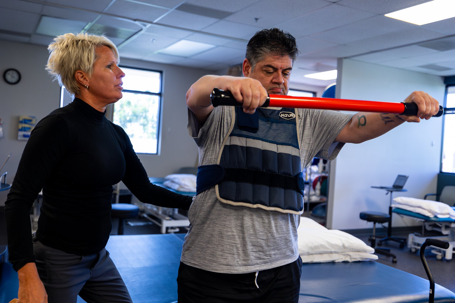
Peoria man shines light on physical therapy journey as a recovering stroke patient

 You can obviously see that this facility doesn't understand patient motivation at all.

You don't understand ONE GODDAMN THING ABOUT SURVIVOR MOTIVATION, DO YOU? You create 100% recovery protocols and your survivor will be motivated to do the millions of reps needed(NO pushing required!) because they are looking forward to 100% recovery. GET THERE! 

The problem is stroke researchers are not motivated to solve stroke. What the fuck is your solution to that failure? We still don't know how to motivate stroke medical 'professionals' to solve stroke to 100% recovery!

Peoria man shines light on physical therapy journey as a recovering stroke patient

Posted

Peoria resident Oscar Martinez always thought he was invincible.

That is, until he suffered a stroke the day after Christmas in 2022.

“I remember, we were waiting for our daughter and grandson to come over so we could give my grandson his Christmas presents,” Martinez, 51, recalls. “That’s when I started to sweat a lot. I called out to my wife for help, and she immediately noticed I wasn’t okay. I couldn’t move or speak. That’s when she realized I was having a stroke.”

Martinez was rushed to the hospital, and when he woke up, he was unsure of what had happened.

“The nurse came into the room and told me that I had a stroke,” Martinez says.

While a setback, Martinez remembers thinking he would bounce back in a few months. However, six months post-stroke, he realized it would be a much longer road to recovery. One he’s still facing today.

“It’s been a rollercoaster of emotions and challenges,” Martinez explains.

The symptoms of a stroke are discussed often, but Martinez and his physical therapist, Stephanie Larson with Banner Physical Therapy, want to shine a spotlight on what the rehabilitation process looks like for someone recovering from a stroke; highlighting some of the trials and tribulations that aren’t usually talked about.

“We focus on helping the patient do things like getting in and out of bed, in and out of the car, sitting down, standing up, balance training, and then of course help our patients, like Oscar, get back to activities they enjoy doing,” Larson said.

Larson added that one of the most surprising activities of rehabilitating a stroke patient is the fall prevention exercises that include practicing getting on and off the floor, as well as practicing falling safely to prevent further injury.

“Another surprising factor of rehab might be how hard we have to push these patients following a stroke in order to regain as much function as possible,” Larson says.

Martinez said the activities and encouragement Larson mentions have helped fast-track his progress.

“Thanks to these activities, I’m able to walk with the assistance of a cane and an ankle foot orthosis (AFO) - which is a device that helps stabilize my ankle,” Martinez said. “When I started physical therapy, I was in a wheelchair.”

However, Larson states that physical therapy recovery for stroke patients isn’t a "one-size fits all."

“The recovery time and process are different for every single patient,” Larson notes. “No two patients with a stroke are the same and they need to be treated with an individualized treatment plan focused on attaining their goals and regaining as much function as possible.”

Martinez is hopeful and remains positive.

“To anyone who might be going through a similar situation right now – keep pushing and believe in yourself,” urges Martinez. “Look for help in family and your community, be okay with your new normal, don’t take anything for granted. Take care of your health and your loved ones. Life really is too short.”

Banner Physical Therapy is part of a joint venture partnership between Banner Health and Select Medical’s Outpatient Division, a nationally prominent, locally driven provider of outpatient physical rehabilitation. Banner Physical Therapy offers 60 convenient locations throughout Arizona. The centers provide a wide range of services to patients, including physical therapy, hand/occupational therapy, pelvic health, cancer rehabilitation, pediatric therapy, sports medicine and work injury prevention and management.

For more information, visit selectmedical.com/outpatient.

Targeting interhemispheric inhibition with neuromodulation to enhance stroke rehabilitation

 In my case there is no interhemispheric inhibition since it is dead brain over there. So what is your dead brain rehab protocol? There must be millions of us out there and I've never seen any research on that problem.

Targeting interhemispheric inhibition with neuromodulation to enhance stroke rehabilitation

L.J. Boddington, J.N.J. Reynolds * Brain Health Research Centre and Brain Research NZ Centre of Research Excellence, Department of Anatomy, University of Otago, New Zealand article info Article history: Received 26 February 2016  
Received in revised form 10 December 2016  
Accepted 10 January 2017 Available online xxx Keywords: Interhemispheric inhibition Stroke Rehabilitation Neuromodulation Transcranial magnetic stimulation Electrical stimulation  
 

abstract  

 
Background/Objectives: 
 
 Interhemispheric inhibition in the brain plays a dynamic role in the production of voluntary unimanual actions. In stroke, the interhemispheric imbalance model predicts the presence of asymmetry in interhemispheric inhibition, with excessive inhibition from the contralesional hemisphere limiting maximal recovery. Stimulation methods to reduce this asymmetry in the brain may be promising as a stroke therapy, however determining how to best measure and modulate interhemispheric inhibition and who is likely to benefit, remain important questions.  
 
Methods: 
 
 This review addresses current understanding of interhemispheric inhibition in the healthy and stroke lesioned brain. We present a review of studies that have measured interhemispheric inhibition using different paradigms in the clinic, as well as results from recent animal studies investigating stimulation methods to target abnormal inhibition after stroke.  
 
Main findings/Discussion: 
 
 The degree to which asymmetric interhemispheric inhibition impacts on stroke recovery is controversial, and we consider sources of variation between studies which may contribute to this debate. We suggest that interhemispheric inhibition is not static following stroke in terms of the movement phase in which it is aberrantly engaged. Instead it may be dynamically increased onto perilesional areas during early movement, thus impairing motor initiation. Hence, its effect on stroke recovery may differ between studies depending on the technique and movement phase of eliciting the measurement. Finally, we propose how modulating excitability in the brain through more specific targeting of neural elements underlying interhemispheric inhibition via stimulation type, location and intensity may raise the ceiling of recovery following stroke and enhance functional return.

Wearable Scanner Maps Children’s Brain Activity

 Our stroke medical 'professionals' should immediately see this as a way to objectively map brain activity in stroke survivors and use that to direct the protocols needed for recovery. Then use the mapping to verify recovery is occurring per the protocol. At least this would occur if we had any leadership or strategy at all.

Wearable Scanner Maps Children’s Brain Activity

Summary: Researchers used a wearable MEG scanner to map brain activity in young children, providing new insights into brain development and conditions like autism. The lightweight, adaptable helmet with quantum technology allows for high-quality, movement-friendly scanning. This breakthrough enables the study of crucial developmental milestones and brain function from a very young age.

Key Facts:

  1. Wearable MEG scanner maps brain activity in children as young as two.
  2. Quantum technology allows for high-quality, movement-friendly scanning.
  3. Study provides insights into developmental milestones and autism.

Source: University of Nottingham

New research has given the clearest ever picture of young children’s developing brains, using a wearable brain scanner to map electrical brain activity. The work opens up new possibilities for tracking how critical developmental milestones, like walking and talking, are underpinned by changing brain function, and how neurodevelopmental conditions like autism emerge.  

The research team, led by scientists from the University of Nottingham’s School of Physics and Astronomy, used a novel design of magnetoencephalography (MEG) scanner to measure brain electrophysiology in children as young as two. 

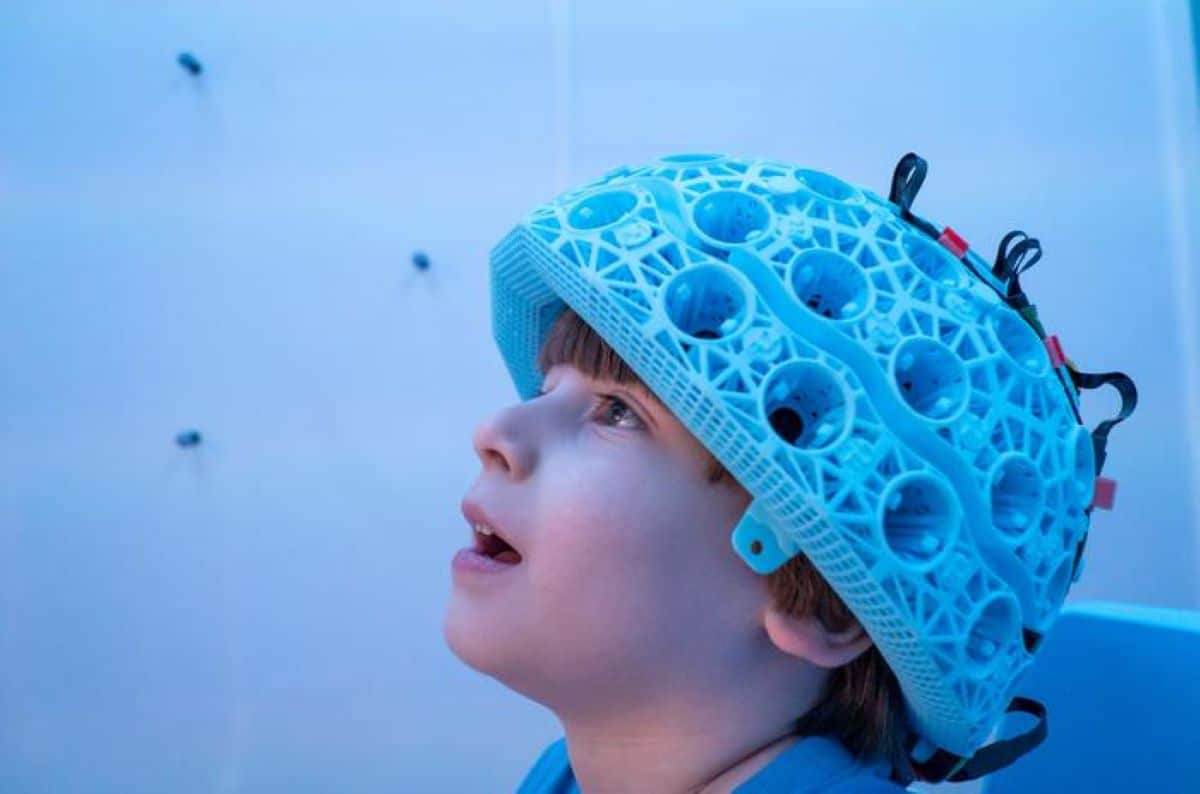
This shows a choild wearing the device.
Child wearing one of the MEG-OPM helmet style brain scanners . Credit: University of Nottingham

The findings have been published in eLife.

Brain cells operate and communicate by producing electrical currents. These currents generate tiny magnetic fields that can be detected outside the head.

Researchers used their novel system to measure these fields, and mathematical modelling to turn those fields into high fidelity images showing, millisecond-by-millisecond, which parts of the brain are engaged when we undertake tasks.

The wearable brain scanner is based on quantum technology, and uses LEGO-brick-sized sensors – called optically pumped magnetometers (OPMs) – which are incorporated into a lightweight helmet to measure the fields generated by brain activity.

The unique design means the system can be adapted to fit any age group, from toddlers to adults. Sensors can be placed much closer to the head, enhancing data quality. The system also allows people to move whilst wearing it, making it ideal for scanning children who find it hard to keep still in conventional scanners.

27 children (aged 2-13 years) and 26 adults (aged 21-34 years) took part in the study, which examined a fundamental component of brain function called ‘neural oscillations’ (or brain waves). Different areas of the brain are responsible for different aspects of  behaviour and neural oscillations promote communication between these regions.

The research team measured how this connectivity changes as we grow up, and how our brains use short, puncutate bursts of electrophysiological activity to inhibit networks of brain regions, and consequently to control how we attend to incoming sensory stimuli.

The work was jointly led by Dr Lukas Rier, and Dr Natalie Rhodes from the University of Nottingham’s School of Physics and Astronomy.

Dr Rier said: “The wearable system has opened up new opportunities to study and understand children’s brains at much younger ages than was previously possible with MEG. 

“There are important reasons for moving to younger participants: from a neuroscientific viewpoint, many critical milestones in development occur in the first few years (even months) of life. If we can use our technology to measure the brain activities that underpin these developmental milestones, this would offer a new understanding of brain function.”

The research, which was funded by the Engineering and Physics Research Council (EPSRC), included academic collaborators from SickKids Hospital in Toronto, Canada, and industry partners from US based atomic device company QuSpin and Nottingham based company Cerca Magnetics Limited.

Dr Rhodes was a University of Nottingham undergraduate student in Physics, and a postgraduate student when the work was carried out.

She has now moved to a postdoctoral position in Toronto, and explains: “This study is the first of its kind using wearable MEG technology and provides a platform to launch new clinical research in childhood disorders. This means that we can begin to explore not only healthy brain development, but also the neural substrates that underlie atypical development in children.”

World renowned neuroscientist Dr Margot Taylor – also an author on the paper – is leading research into autism in Toronto.

She said: “Our work is dedicated to studying brain function in young children with and without autism. This study is the first to demonstrate that we can track brain development from a very young age. This is hugely exciting for possible translation to clinical research and work such as this help us understand how autism develops.”

The University launched a spin-out company Cerca Magnetics in 2020 to commercialise OPM-MEG scanners and related technologies. The wearable system has been installed in a number of high profile research institutions across the globe, including SickKids hospital in Toronto.

The research teams in both institutions are now working together to expand the amount of neurodevelopmental data, on both healthy and atypical brain function.

About this neurotech and neurodevelopment research news

Author: Emma Thorne
Source: University of Nottingham
Contact: Emma Thorne – University of Nottingham
Image: The image is credited to University of Nottingham

Heart disease and stroke could affect at least 60% of adults in U.S. by 2050

 Which means the AHA/ASA had better get cracking on solving stroke to 100% recovery! Even the AHA/ASA doesn't know or remember that stroke has been called neurological disease by the WHO since 2006 and no one seems to know that.

Heart disease and stroke could affect at least 60% of adults in U.S. by 2050

By American Heart Association News

Carol Yepes/Moment via Getty Images
(Carol Yepes/Moment via Getty Images)

At least 6 in 10 adults in an older, more diverse U.S. population could be affected by cardiovascular disease within the next 30 years, according to two new science reports.

The projected rise in heart disease and stroke – along with several key risk factors, including high blood pressure and obesity – is likely to triple related costs to $1.8 trillion by 2050, according to two American Heart Association presidential advisories published Tuesday in the AHA journal Circulation. One report looks at the projected increase in cardiovascular disease rates in the decades ahead, while the other projects their total related costs.

"The landscape of cardiovascular disease in the U.S. is seeing the arrival of a near-perfect storm," Dr. Dhruv S. Kazi, vice chair of the advisory writing group, said in a news release. Kazi is head of health economics and associate director of the Richard A. and Susan F. Smith Center for Outcomes Research in Cardiology and director of the cardiac critical care unit at Beth Israel Deaconess Medical Center in Boston.

"The last decade has seen a surge of cardiovascular risk factors such as uncontrolled high blood pressure, diabetes and obesity, each of which raises the risks of developing heart disease and stroke," he said. "It is not surprising that an enormous increase in cardiovascular risk factors and diseases will produce a substantial economic burden."

Cardiovascular disease refers to conditions such as coronary heart disease, including heart attacks; heart failure, which is when the heart doesn't pump as well as it should; heart arrhythmias, including a type of irregular heart rhythm called atrial fibrillation; vascular disease, which affects the veins and arteries; congenital heart defects; stroke; and high blood pressure.

Because high blood pressure, also known as hypertension, contributes to nearly all types of heart disease and stroke, researchers made separate predictions for this condition. According to their projections, from 2020 to 2050, high blood pressure is expected to rise from 51.2% to 61% of the U.S. population.

Excluding people with high blood pressure, researchers project 15% of the population will have developed cardiovascular disease by 2050, up from 11.3% in 2020. This includes a doubling of stroke rates.

Obesity rates are expected to climb from 43.1% to 60.6% of the population, with adults ages 20 to 64 expected to see the highest growth because of unhealthy diets.

The diabetes rate is expected to rise from 16.3% to 26.8% of the population.

Risk factors are rising even among children and young adults.

By 2050, obesity is expected to affect a third of all children, up from 20% in 2020. The highest increases are expected among children 2 to 5 years old and 12 to 19. Contributing factors, such as insufficient physical activity and a poor diet, are expected to remain as high as 60% of all children over the next three decades.

Kazi said disease rates are climbing at a time of pivotal demographic shifts.

"The last of the Baby Boomers will hit 65 in 2030, so about 1 in 5 people in the U.S. will be over 65, outnumbering children for the first time in U.S. history," he said. "Since cardiovascular risk increases with age, the aging population increases the total burden of cardiovascular disease in the country."

Researchers anticipate the Hispanic, Asian and multiracial populations in the U.S. will more than double in the coming decades.

"By 2060, more than two-thirds of children will belong to underserved, disenfranchised populations, which traditionally have higher rates of cardiovascular disease and risk factors," Kazi said.

According to the projections, the biggest rise in the total number of people with cardiovascular disease will be among Hispanic adults, with Asian adults also seeing a large increase. Black adults are projected to have the highest rates of high blood pressure, diabetes and obesity, as well as the highest rates of inadequate sleep and poor diets. Asian adults are projected to have the highest rates of inadequate physical activity, with rates also high among Asian and Hispanic children.

Hispanic children are projected to have the highest obesity rates, along with the greatest projected growth in obesity, high blood pressure and diabetes. Black children are expected to have the highest overall rates of high blood pressure and diabetes.

"We found larger increases in the prevalence of cardiovascular disease and risk factors, and in the number of people with these conditions, among people from racially and ethnically diverse backgrounds," Dr. Karen E. Joynt Maddox, chair of the writing committee, said in the release. She is an associate professor of medicine at Washington University School of Medicine in St. Louis. While some of the increase is due to U.S. demographic shifts, much of the inequity in cardiovascular disease and risk factors "remains attributed to systemic racism, as well as socioeconomic factors and access to care," she said.

The researchers did find some positive trends. Overall, the percentage of people who don't get enough physical activity is expected to drop, from 33.5% to 24.2% of the population. And smoking rates are expected to drop by nearly half, from 15.8% to 8.4% of the population.

The writing committee noted that aggressive approaches to reducing cardiovascular risk factors could alter some of these predictions. Any reductions in disease could help lower costs, Kazi said.

"We can bend the cost curve on cardiovascular disease, but this will require strategic investments in cardiovascular prevention and treatment," he said. "It will take all of us working together to make this happen."

Intracerebral hemorrhage stroke outcomes improve with early minimally invasive surgery evaluated in clinical trial

 You'll have to hope your competent? hospital is closely following this research and will implement the protocol as soon as it comes out. Two flaws in that plan;

  1. Your hospital probably doesn't have a research analyst whose only job is to follow research and get it implemented in your hospital.

  2. I have never seen any research that points to a protocol that was written up as a result of that research.

Intracerebral hemorrhage stroke outcomes improve with early minimally invasive surgery evaluated in clinical trial

Peer-Reviewed Publication

Indiana University School of Medicine

INDIANAPOLIS – Patients with an intracerebral hemorrhage have better medical outcomes when surgeons perform an early minimally invasive removal of a hematoma compared to those receiving the standard of care, according to a study published in the New England Journal of Medicine.

Indiana University School of Medicine faculty clinicians Mitesh Shah, MD, Bradley Bohnstedt, MD, Regg Singh, MD and Jason Allen, MD, PhD are co-authors on a paper that assessed the findings of the ENRICH trial, a randomized clinical trial aimed at evaluating the effectiveness, safety and economics of the intervention of a minimally invasive neurosurgery technique.

The study found that patients with an intracerebral hemorrhage — a type of stroke caused by bleeding inside the brain — who had minimally invasive surgical evacuation of a brain blood clot and guideline-based medical management within 24 hours had better survival rates and functional outcomes at 180 days compared to patients who only received medical management. 

Neurosurgeons used BrainPath and Myrid devices developed by the NICO Corporation, based out of Carmel, Indiana, to perform the surgery. Surgeons create a path to the site of bleeding by gently moving aside brain tissue. Once the BrainPath device was placed, an automated suction and resection tool — the Myrid device — was used to remove the hematoma in the brain.

“We can offer early clot removal in patients with an intracerebral hemorrhage and tell our patients that they have better outcomes and survival. The families of the patients can ultimately see patients leave the hospital sooner,” said Shah, chair of the Department of Neurological Surgery.

According to the World Stroke Organization, there are more than 3.4 million new intracerebral hemorrhages each year — and over 28% of all incident strokes are intracerebral hemorrhages. In the United States, more than half of patients die within 30 days of a hemorrhage.

The 300 patients in the ENRICH trial, set at 37 centers across the United States, either had a lobar (208 patients) or anterior basal ganglia hemorrhage (92 patients) with a hematoma volume of 30-80 mL and were randomized between the two surgical groups — surgery/medical management and medical management. IU School of Medicine and its health partner IU Health was one of the sites in the trial, where 14 patients received care.

Nearly three-fourths of the surgical group in the clinical trial saw the volume of their blood clot decrease to 15 mL or less after surgery was performed within 24 hours. At 30 days post-hemorrhage, 90.7% of surgery patients survived, while 82% of medical management patients survived.

“By evacuating the clot with this early intervention, we can increase survivability and reduce the number of days spent in critical care and reduce health care costs,” Bohnstedt said.

About IU School of Medicine

The IU School of Medicine is the largest medical school in the U.S. and is annually ranked among the top medical schools in the nation by U.S. News & World Report. The school offers high-quality medical education, access to leading medical research and rich campus life in nine Indiana cities, including rural and urban locations consistently recognized for livability. According to the Blue Ridge Institute for Medical Research, the IU School of Medicine ranks No. 13 in 2023 National Institutes of Health funding among all public medical schools in the country.