Changing stroke rehab and research worldwide now.Time is Brain! trillions and trillions of neurons that DIE each day because there are NO effective hyperacute therapies besides tPA(only 12% effective). I have 523 posts on hyperacute therapy, enough for researchers to spend decades proving them out. These are my personal ideas and blog on stroke rehabilitation and stroke research. Do not attempt any of these without checking with your medical provider. Unless you join me in agitating, when you need these therapies they won't be there.

What this blog is for:

My blog is not to help survivors recover, it is to have the 10 million yearly stroke survivors light fires underneath their doctors, stroke hospitals and stroke researchers to get stroke solved. 100% recovery. The stroke medical world is completely failing at that goal, they don't even have it as a goal. Shortly after getting out of the hospital and getting NO information on the process or protocols of stroke rehabilitation and recovery I started searching on the internet and found that no other survivor received useful information. This is an attempt to cover all stroke rehabilitation information that should be readily available to survivors so they can talk with informed knowledge to their medical staff. It lays out what needs to be done to get stroke survivors closer to 100% recovery. It's quite disgusting that this information is not available from every stroke association and doctors group.

Monday, October 3, 2022

“Use It or Lose It:” What This Popular Neurorehab Phrase Means by Flint Rehab

A couple points I'd like to make on this:

1. I disagree on 'Use it or lose it' existing for stroke survivors. You can read all about my reasons for that in these 11 posts.

2. Exercising the dominant side increases recovery of the affected side. Post here:

Compensatory rehabilitation limits motor recovery after stroke

But I'm not medically trained so my points should not be listened to.  

The latest here:

“Use It or Lose It:” What This Popular Neurorehab Phrase Means by Flint Rehab
Medically reviewed by Andrew Tran PT, DPT, NCS, CSCS — written by Flint Rehab.

Last updated on December 7, 2021

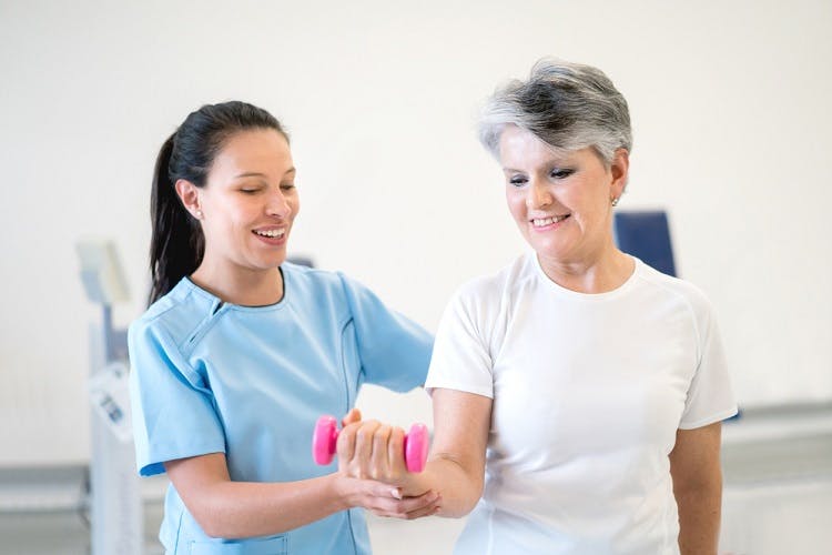
therapist teaching client about "use it or lose it" to promote recovery after neurological injury

If you’re working on recovery after a neurological injury, you’ve likely heard the phrase “use it or lose it.” This is one of the main principles of neuroplasticity, which is the central nervous system’s ability to make adaptive changes based on the behaviors you repetitively perform.

To help you understand this popular neurorehabilitation phrase, this article will discuss:

What “Use It or Lose It” Means           

To minimize losses after neurological injury, individuals must focus on promoting neuroplasticity to reorganize the central nervous system’s neural circuitry and restore compromised functions. One of the most effective ways to do this is to think “use it or lose it.” It simply means that in order to retain proficiency over a function, you must practice it regularly.

Every function you perform activates a unique set of neural pathways in the central nervous system (the brain and spinal cord). The most frequently activated neural pathways are strengthened and maintained, while those less frequently activated become neglected and forgotten.

This occurs because the central nervous system no longer senses a demand for those functions. Therefore, to be as efficient as possible and save energy for more in-demand functions, it will start to forget how to perform unused functions.

Consequently, prolonged disuse can lead to learned non-use, which refers to the conditioned suppression of affected body parts. For example, when the left arm is weakened by a stroke, individuals tend to compensate by using their unaffected right arm. Consistently using the unaffected arm leads to disuse of the affected arm until eventually, individuals forget how to use their affected arm.

The only way to prevent functions from worsening and becoming useless after a neurological injury is to use them. Repetitively practicing functions affected by neurological injury reinforces demand for them and encourages the central nervous system to reorganize those functions to unaffected regions of the brain/spinal cord. The more you practice affected functions, the stronger the newly rewired functions become.

Now that you understand what “use it or lose it” means, let’s discuss some other principles of neuroplasticity.


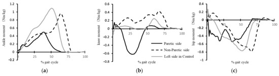
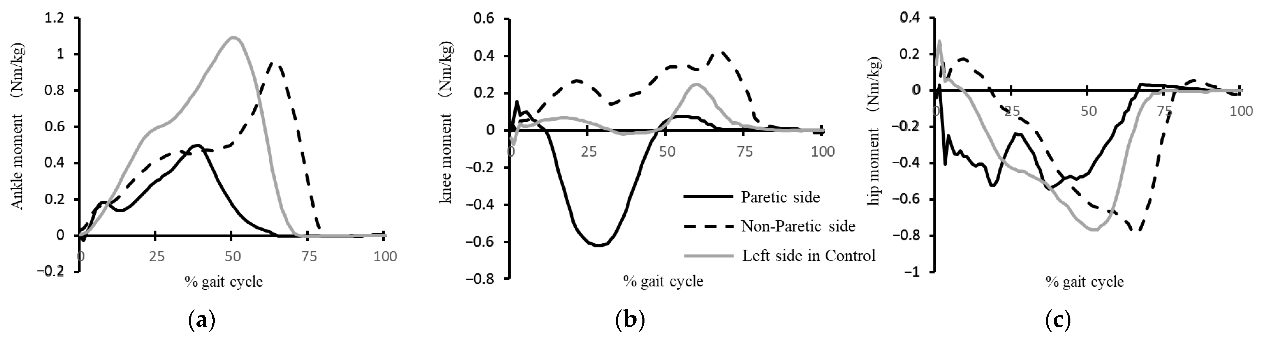
Kinetic Interjoint Coordination in Lower Limbs during Gait in Patients with Hemiparesis

So you described something but didn't give us a protocol on what EXACTLY NEEDS TO BE DONE. Useless.  No objective damage diagnosis which should lead directly to an EXACT protocol.  Does no one know how to do research that actually helps survivors? Obviously the mentors and senior researchers here don't.


Kinetic Interjoint Coordination in Lower Limbs during Gait in Patients with Hemiparesis

1
Department of Physical Medicine and Rehabilitation, Graduate School of Medicine, Tohoku University, Sendai 980-8575, Japan
2
Department of Robotics, Graduate School of Engineering, Tohoku University, Sendai 980-8579, Japan
3
Graduate School of Biomedical Engineering, Tohoku University, Sendai 980-8575, Japan
*
Author to whom correspondence should be addressed.
Academic Editors: Seifollah Gholampour and Mohammad Reza Safaei
Biomechanics 2022, 2(3), 466-477; https://doi.org/10.3390/biomechanics2030036
Received: 5 July 2022 / Revised: 7 September 2022 / Accepted: 8 September 2022 / Published: 11 September 2022
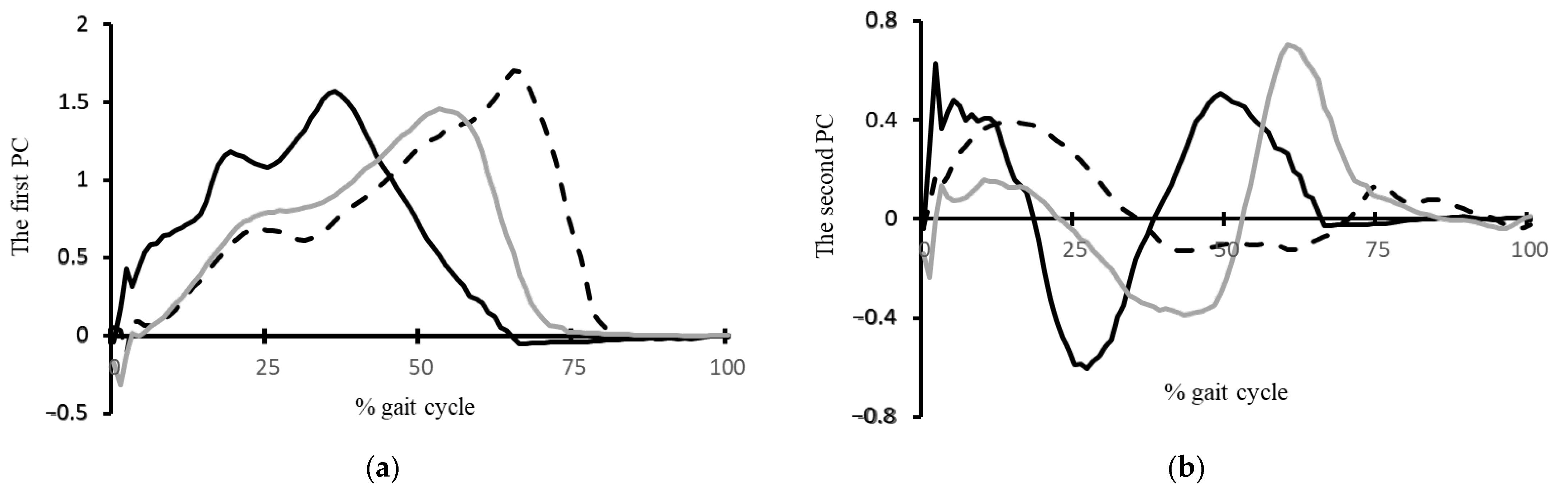
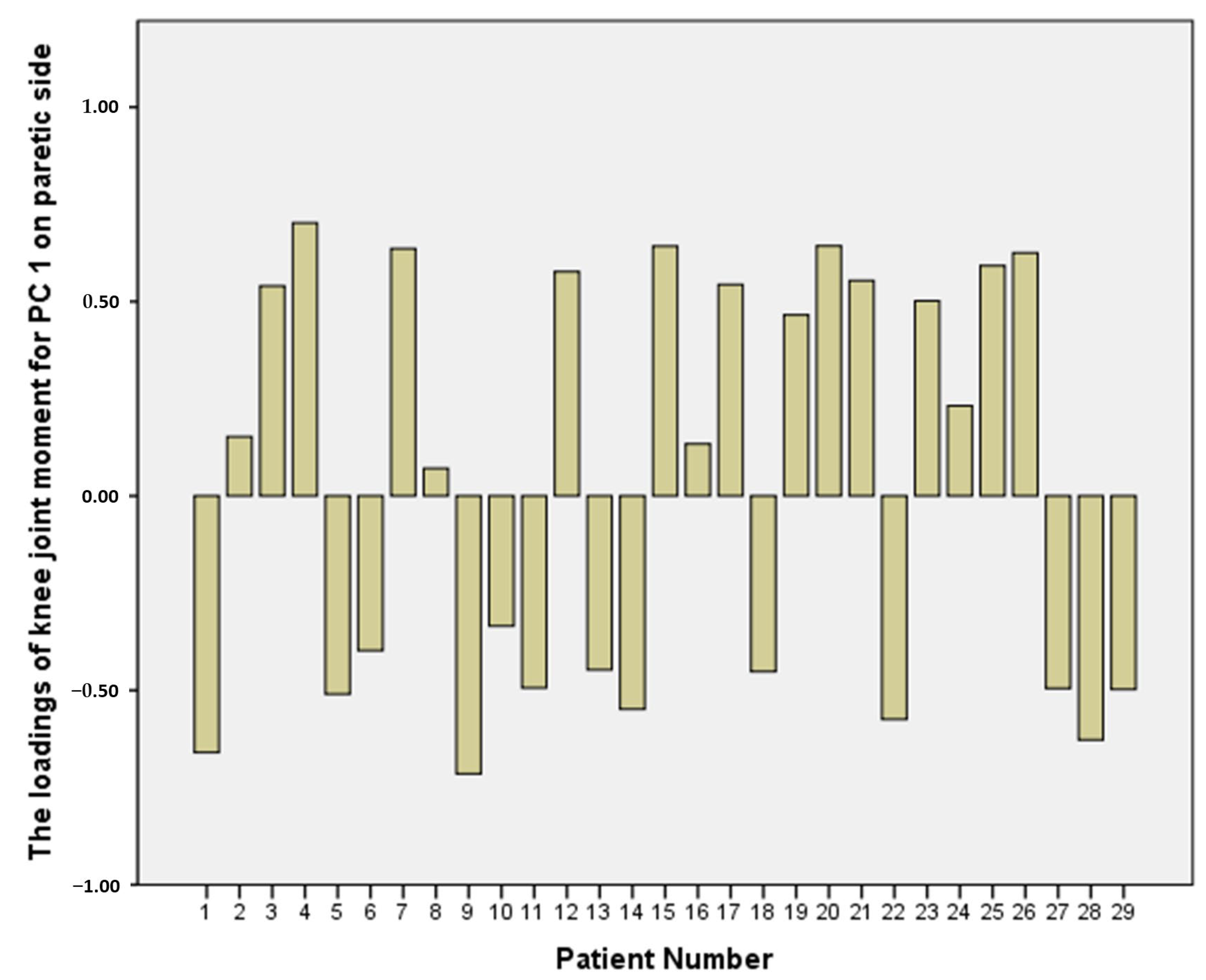
The coordination of joint moments in the same limb—otherwise known as kinetic interjoint coordination—during gait in patients with hemiparesis remains unclear. This study clarifies the characteristics of kinetic interjoint coordination in the lower limbs using a principal component analysis (PCA). Using a three-dimensional motion analysis system and force plates, the kinematic and kinetic data from 29 patients with hemiparesis and 12 healthy controls were measured when they walked along a 7 m walkway. The spatiotemporal principal components (PCs) of the hip, knee, and ankle joint moments were calculated using a PCA and the motor modules during gait were identified. We adopted a case–control study design to clarify the kinetic interjoint coordination characteristics during gait in patients with hemiplegia. As the results of comparisons between the patients and controls showed, the peak timing of the first PC, which had high loadings of hip and ankle joint moments on the paretic side, was significantly earlier than that on the other sides. The loading of the knee joint moment for the first PC on the paretic side was significantly lower than that on the non-paretic side (p < 0.05), which was highly variable with negative and positive values. The results demonstrated that the first motor module comprising hip and ankle joint moments on the paretic side during gait in patients with hemiparesis may be merged with knee joint flexion or the extension moment, and may have an atypical temporal component. The index of kinetic interjoint coordination would be a useful tool for robotic-based systems for effective rehabilitation, which would significantly contribute to the acceleration of collaborative research in the fields of engineering and rehabilitation medicine. View Full-Text
Show Figures

Figure 1

Sunday, October 2, 2022

Evaluation of Motion Segment Size as a New Sensor-based Functional Outcome Measure in Stroke Rehabilitation

I have zero understanding of what the hell this does for getting survivors recovered.

Evaluation of Motion Segment Size as a New Sensor-based Functional Outcome Measure in Stroke Rehabilitation

Hyung Seok Nam 1,2,3,4, Woo Hyung Lee 1,2,Han Gil Seo 2, Matthew W. Smuck 3 Sungwan Kim AbstractObjective:  
 
To evaluate a novel parameter, motion segment size (MSS), in stroke patients with upper limb impairment and validate its clinical applicability by correlating results with a standard clinical task-based functional evaluation tool.
Methods:
In this cross-sectional study, patients with hemiplegia and healthy controls equipped with multiple inertial measurement unit (IMU) sensors performed Action Research Arm Test(ARAT) and activities of daily living (ADL) tasks. Acceleration of the wrist and Euler angles of each upper limb segment were measured. The average and maximum MSS, accumulated motion, total performance time, and average motion speed (AMS) were extracted for analysis.
Results:
 
Data from nine patients and 10 controls showed that the average MSS of forearm
supination/pronation and elbow flexion/extension during full ARAT tasks showed a significant difference between patients and controls and a significant correlation with ARAT scores.
 
Conclusions:  
 
We suggest that MSS may provide clinically relevant information regarding upper limb functional status in stroke patients.
1 Department of Biomedical Engineering, Seoul National

Dual therapy for lower target LDL after stroke reduces risk for events at 5 years

 For discussion with your doctor. If your doctor is competent s/he will bring this up before you mention it.

Dual therapy for lower target LDL after stroke reduces risk for events at 5 years 

An LDL target of less than 70 mg/dL with ezetimibe plus statin therapy was associated with lower risk for subsequent events after stroke/transient ischemic attack at 5 years vs. a target between 90 mg/dL and 110 mg/dL, researchers reported.

In addition, ezetimibe plus statin therapy — dual therapy — for a lower target LDL was not associated with increased risk for intracranial bleeding, according to findings of a post hoc analysis of the Treat Stroke to Target trial published in Stroke.

Heart Brain 2019 Adobe
Source: Adobe Stock

“In the lower target group, dual therapy with statin and ezetimibe significantly reduced major vascular events, and the reduction was not significant on the statin monotherapy, as compared with all patients in the higher target group,” Pierre Amarenco, MD, chairman of the department of neurology and the Stroke Center at Bichat Hospital and professor of neurology at Xavier Bichat Medical School and Denis Diderot University in Paris, and colleagues wrote. “This difference was observed although the mean LDL cholesterol achieved was very similar in both groups.”

The Treat Stroke to Target trial

Treat Stroke to Target was a parallel-group trial conducted in France and South Korea and included 2,860 patients with stroke or TIA and evidence of cerebrovascular or coronary artery atherosclerosis (mean age, 67 years; 68% men; mean LDL at baseline, 135 mg/dL). Participants received dual therapy or statin monotherapy and were assigned to an LDL target of less than 70 mg/dL or 90 to 110 mg/dL. The primary endpoint was subsequent stroke, MI, urgent revascularization or CV death at a median follow-up of 3.5 years.

As Healio previously reported, patients with signs of atherosclerosis after stroke or TIA who achieved a LDL level of less than 70 mg/dL had lower risk for subsequent CV events compared with patients achieving LDL between 90 mg/dL and 110 mg/dL (adjusted HR = 0.78; 95% CI, 0.61-0.98).

Dual vs. monotherapy and lower target LDL

For the post hoc analysis, researchers evaluated whether dual therapy or statin monotherapy reduced risk for the primary outcome in patients who achieved a lower target LDL compared with a higher target LDL.

In the group assigned to the lower LDL target, those on dual therapy had higher mean LDL at baseline compared with patients on statin monotherapy (141 vs. 131 mg/dL; P < .001).

Mean achieved LDL was 66.2 mg/dL in the dual therapy group and 64.1 mg/dL in the statin monotherapy group.

Amarenco and colleagues reported that dual therapy for a lower target LDL was associated with lower risk for the primary outcome compared with a higher target LDL (HR = 0.6; 95% CI, 0.39-0.91; P = .016).

However, there was no association between statin monotherapy and lower risk for the primary outcome in the lower LDL target group compared with the higher target (HR = 0.92; 95% CI, 0.7-1.2; P = .52).

Risk for intracranial bleeding was also lower in patients on dual therapy with an LDL target of less than 70 mg/dL compared with all patients with higher target LDL (HR = 0.62; 95% CI, 0.41-0.94; P = .023).

“Explanation for such a different effect between dual therapy and statin monotherapy groups may be a higher baseline mean LDL cholesterol level in the dual therapy group, with consequently greater reduction in LDL cholesterol from baseline,” the researchers wrote. “Indeed, the effect of LDL-lowering therapy has always been associated with the magnitude of the reduction in LDL cholesterol from baseline.”


An update on predicting motor recovery after stroke

Everything you're doing here is WRONG,WRONG WRONG! Damn it all, survivors want recovery, not these useless predictions of failure to recover! Does anybody in stroke actually think?

I doubt anything has changed for the better in 8 years. I'd have you all fired.


An update on predicting motor recovery after stroke

2014, Annals of physical and rehabilitation medicine
Nouveaute´ ssurlare´ cupe´ rationmotriceapre`sAVC C.M.Stinear a,b,*,1W.D.Byblow b,c,1,S.H.Ward c,
a Clinical Neuroscience Laboratory,Department of Medicine,University of Auckland,Private Bag,92019Auckland, New Zealand b Centre for Brain Research,University of Auckland,Private Bag,92019Auckland, New Zealand c Movement Neuroscience Laboratory, Department of Sport and Exercise Science,University of Auckland,Private Bag,92019Auckland, New Zealand Received 9 August 2014;accepted 9 August 2014

Abstract

Being able to predict an individual’s potential for recovery of motor function after stroke may facilitate the use of more effective targeted rehabilitation strategies, and management of patient expectations and goals.This review summarises developments since 2010 of approaches based on clinical, neurophysiological and neuroimaging measures for predicting individual patients’ potential for upper limb recovery. Clinical assessments alone have low prognostic accuracy. Transcranial magnetic stimulation can be used to assess the functional integrity of the corticomotor pathway, and has some predictive value but is not superior when used in isolation due to its low negative predictive value. Neuroimaging measures can be used to assess the structural integrity of descending white matter tracts. Recent studies indicate that the integrity of corticospinal and alternate motor tracts in both hemispheres may be useful predictors of motor recovery after stroke. The PREP algorithm is currently the only sequential algorithm that combines clinical, neurophysiological and neuroimaging measures at the subacute stage to predict the potential for subsequent recovery of upper limb function. Future research could determine if a similar algorithmic approach may be useful for predicting the recovery of gait after stroke.(Everything you're doing here is WRONG,WRONG WRONG! Damn it all, survivors want recovery, not these useless predictions of failure to recover! Does anybody in stroke actually think?)
#2014Elsevier MassonSAS.
All rights reserved.

Predictors of post-stroke cognitive impairment using acute structural MRI neuroimaging: A systematic review and meta-analysis

Are your mentors and senior researchers that clueless that they don't know that predicting cognitive impairment is totally fucking useless for survivors?  If we had ANY KIND OF STROKE STRATEGY AT ALL, every piece of stroke research would lead directly to creation of stroke protocols that fix stroke problems.  I'd fire you all for being a complete waste.

Predictors of post-stroke cognitive impairment using acute structural MRI neuroimaging: A systematic review and meta-analysis

Abstract

Background:

Stroke survivors are at an increased risk of developing post-stroke cognitive impairment and post-stroke dementia; those at risk could be identified by brain imaging routinely performed at stroke onset.

Aim:

This systematic review aimed to identify features which are associated with post-stroke cognitive impairment (including dementia) on magnetic resonance imaging (MRI) performed at stroke diagnosis.

Summary of review:

We searched the literature from inception to January 2022 and identified 10,284 records. We included studies that performed MRI at the time of stroke (0–30 days after a stroke) and assessed cognitive outcome at least 3 months after stroke. We synthesized findings from 26 papers, comprising 27 stroke-populations (N = 13,114, average age range = 40–80 years, 19–62% female). When data were available, we pooled unadjusted (ORu) and adjusted (ORa) odds ratios.
We found associations between cognitive outcomes and presence of cerebral atrophy (three studies, N = 453, ORu = 2.48, 95% CI = 1.15–4.62), presence of microbleeds (two studies, N = 9151, ORa = 1.36, 95% CI = 1.08–1.70), and increasing severity of white matter hyperintensities (three studies, N = 704, ORa = 1.26, 95% CI = 1.06–1.49). Increasing cerebral small vessel disease score was associated with cognitive outcome following unadjusted analysis only (two studies, N = 499, ORu = 1.34, 95%CI = 1.12–1.61; three studies, N = 950, ORa = 1.23, 95% CI = 0.96–1.57). Associations remained after controlling for pre-stroke cognitive impairment. We did not find associations between other stroke features and cognitive outcome, or there were insufficient data.

Conclusion:

Acute stroke MRI features may enable healthcare professionals to identify patients at risk of post-stroke cognitive problems. However, there is still substantial uncertainty about the prognostic utility of acute MRI for this.

Introduction

Cognitive problems after stroke are of major concern to stroke survivors and their families.1(Yep, AND YOU'RE DOING NOTHING TO SOLVE THEM!) Identifying who is at risk at the time of stroke may enable healthcare professionals to arrange appropriate follow-up, inform patients and their carers, and plan for possible future health outcomes. Individuals at risk of post-stroke cognitive problems could also be targeted for clinical trials with cognitive endpoints.
The cognitive consequences of stroke are conventionally described as post-stroke cognitive impairment (PSCI—impaired performance on a structured cognitive assessment) and the subcategory of post-stroke dementia (PSD—a clinical diagnosis of a cognitive change sufficient to interfere with daily life).
International guidelines for PSCI highlight that there are currently no prediction tools suitable for clinical practice.2 A survey of 60 UK healthcare professionals reported that respondents were aware that imaging features could predict PSCI, but they did not use these features in clinical practice.3 Acute stroke neuroimaging could help healthcare professionals to identify who is at risk of PSCI.
Acute stroke computed tomography (CT) brain imaging is routinely performed in clinical practice to determine the cause of stroke. CT brain imaging is inexpensive and quick to perform but has lower resolution than magnetic resonance imaging (MRI). Recently, MRI has become more available for stroke diagnosis in clinical practice. MRI also allows the identification of neuroimaging features such as cerebral microbleeds (CMB) that are rarely visible on CT brain scans. MRI may help identify neuroimaging features associated with post-stroke cognitive problems.
Cerebral small vessel disease (cSVD) is commonly associated with stroke and dementia.4 Neuroimaging features include white matter hyperintensities (WMH), CMB, lacunes, perivascular spaces (PVS), recent small subcortical infarcts, and cerebral atrophy.5 Three systematic reviews have described the associations between neuroimaging features and PSD/PSCI.68 One review found that stroke survivors with moderate to severe WMH had a two-to-three-fold increased risk in PSD/PSCI.7 Another review reported that medial temporal lobe atrophy (MTLA) and global atrophy were associated with increased risk of PSCI,6 and the third review highlighted an association between MTLA, WMH, and PSCI.8 These reviews included studies that performed brain imaging up to several months after a stroke, which does not reflect what happens in clinical practice. Only one review performed a sensitivity analysis comparing the association between severity of WMH and PSD when identified on CT versus MRI.7 The reviews did not report the association between acute stroke lesions and post-stroke cognitive outcome. However, a multicohort study of 2950 stroke survivors reported that infarcts in the left thalamus, left frontotemporal lobes, and right parietal lobe were associated with PSCI.9 Our previous systematic review focused on the prognostic utility of acute stroke CT finding that presence of atrophy, WMH, and pre-existing stroke lesions were associated with a two-to-three-fold increase in risk of PSD, and WMH was associated with a three-fold increased risk in PSCI.10 MRI is increasingly being used in clinical practice and is recommended for suspected TIA.11 A similar review focusing on MRI was needed.
 
More at link.

Young brain fluid improves memory in old mice

 But is young blood or bone marrow better? And what is the exact definition of young in humans?

Young Blood Revitalizes the Aging Brain June 2014 

Characterizing the Mechanism of Young Bone Marrow Derived Microglia-Like Cells on Restoring Cognitive Function in the Aged Brain and After Ischemic Injury

March 2022

The latest here:

Young brain fluid improves memory in old mice

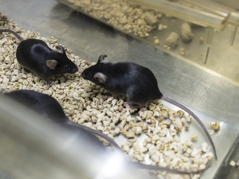
ice sit in a container at a Cyagen Biosciences Inc. facility in Taicang, Jiangsu province, China.

Young cerebrospinal fluid probably improves the conductivity of the neurons in ageing mice.Credit: Qilai Shen/Bloomberg/Getty

Scientists have been trying to unravel the mysteries of why memory diminishes with age for decades. Now they have discovered a possible remedy — cerebrospinal fluid from younger brains1.

Cerebrospinal fluid (CSF) from young mice can improve memory function in older mice, researchers report today in Nature. A direct brain infusion of young CSF probably improves the conductivity of the neurons in ageing mice, which improves the process of making and recalling memories. The team also suggests that the improvements are largely due to a specific protein in the fluid.

“This is super exciting from the perspective of basic science, but also looking towards therapeutic applications,” says Maria Lehtinen, a neurobiologist at Boston Children’s Hospital in Massachusetts.

CSF is the central nervous system’s version of plasma: a soup of essential ions and nutrients that cushions the brain and spinal cord and is essential for normal brain development. Physicians frequently use it as an indicator of brain health, and a biomarker of neurological diseases. But as mammals age, CSF loses some of its punch. Those changes might affect cells related to memory, says co-author Tal Iram, a neuroscientist at Stanford University in California. “Could we do something about it by re-exposing these cells to younger CSF?” she asks. “That was the overarching question.”

Testing memory

The first step for Iram and her team was to give ageing mice an experience they would remember. The team gave 20-month-old mice three small electric shocks on their foot in tandem with several flashes of light and sound, to create an association between the lights and the shock. The researchers then infused the brains of one group of 8 mice with CSF from 10-week-old mice, while a control group of 10 mice were given artificial CSF.

After three weeks the mice faced the same sounds and lights, but this time without a shock — recreating the context of the fear without the actual fear-inducing action. Mice that receive young CSF remembered the shock and froze in fear almost 40% of the time, but that happened only around 18% of the time in mice given artificial CSF. The findings suggest that young CSF can restore some declines in ageing-brain abilities. “The broader implication is that the brain is still malleable and there are ways to improve its function,” says co-author Tony Wyss-Coray, a neuroscientist at Stanford. “It’s not all lost.”

The work on CSF is inspired by Wyss-Coray’s past work showing that plasma from young mice could restore memory function in older rodents2,3. A start-up co-founded by Wyss-Coray, Alkahest in San Carlos, California, has conducted small trials suggesting some cognitive benefits in mice and people with dementia given the company’s plasma-derived products. Other groups are exploring different methods for using young plasma, but the field is still in its infancy.

The brain’s wiring

The hippocampus is the brain’s memory control centre: it is responsible for creating, retaining and recalling memories. The team therefore looked at this seahorse-shaped structure to get a better understanding of how young CSF might improve the memory function of ageing mice. The researchers found that the structure upregulated genes related to a cell called an oligodendrocyte. Oligodendrocytes produce the myelin sheath around neurons’ tails, essentially “the plastic coating over the wires in the brain”, says Wyss-Coray. And like wire insulation, that sheathing helps with conductivity. Specifically, the CSF helps to generate more of the early-stage oligodendrocytes known as oligodendrocyte progenitor cells. Generating more cells that insulate nerve connections helps to maintain brain function, Wyss-Coray adds.

The researchers also isolated a protein from the CSF cocktail that another analysis had suggested was a compelling candidate for improving memory: fibroblast growth factor 17 (Fgf17). Infusion of Fgf17 had a similar memory-restoring effect to infusing CSF. Furthermore, giving the mice an antibody that blocked Fgf17’s function impaired the rodents’ memory ability. Wyss-Coray and Iram have applied for a patent on their findings around Fgf17.

More at link.

Stroke Symposium planned at St. Mary Medical Center - Hobart,IN

Go there and keep asking the same questions.

1. How do I get to 100% recovery?

2. What are you SPECIFICALLY DOING  to get survivors to 100% recovery? Laugh maniacally if they suggest guidelines, guidelines are worthless. You need EXACT PROTOCOLS.

3. How are you getting to 100% recovery when you are the 1 in 4 per WHO that has a stroke.  Make it personal to the speakers.


Hemming and hawing are not allowed. Neither is the craptastic saying; 'All strokes are different, all stroke recoveries are different'. Do not let them off the hook until they answer.

Stroke Symposium planned at St. Mary Medical Center - Hobart,IN

Stroke Symposium planned at St. Mary Medical Center

St. Mary Medical Center in Hobart

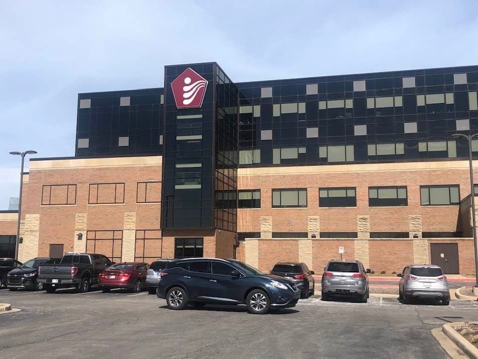
St. Mary Medical Center will host a symposium next month on the fifth leading cause death in the United States. The “Time is Brain” stroke symposium is scheduled for 8 a.m. to noon Oct. 13 at Community Healthcare System's medical center at 1500 S. Lake Park Ave., Hobart.

Free health screenings, vendor booths and continental breakfast will be offered at 8 a.m. Then, emergency medicine physician Lauren Rutili, neurologist Mark Simaga, and cardiologist Kais Yehyawi will make presentations on various topics and stroke patients will share personal stories of defeating the disease, which is the leading cause of disability in the United States.

The American Heart Association estimates 80% of strokes can be prevented by maintaining a healthy lifestyle and controlling risk factors like quitting smoking.

"If you or someone you care for smokes or vapes, please seek help. St. Mary Medical Center offers cessation classes, and QuitNowIndiana.com has free tools to help you be successful," Comunity Health said in a press release. "Move more. Active adults are 25 to 30% less likely to suffer a stroke. Keep blood pressure in control. High blood pressure, or hypertension, is the leading cause of strokes."

Saturday, October 1, 2022

Efficacy and Safety of Vagus Nerve Stimulation in Stroke Rehabilitation: A Systematic Review and Meta-Analysis

Well you blithering idiots it has been proven to work a long time ago.  All your mentors and senior researchers had to do was to tell you to create a protocol on this and get it distributed to all 10 million yearly stroke survivors  now and into the future. And they completely failed at their job, thus wasting all this time and money.

Efficacy and Safety of Vagus Nerve Stimulation in Stroke Rehabilitation: A Systematic Review and Meta-Analysis

Abstract

Introduction: Recent randomized controlled trials (RCTs) have assessed the role of vagus nerve stimulation (VNS) when paired with standard rehabilitation in stroke patients. This review aimed to evaluate the efficacy and safety of VNS as a novel treatment option for post-stroke recovery.  

Methods: 

We searched PubMed, EMBASE, Cochrane Database of Systematic Reviews, Cochrane Central Register of Controlled Trials (CENTRAL), and CINAHL Plus for articles published from their date of inception to June 2021. RCTs investigating the efficacy or safety of VNS on post-stroke recovery were included. The outcomes were upper limb sensorimotor function, health-related quality of life, level of independence, cardiovascular effects, and adverse events. The risk of bias was assessed using the Cochrane risk-of-bias tool, while the certainty of the evidence was assessed using the Grading of Recommendations, Assessment, Development, and Evaluations (GRADE) criteria. Review Manager 5.4 was used to conduct the meta-analysis.  

Results: 

Seven RCTs (n = 236 subjects) met the eligibility criteria. Upper limb sensorimotor function, assessed by the Fugl-Meyer Assessment for Upper Extremity (FMA-UE), improved at day 1 (n = 4 RCTs; standardized mean difference [SMD] 1.01; 95% confidence interval [CI]: 0.35–1.66) and day 90 post-intervention (n = 3 RCTs; SMD 0.64; 95% CI: 0.31–0.98; moderate certainty of evidence) but not at day 30 follow-up (n = 2 RCTs; SMD 1.54; 95% CI: −0.39 to 3.46). Clinically significant upper limb sensorimotor function recovery, as defined by ≥6 points increase in FMA-UE, was significantly higher at day 1 (n = 2 RCTs; risk ratio [RR] 2.01; 95% CI: 1.02–3.94) and day 90 post-intervention (n = 2 RCTs; RR 2.14; 95% CI: 1.32–3.45; moderate certainty of the evidence). The between-group effect sizes for upper limb sensorimotor function recovery was medium to large (Hedges’ g 0.535–2.659). While the level of independence improved with VNS, its impact on health-related quality of life remains unclear as this was only studied in two trials with mixed results. Generally, adverse events reported were mild and self-limiting.  

Conclusion: 

VNS may be an effective and safe adjunct to standard rehabilitation for post-stroke recovery; however, its clinical significance and long-term efficacy and safety remain unclear.(Really?)

© 2022 The Author(s). Published by S. Karger AG, Basel


Introduction

Stroke is often associated with a significant disease burden requiring substantial treatment, especially post-stroke care [1]. Most survivors suffer from neurological deficits and require around-the-clock care or institutionalization due to limited functional independence [2]. Rehabilitation is an important component of post-stroke care. The goal of rehabilitation is to provide extensive education and task-specific training to maximize functional abilities, thus improving the level of independence. Physical rehabilitation facilitates synaptic plasticity and cortical reorganization within the motor cortex [3].

Over the years, several large randomized controlled trials (RCTs) of increased rehabilitation regime, the use of rehabilitation devices, and brain stimulation therapies devices have not demonstrated clinically meaningful motor recovery compared to standard rehabilitation therapy only [4, 5]. Several preclinical trials have reported favourable post-stroke recovery following combined vagus nerve stimulation (VNS) and rehabilitation therapy, and several clinical trials have shown promising results [6-8]. VNS is a technique that provides stimulations to the vagus nerve via an implantable device or a non-implantable device attached to the skin overlying the vagus nerve [9, 10]. The enhanced rehabilitation model is postulated to induce a brain environment that might increase the potential for experience-dependent plasticity [11]. A recent meta-analysis of animal studies highlighted that combined VNS and rehabilitation therapy facilitated clinically significant motor function recovery following neurological injuries [10]. This study aimed to systematically review the efficacy and safety of VNS on post-stroke recovery.

Ananda R.a,b · Roslan M.H.B.a · Wong L.L.a · Botross N.P.a · Ngim C.F.a · Mariapun J.a

Author affiliations

Corresponding Authors


More at link.


The use of self-management strategies for stroke rehabilitation: a scoping review

WHY, WHY, WHY? Why should there be any self management? Survivors expect that their therapists and doctors have created EXACT 100% RECOVERY PROTOCOLS. And all survivors have to do is follow them to get recovered.

The use of self-management strategies for stroke rehabilitation: a scoping review


Received 30 Jun 2022, Accepted 18 Sep 2022, Published online: 27 Sep 2022
 

Introduction

Self-management is generally considered a dynamic and collaborative process by individuals and caregivers to manage a chronic condition. Self-management has recently emerged as a promising strategy for stroke rehabilitation. This scoping review aims to examine and summarize self-management strategies utilized by stroke survivors for stroke rehabilitation.

Methods

PubMed, Scopus, CINAHL (EBSCO), Embase, and ProQuest were searched for articles published between January 2010 and December 2021. Studies were selected if they were published in English in a peer-reviewed journal, utilized a non-experimental research design, and focused on adult stroke survivors. All relevant information from the included articles was extracted in a systematic way using a pre-developed data extraction form. Two authors performed data extraction and quality evaluation independently. All issues were resolved through discussion among the authors.

Results

We narratively summarized the findings of 15 quantitative, qualitative, and mixed-method studies, including a total of 1,494 stroke survivors. The stroke survivors used a range of self-management strategies for their stroke rehabilitation, including domains related to lifestyle, social support, communication, knowledge and information, and goal-setting. Gender, age, stroke-related disability, fatigue, self-management education, social support, and communication with others were found to be associated with self-management use in stroke rehabilitation.

Conclusions

This scoping review provides an important overview on stroke survivors’ use of self-management strategies and their experience. Their use of self-management strategies is complicated and multifaceted, comprising several domains and involving a diverse range of approaches and personal experiences. However, we identified several gaps in the literature and more research is required.

Mechanisms of Short-Term Training-Induced Reaching Improvement in Severely Hemiparetic Stroke Patients: A TMS Study

Did your doctors or hospital implement anything from this in the ensuing 11 years?

Do you prefer your doctor and hospital incompetence NOT KNOWING? OR NOT DOING?


Mechanisms of Short-Term Training-Induced Reaching Improvement in Severely Hemiparetic Stroke Patients: A TMS Study

2011, Neurorehabilitation and Neural Repair
 
Michelle L. Harris-Love, PhD 1,
Susanne M. Morton, PhD 2,
Monica A. Perez, PhD 3, and
Leonardo G. Cohen, MD 4
1 National Rehabilitation Hospital, Georgetown University, Washington, DC, USA
2 University of Iowa, Iowa City, IA, USA
3 University of Pittsburgh, Pittsburgh, PA, USA
4 Human Cortical Physiology & Stroke Neurorehabilitation Section, National Institute ofNeurological Disorders and Stroke, National Institutes of Health, Bethesda, MD, USA

Abstract

Background—
 
The neurophysiological mechanisms underlying improved upper-extremity motorskills have been partially investigated in patients with good motor recovery but are poorlyunderstood in more impaired individuals, the majority of stroke survivors.
Objective—
 
The authors studied changes in primary motor cortex (M1) excitability (motorevoked potentials [MEPs], contralateral and ipsilateral silent periods [CSPs and ISPs] usingtranscranial magnetic stimulation [TMS]) associated with training-induced reaching improvementin stroke patients with severe arm paresis (n = 11; Upper-Extremity Fugl-Meyer score (F-M) = 27± 6).
Methods—
 
All patients underwent a single session of reaching training focused on moving the affected hand from a resting site to a target placed at 80% of maximum forward reaching amplitude in response to a visual “GO” cue. Triceps contribute primarily as agonist and biceps primarily as antagonist to the trained forward reaching movement. Response times were recorded for each reaching movement.
Results—
 
Preceding training (baseline), greater interhemispheric inhibition (measured by ISP) in the affected triceps muscle, reflecting inhibition from the nonlesioned to the lesioned M1, was observed in patients with lower F-M scores (more severe motor impairment). Training induced improvements in reaching were greater in patients with slower response times at baseline.Increased MEP amplitudes and decreased ISPs and CSPs were observed in the affected triceps but not in the biceps muscle after training.
Conclusion—
 
These results indicate that along with training-induced motor improvements,training-specific modulation of intrahemispheric and interhemispheric mechanisms occurs after reaching practice in chronic stroke patients with substantial arm impairment.

Corresponding Author:
 Leonardo G. Cohen, MD, 10 Center Dr, MSC 1428, Bethesda, MD 20892, USA cohenl@ninds.nih.gov.Declaration of Conflicting InterestsThe author(s) declared no potential conflicts of interest with respect to the authorship and/or publication of this article.


Why thinking hard makes us feel tired

 Is this part of the reason there is so much fatigue in stroke survivors? Initially I had to think very hard just to walk semi-properly mainly because my premotor cortex was mostly dead and no longer could coordinate the timing of firing all the different muscles used in walking.

At least half of all stroke survivors experience fatigue 

Or is it 70%?

Or is it 40%?

WHOM will do the research to see if this is true for stroke survivors AND then do the research to solve that fatigue problem?

Why thinking hard makes us feel tired

Asian businesswoman online working at the night. Busy and exhausted of work overtime at home.

Mental strain can lead to changes in brain physiology that cause feelings of tiredness.Credit: Getty

It’s not just in your head: a desire to curl up on the couch after a day spent toiling at the computer could be a physiological response to mentally demanding work, according to a study that links mental fatigue to changes in brain metabolism.

The study, published on 11 August in Current Biology1, found that participants who spent more than six hours working on a tedious and mentally taxing assignment had higher levels of glutamate — an important signalling molecule in the brain. Too much glutamate can disrupt brain function, and a rest period could allow the brain to restore proper regulation of the molecule, the authors note. At the end of their work day, these study participants were also more likely than those who had performed easier tasks to opt for short-term, easily won financial rewards of lesser value than larger rewards that come after a longer wait or involve more effort.

The study is important in its effort to link cognitive fatigue with neurometabolism, says behavioural neuroscientist Carmen Sandi at the Swiss Federal Institute of Technology in Lausanne. But more research — potentially in non-human animals — will be needed to establish a causal link between feelings of exhaustion and metabolic changes in the brain, she adds. “It’s very good to start looking into this aspect,” says Sandi. “But for now this is an observation, which is a correlation.”

Tired brain

Previous research has demonstrated effects of mental strain on physiological parameters such as heart-rate variability and blood flow, but these tend to be subtle, says Martin Hagger, a health psychologist at the University of California, Merced. “It’s not like when you’re exercising skeletal muscle,” he says. “But it is perceptible.”

Cognitive neuroscientist Antonius Wiehler at the Paris Brain Institute and his colleagues thought that the effects of cognitive fatigue could be due to metabolic changes in the brain. The team enrolled 40 participants and assigned 24 of them to perform a challenging task: for example, watching letters appear on a computer screen every 1.6 seconds and documenting when one matched a letter that had appeared three letters ago. The other 16 participants were asked to perform a similar, but easier task. Both teams worked for just over six hours, with two ten-minute breaks.

While the study participants focused on their work, Wiehler and his team used a technique called magnetic resonance spectroscopy to measure levels of glutamate in a region of the brain called the lateral prefrontal cortex.

The prefrontal cortex is the home of cognitive control — the part of the brain that allows people to suppress their impulses. “If you get stung by an insect, you want to scratch,” says Wiehler. “If you’re stopping this reflex, that would be cognitive control.” It’s also the system that humans rely on to choose tempting short-term rewards, such as an unhealthy snack, over long-term gains.

The researchers found that participants who laboured on the more difficult task had accumulated more glutamate in this region of the brain by the end of the day than had those who worked on the easier task. And, given a choice between an immediate cash reward and a larger reward that would come months later, they were more likely to choose the smaller, short-term reward than they were at the start of the day.

Wiehler now hopes to use this system to learn more about how to recover from mental exhaustion. “It would be great to find out more about how glutamate levels are restored,” he says. “Is sleep helpful? How long do breaks need to be to have a positive effect?” Studies of cognitive fatigue could also be key to understanding how workers react to — and recover from — high-stakes mental work such as air-traffic control, in which even a brief loss of focus can cost lives.

And now that a system has been established to measure metabolic changes in response to mental fatigue, Hagger hopes that other researchers will try the approach. “Means to detect this have hitherto not been sensitive enough, so this research paves the way for future researchers to explore cognitive fatigue,” he says.

That research — and particularly animal studies, in which glutamate levels can be experimentally altered — could unpick the molecular mechanisms that cause the molecule to accumulate during difficult mental work and how that affects brain activity, says Sandi. “This is the tricky part.”

doi: https://doi.org/10.1038/d41586-022-02161-5


doi: https://doi.org/10.1038/d41586-022-02161-5