Changing stroke rehab and research worldwide now.Time is Brain! trillions and trillions of neurons that DIE each day because there are NO effective hyperacute therapies besides tPA(only 12% effective). I have 523 posts on hyperacute therapy, enough for researchers to spend decades proving them out. These are my personal ideas and blog on stroke rehabilitation and stroke research. Do not attempt any of these without checking with your medical provider. Unless you join me in agitating, when you need these therapies they won't be there.

What this blog is for:

My blog is not to help survivors recover, it is to have the 10 million yearly stroke survivors light fires underneath their doctors, stroke hospitals and stroke researchers to get stroke solved. 100% recovery. The stroke medical world is completely failing at that goal, they don't even have it as a goal. Shortly after getting out of the hospital and getting NO information on the process or protocols of stroke rehabilitation and recovery I started searching on the internet and found that no other survivor received useful information. This is an attempt to cover all stroke rehabilitation information that should be readily available to survivors so they can talk with informed knowledge to their medical staff. It lays out what needs to be done to get stroke survivors closer to 100% recovery. It's quite disgusting that this information is not available from every stroke association and doctors group.

Wednesday, July 1, 2026

New Artificial Neurons Cause Living Brain Cells to Fire

 If your competent? doctor can't figure out how to get dendritic branching/neurite outgrowth and axon pathfinding to work to connect up gray matter again then maybe this could work.

New Artificial Neurons Cause Living Brain Cells to Fire

A team at Northwestern University has printed artificial neurons from molybdenum disulfide (MoS₂) — a semiconducting mineral — on flexible plastic that produce spiking waveforms closely matching biological action potentials in shape, width, and timing. When delivered to living Purkinje cells in mouse cerebellar tissue, the artificial spikes drove the cells to fire — the first demonstration that a printed device can produce electrical signals a real brain cell accepts and responds to.

The result, recently published in Nature Nanotechnology, could lay groundwork for a new generation of neural interfaces — prosthetic limbs that deliver realistic sensation, spinal cord bridges that relay motor commands, and benchtop disease models with tunable parameters.

“There’s this white space — organic devices are too slow, metal oxides are too fast — and biology lives in between,” said Mark C. Hersam, PhD, the study’s senior author and Walter P. Murphy Professor of Materials Science and Engineering at Northwestern University in Evanston, Illinois.
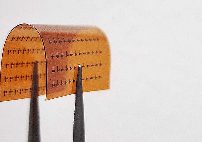
photo of Flexible polymide
An array of printed artificial neurons on flexible polyimide held by tweezers to show the substrate bending.

“We got these devices working at that timescale,” he said, “and when you have the right timescale and the right spike shape, you can directly interface with living cells.”

Key Points
  • Printed MoS2 artificial neurons generated action-potential-like spikes.
  • Spike shape/timing matched biological APs; duration ≈0.7-2 ms.
  • Mouse Purkinje cells fired to artificial spikes at <200 Hz.
  • 740 Hz output failed; neuronal firing capacity limits response.
  • Potential uses: neural interfaces, spinal bridges, tunable disease models.
How do printed neural interfaces compare with metal oxide devices?
What limits long-term stability of MoS2 neural circuits?
Which cerebellar diseases could benefit from tunable circuit models?

Engineers Interface With Neuroscientists

The collaboration began not in biology but in electrical engineering. Hersam’s National Science Foundation grant aimed to build computing hardware that mimics the brain’s energy efficiency. The human brain runs on about 20 watts, whereas modern AI training runs on megawatts — a millionfold difference — and is extravagantly wasteful and potentially harmful to the environment.

The grant’s challenge: build computing hardware that mimics the brain’s efficiency.

“Most of the artificial neurons in the literature, if you actually look at their spiking profiles, they look more like a sine wave or just an oscillator, not a sharp action potential,” Hersam said. “They don’t achieve things like bursts of spikes, which is one of the things we demonstrate.”

photo of Meghana Holla, PhD
Meghana Holla, PhD

But to mimic the brain, Hersam needed people who study it. He teamed up with Indira M. Raman, PhD, a neurophysiologist in the Department of Neurobiology at Northwestern University, and her lab. Her doctoral students, Spencer Brown, PhD, and Meghana Holla, PhD, visited Hersam’s lab to see what the engineers were up to.

The engineers in Hersam’s lab showed them the device output, with waveforms spiking at 3000 times per second, much too fast to mimic a neural cell. Purkinje cells may be among the fastest-firing neurons in the brain, but they only reach about 100 spikes per second.

“That’s not a neuron,” Brown, an incoming assistant professor of neuroscience at Brandeis University in Waltham, Massachusetts, recalled. “They can’t do that.”
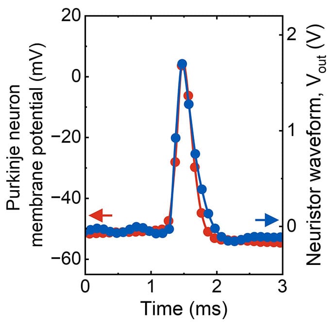
photo of Purkinje neuron Chart
The artificial neuron’s output (blue) overlaid with a living Purkinje cell’s action potential (red). Both spikes match closely in shape and duration, completing within about 2 milliseconds.

Over the following year, both fields discovered they used identical terminology, such as long-term potentiation, memory, and synapse weight, to mean different things.

photo of Spencer T. Brown, PhD
Spencer Brown, PhD

“We thought we were talking about the same things, but we weren’t,” Brown said.

Brown and Holla provided “ground truth”: Each spike had to last between a fraction of a millisecond and a few milliseconds, matching a real action potential.

And the firing rate — the number of spikes per second — had to fall between single digits and low hundreds, not the thousands the engineers’ devices had been producing.

The engineers took the neuroscientists’ advice and successfully reconfigured the circuit to match.

The Glue That Makes Artificial Neurons Fire

The artificial neurons are built from a liquid, a custom ink formulated for a specialized printer. MoS2, a semiconducting mineral, is peeled into flakes that are just a few atoms thick and suspended in ethanol. Without that suspension, the flakes clump together and settle out.

To keep them suspended, the researchers add ethylcellulose, a polymer derived from wood pulp, which coats each MoS2 flake and holds it apart from its neighbors, kind of like glue. The resulting ink is a stable suspension of semiconductor particles in solvent, and it flows through an aerosol jet printer that deposits it as a fine mist onto flexible plastic.
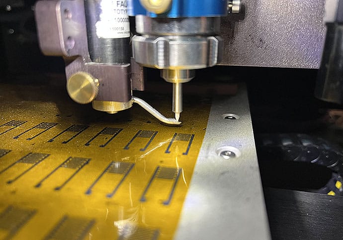
photo of inkjet printer
An aerosol jet printer deposits molybdenum disulfide ink onto flexible plastic. The nozzle sprays the ink as a fine mist, printing rows of artificial neurons without a cleanroom.

The ethylcellulose scaffolding is essential for the artificial neurons to communicate like a network. When the printed film is baked at 350 °C, the ethylcellulose partially decomposes into carbon residue that settles into tiny, nanometer-sized gaps between flakes to form conductive bridges. Once fabricated, the device operates at room temperature.

Then comes what’s called electroforming. The first time a large current passes through the device, it doesn’t flow evenly. Some pathways are slightly more conductive, such as wherever carbon residue accumulated more thickly, or wherever flakes overlapped. The more conductive pathways carry more current, and more current generates more heat. That heat decomposes more polymer residue into carbon along the same route, making it more conductive and drawing still more current toward it.

photo of Mark C. Hersam, PhD
Mark C. Hersam, PhD

The result is a single dominant channel — a filament — burned through the thickness of the film. “This occurs in a spatially inhomogeneous manner, leading to the formation of a conductive filament…all the current constricted into a narrow region,” said Hersam.

The filament has two states: hot and conducting, or cool and nonconducting.

On its own, that’s just a switch. What turns it into something that fires like a neuron is the circuit around it.

“This is a random network of flakes with gaps of a few nanometers,” said Vinod K. Sangwan, PhD, co-corresponding author and research associate professor of materials science and engineering at Northwestern University. “You cannot have atoms going from one place to another across that vacuum. The only mechanism left is thermal.”

In the full artificial neuron, the printed switch sits alongside a capacitor, which is a component that stores electrical charge. A steady input current slowly charges the capacitor, the way a biological neuron gradually accumulates signals from its neighbors.

The filament heats up and becomes conductive, and the capacitor rapidly discharges through it. That sudden discharge is the spike — a sharp, fast voltage pulse. Then the filament cools, the switch resets, and the capacitor begins slowly charging again. The cycle repeats: slow accumulation, sudden firing, reset.

And because the filament heats and cools on a millisecond timescale, the spikes fall within the same timing window as a real neuronal action potential.

How Real Brain Cells Respond to Artificial Neurons

Holla, who completed her PhD in Raman’s lab and is now a postdoctoral researcher studying memory at New York University in New York City, designed and ran experiments in mouse cerebellar slices. She positioned a stimulation electrode on the parallel fibers, the main pathway that excites Purkinje cells, and a recording electrode on the Purkinje cells themselves.

She played recordings of the artificial neurons’ waveforms into the tissue through a standard stimulation electrode at four different speeds: 7, 60, 218, and 740 spikes per second.

At every speed below 200 spikes per second, the Purkinje cells fired in response. The strongest results came at 60 spikes per second, where each artificial spike lasted 0.7 milliseconds, which is fast enough to trigger the cell but brief enough to avoid flooding the tissue with unnecessary current.

Above 200 spikes per second, the cells stopped responding. They simply cannot fire that fast. The team included the 740-spikes-per-second condition on purpose to directly challenge the many engineering groups building artificial neurons that operate at those speeds. “We had to show them [740 spikes] wasn’t sufficient,” Brown said. “You can’t work that fast.”

“You can see the living neurons respond to our artificial neuron,” Hersam said. But he is careful to note a caveat: The printed artificial neurons were not touching the brain tissue. The waveforms they generated were recorded and then played back into the slice through standard laboratory stimulation equipment.

The next step is to prove the printed device itself can interface with living tissue.

Clinical Possibilities

Ian Gaudet, PhD, a neuroscientist at Florida Atlantic University in Boca Raton, Florida, who was not involved in the study, sees multiple clinical possibilities from this work.

photo of Ian Gaudet, PhD
Ian Gaudet, PhD

“I’ve been waiting for [work like this] for years,” Gaudet said. “The signals coming off of these devices are the right shape, the right speed, and the right language for real neurons to be properly affected by them.”

The printed artificial neuron, he argues, is like a translator, converting digital information into electrical patterns neurons accept as input. And in prosthetic limbs, it could replace the rectangular pulses that give amputees a buzzing sensation with signals that peripheral nerves evolved to receive. A crude approximation of sensation could become something much closer to the real feeling.

“The idea would be to have this system where you’re controlling your prosthetic limb and you are feeling your prosthetic limb using the existing neuronal systems of your peripheral nervous system,” Gaudet said.

In spinal cord injury, it could convert decoded motor intentions into biologically shaped signals that motor neurons below a lesion treat as natural commands.

“If you can make that signal seamless,” Gaudet said, “people with spinal cord injuries could walk again one day.”

But where these artificial neurons may prove most valuable first is not as replacements for any damaged brain tissue but as test models. Researchers could build a small artificial cerebellar circuit on a benchtop, configure each element to fire like a different cell type, then deliberately break it to change the firing rate, and, in turn, simulate Purkinje cell loss in spinocerebellar ataxia.

Or one could remove an element to model a cerebellar stroke to see what happens — a disease model that could lead to novel treatments.

“You can turn this on, or turn this off,” Gaudet explained. “What happens if we mimic the patterns that we see in people who have a certain disease?”

Gaudet suggests researchers may use these devices as a physical disease model with real electrical dynamics.

What the Artificial Neuron Cannot Do

Hersam’s next goal is a small circuit — perhaps 10 artificial neurons — where each one fires differently, and together they accomplish what would require thousands of conventional transistors.

Silicon achieves complexity by having billions of identical devices,” Hersam said. “The brain is the opposite. It’s heterogeneous. The complexity is at the device level.”

But Gaudet sees a gap no circuit design can yet fill: Biological neurons grow new connections and prune old ones, strengthening pathways that are used and weakening those that aren’t. Hersam’s lab’s printed neurons — or any other neuromorphic technology that mimics neuronal dynamics — can’t achieve that level of complexity yet.

A Common Smartphone Metric May Be Brain Health’s Next Frontier

 How will your competent? doctor use this to help your memory and glymphatic clearance  post stroke? Oh sorry, DOING NOTHING LIKE ALL RESEARCH IS TREATED?

A Common Smartphone Metric May Be Brain Health’s Next Frontier

Memory researcher Sara Mednick, PhD, had been chasing a mysterious signal for years when she came across a paper that changed everything.

photo of Sara Mednick
Sara Mednick, PhD

As a cognitive scientist at the University of California, Irvine, Mednick had been studying how the brain creates memories during sleep. Her research kept pointing to the autonomic nervous system: In 2016, she reported that autonomic activity — as measured by heart rate variability (HRV), the variation in time between heartbeats — enhances brain plasticity during REM sleep, improving memory consolidation. Her 2019 study showed that autonomic and central nervous system activity together benefit memory.

“I kept saying, there’s this autonomic signal that’s really, really important, and I don’t know what it is,” Mednick said.

Things started to click when she found a 2022 study revealing a pattern in the brains of sleeping mice. Every 50 seconds, the locus coeruleus — a small nucleus in the pons of the brainstem — generated an infraslow oscillation of norepinephrine, the key neurotransmitter of the locus coeruleus. A drop in norepinephrine followed, creating spindles (bursts of brain wave activity). The rhythm determined effective memory consolidation.

Key Points
  • Very low-frequency HRV may index locus coeruleus norepinephrine activity during sleep.
  • HRV correlated with sleep spindle timing and memory consolidation in humans and mice.
  • Non-REM norepinephrine oscillation frequency predicted glymphatic clearance in mice.
  • Sleep disruption, aging, stress, depression, CV disease may impair glymphatic waste removal.
  • Smartphone/smartwatch HRV could become a noninvasive biomarker for neurodegeneration risk.
How does locus coeruleus activity regulate sleep-dependent glymphatic flow?
Which HRV frequency bands best predict glymphatic clearance?
Can HRV distinguish normal aging from early neurodegeneration?

Mednick realized, “Oh, it’s the locus coeruleus. That’s what this signal that I’ve been measuring is.”

One of the study authors was Maiken Nedergaard, MD, DMSc, a neuroscientist at the University of Rochester Medicine in Rochester, New York, who in 2012 discovered the glymphatic system, the brain mechanism that clears away waste while we sleep. Nedergaard and her colleagues had put biosensors in the rodents’ prefrontal cortex, allowing them to measure norepinephrine. “If you couldn’t measure norepinephrine activity, it just looked like the locus coeruleus was kind of dead” during sleep, Mednick said. This study showed that it’s not.

photo of Maiken Nedergaard
Maiken Nedergaard, MD, DMSc

In 2025, the same researchers discovered a new insight: The frequency of norepinephrine oscillation during non-REM sleep predicted glymphatic clearance in mice. “You could see that the amount of waste clearance correlated to this infraslow rhythm,” said co-author Celia Kjaerby, PhD, an associate professor of neuroscience at the University of Copenhagen in Copenhagen, Denmark.

Since then, Kjaerby, Nedergaard, and Mednick have teamed up on new research — a reviewed preprint published in eLife in March — connecting HRV to glymphatic clearance. The finding suggests HRV could be used to measure how well the glymphatic system is working.

That would be a “massive breakthrough,” Mednick said. Such an accessible biomarker (standard on smartwatches and smartphones) could help identify patients at risk for neurodegenerative disease and give researchers a powerful tool to test treatments aimed at repairing a broken glymphatic system and slowing cognitive decline.

Making the Connection

Of course, none of this was on Mednick’s radar as she immersed herself in that paper in 2022. Determined to collaborate with the neuroscientists, she packed her bags for a 6-month sabbatical in Copenhagen. It was clear to her that they were all working on the same thing: “Me in my human models, and Celia Kjaerby and Maiken Nedergaard in their rodent models,” she said.

photo of Celia Kjaerby
Celia Kjaerby, PhD

She and Kjaerby, who was starting her own research group within Nedergaard’s international lab, gradually learned the ins and outs of each other’s work.

“None of this was really related to [HRV] until I came,” Mednick said. “They had heart rate in their signals, but nobody was measuring it.”

In her lab, Mednick had been recording a very slow frequency heart rhythm in humans that changed whenever memory-consolidating “big infraslow spindles” appeared on their EEGs. It looked like an exact match to the rodent rhythm.

“We were sort of like, no way. This fits perfectly together,” Kjaerby said. “What you’re seeing must be what we are seeing.”

When Mednick applied her HRV algorithms to Kjaerby and Nedergaard’s rodent data, the connection was undeniable: The tiny fluctuations occurring every 50 seconds corresponded exactly to the rodents’ locus coeruleus and norepinephrine activity and predicted the same sleep spindles she’d seen in humans.

Why HRV Reflects Glymphatic Clearance

While you sleep, cerebrospinal fluid is being pumped through perivascular channels in your brain, flushing out metabolic waste. The process accelerates the longer you slumber and slows as you wake up.

When sleep is disrupted or the system falters, it increases the risk for neurodegenerative diseases such as Alzheimer’s disease, Parkinson’s disease, and frontotemporal dementia. Stress, depression, cardiovascular disease, and aging can all disrupt sleep — potentially interrupting the process and raising risk for cognitive decline, as noted in a May review conducted by Nedergaard and published in Science.

The vascular movement helping to pump out waste is closely tied to heart rate fluctuations, explaining why HRV may provide a window into the process.

“It’s a very important observation, that it’s locus coeruleus and norepinephrine that drives [HRV],” Nedergaard said. “We know that locus coeruleus and norepinephrine is a major driver, or basically pump, of the glymphatic system.”

Measuring glymphatic flow has previously been invasive and expensive, requiring a lumbar drain and contrast MRI scans. Noninvasive glymphatic imaging platforms have seen “an explosion of studies” in recent years, Nedergaard said, but these still require specialized equipment.

HRV is already tracked on most smartwatches. If the right algorithm applied to a smartwatch could pick up the very low frequency HRV range, it could potentially provide consumers with a measure of glymphatic clearance — right from their smartphones, Mednick said.

Limitations and What’s Next

The research leaves open questions about the relationship between HRV and the autonomic nervous system.

“Our study has just done a rough correlation between the heart rate and the arousal system,” Kjaerby said. “It seems there is a link — and we have not fully characterized the link.”

Previous research suggests a strong link between the locus coeruleus and parasympathetic nervous system activity, which could explain why the heart slows when the locus coeruleus is suppressed. Or locus coeruleus activity could be coupled to the sympathetic nervous system, leading to heart rate acceleration with locus coeruleus arousal. But “these are speculations based on our paper,” Kjaerby said.

The next step is developing a robust HRV biomarker to noninvasively measure what the brainstem is doing, Mednick said. Meanwhile, all those emerging glymphatic imaging platforms could provide more evidence. “What I’m most excited about is to use it as a way to see whether treatment actually improves glymphatic function,” Nedergaard said.

For example, reduced HRV in the very low frequency range could hint at memory consolidation problems. But Kjaerby wonders if vagus nerve stimulation could restore an optimal rhythm — and an HRV measure could reveal how well that manipulation works.

For Mednick, knowing that the locus coeruleus drives HRV represents a starting point for a much deeper dive. “Every single thing that our body and minds need requires some amount of recruitment from the heart,” she said. “If we did a really good analysis of the HRV, I think we could find a lot more information.”

Nedergaard reported serving as a consultant for CNS2 Inc. for unrelated work. Kjaerby and Mednick reported having no relevant disclosures.

Is Creatine Supplementation for Brain Health Worth a Try?

 Your competent? doctor better be very familiar with this.

The earliest one from 2015 is:

Not knowing of this is PURE INCOMPETENCE!

Is Creatine Supplementation for Brain Health Worth a Try?

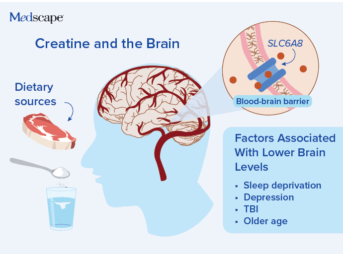
Creatine supplements have been recently examined for their potential to improve brain health and prevent dementia. Given the lack of effective therapies for dementia, it is worth considering whether these supplements are safe and effective.

What Role Does Creatine Play in the Body? 

Creatine is endogenously synthesized in the liver and kidneys, and its amino acid precursors are also provided by a variety of foods, including meat and vegetables. Most of the body’s creatine is stored in muscle cells as phosphocreatine; creatine serves to help replenish adenosine triphosphate during physical activity, which is why we have seen creatine supplements increasingly used in the setting of weight training and exercise over the past decade. 

With the emerging understanding of its role in the brain, research on creatine supplementation has moved from the gym to the brain lab. 

How Creatine May Affect Brain Health 

Creatine is produced in the brain and retina through a process that involves the enzyme guanidinoacetate methyltransferase, which is highly expressed in glial cells. In addition to information on markers of creatine synthesis and activity, evidence is accumulating regarding the action of creatine in the brain

Key Points
  • Creatine synthesized in liver/kidneys; brain/retina synthesis also occurs.
  • Brain creatine transport defects (SLC6A8) cause severe neurologic dysfunction; no treatment.
  • Small studies: supplementation ↑ brain creatine, but less than skeletal muscle.
  • Healthy adults: possible short-term gains in mood, cognition, sleep; women may benefit more.
  • Evidence for dementia/brain disorders remains limited; safety/dosing not fully established.
What mechanisms link creatine to cognitive performance?
Which populations benefit most from creatine supplementation?
How does creatine affect brain creatine on imaging?

Interest in creatine for brain health grew out of the knowledge that creatine deficiency or dysfunction may adversely affect cognition. Defects in the creatine transporter (CRT) gene (SLC6A8) result in low brain creatine levels, along with severe neurologic symptoms, defined as CRT deficiency syndrome. The mechanism of neurologic dysfunction is still unclear, and currently there is no treatment for CRT deficiency syndrome.

Suggested for you
There are different creatine formulations. The most common one used in studies is powder mixed with water, which has a more established bioavailability. Other commercial forms include pills, capsules, and gummies. 

The first step is to verify that creatine supplementation affects brain creatine levels. This is supported by a few small-scale human studies, but the increase is less than that seen in skeletal muscle. 

Authors of a review noted that creatine supplementation may help improve mood, cognition, and sleep in healthy adults. The observed improvements were generally more notable among women than men, which the authors attributed to lower baseline endogenous creatine stores in women. 

creatine and brain

In a 2026 study in 29 sleep-deprived but otherwise healthy adults, 0.2 g/kg creatine monohydrate supplementation was associated with up to a 12% improvement of sleep deprivation-induced deterioration in cognitive skills. For a 150-lb adult, this dosage would be about 14 g of creatine per day. 

Creatine Supplementation for Treating Brain Disorders 

While experimental evidence suggesting that creatine supplementation may help healthy people does not automatically imply that extra creatine would be beneficial in the context of brain disease, there appears to be promise in the use of creatine supplements as an adjunctive treatment in some brain disorders.

According to a 2022 review in Nutrients, creatine supplementation may help reduce some symptoms of traumatic brain injury and concussion.

Creatine also has been explored as a possible treatment for Alzheimer’s disease symptoms or as a disease-modifying therapy. A single-arm pilot trial in 20 patients with Alzheimer’s disease found that 20 g/d creatine monohydrate for 8 weeks was associated with increased total brain creatine and modest improvements on cognitive tests. 

Is There a Place for Creatine Supplementation for Healthy Adults? 

The evidence is still not solid regarding potential benefits of creatine supplements for healthy individuals or for people who have dementia. And while creatine appears to be safe when used as a supplement for exercise and for building muscle, safety concerns have not been entirely ruled out. Furthermore, established dosing and dietary references are lacking. 

Despite these shortcomings, it may be worthwhile for healthy adults or those with cognitive symptoms to try creatine supplements. So far, this supplement shows promise (albeit inconsistent) for short-term cognitive benefits, rather than a lasting impact on brain health. It is the short-lived nature of the effects that makes it worth trying. Many people use other supplements for brain health, like ginkgo biloba or ginseng, and can judge for themselves whether they see any benefits. The same principle applies to creatine – anyone taking it can assess if it improves their cognition or mood, without bearing the burden of deciphering whether a clinically undetectable effect is taking place. 

Heidi Moawad, MD, is a neurologist and medical editor. She is on the teaching faculty at Case Western Reserve University. Dr Moawad resides in Cleveland, Ohio, and enjoys traveling, cooking, and taking exercise classes. 

Popular brain supplement linked to shorter lifespans in men, reports research

 What is your competent? doctor's take on this?

Popular brain supplement linked to shorter lifespans in men, reports research

FDA clears Bioness Medical’s neuromodulation system for stroke rehabilitation


Will your competent? doctor and hospital be DOING ANYTHING WITH THIS?

Do you prefer your doctor, hospital and board of director's incompetence NOT KNOWING? OR NOT DOING? Your choice; let them be incompetent or demand action!

FDA clears Bioness Medical’s neuromodulation system for stroke rehabilitation

Bioness’s PoNS system is designed to improve gait impairment in stroke patients by stimulating the trigeminal and facial cranial nerves.
Bioness Medical’s oral neuromodulation system for rehabilitating stroke patients with gait deficits has secured clearance from the US Food and Drug Administration (FDA). The California-based company’s portable neuromodulation stimulator (PoNS) system delivers neurostimulation through a mouthpiece placed on the tongue. Designed for at-home use in conjunction with physical rehabilitation, the prescription-only device stimulates branches of the trigeminal and facial cranial nerves, activating direct connections to the brainstem. This treatment approach promotes the development of new neural networks to compensate for impaired corticospinal pathways, with the aim of improving walking function in individuals following a stroke, Bioness stated. The device’s clearance for stroke rehabilitation was supported by Bioness’s three-study Stroke Registrational Program (SRP) that evaluated PoNS’ safety and efficacy in conjunction with physical rehabilitation therapy as compared to physical therapy (PT) alone over a 12-week treatment period. Bioness acquired the PoNS system product line from original developer Solana Company (formerly known as Helius Medical Technologies) in April 2026. Prior to the transaction, for which financial terms were not publicly disclosed, PoNS held an FDA indication for multiple sclerosis (MS) treatment.&mThe 159-patient SRP met its primary endpoint, with statistically significant improvements on the Functional Gait Assessment (FGA) scale. Treatment with PoNS plus PT led to an adjusted mean change in FGA of 5.37 points at week 12, versus a non-clinically meaningful change of 3.31 points in the control group. Meanwhile, 45% more subjects treated with PoNS responded to treatment on the basis of a six-point FGA increase threshold, with at least 30% more on the basis of a ≥4-point or ≥5-point improvement threshold. Bioness’ CMO, Antonella Favit-Van Pelt, commented: “The totality of data in chronic stroke survivors with gait deficits confirms the broader evidence of PoNS therapeutic effect in improving walking disability by transitioning the outcome of physical therapy alone to a clinically meaningful effect with a 45.5% increased response rate to PoNS treatment as compared to PT alone.”

Neuromodulation is having a significant impact in the healthcare space for the treatment of conditions ranging from sleep apnoea and chronic migraine to major depressive disorder (MDD). According to GlobalData analysis, the global neuromodulation device market is projected to reach a valuation of over $13bn in 2035, up from around $6.8bn in 2025.

Stroke Recovery: How Patients and Caregivers Can Improve Quality of Life

 First of all you SCREAM at all your stroke providers FOR NOT HAVING  100% RECOVERY PROTOCOLS!  And don't stop screaming until the president AND board of directors gets called in! Then ask how fucking long they will continue incompetence in not having 100% recovery protocols! Suggest a lawsuit for $1000 a dead neuron past the clot buster treatment or stopping of the hemorrhage! Their complacency in accepting recovery failure as a matter of course is PURE INCOMPETENCE!

Notice the word 'care' NOT RECOVERY! That is accepting failure in recovery as a normal matter! Which is why screaming is required if you want change and have your children and grandchildren recover completely from their strokes!

Stroke Recovery: How Patients and Caregivers Can Improve Quality of Life

Recovering from a stroke can be a long and challenging journey. Many survivors work hard to rebuild strength, mobility, speech, and independence. But physical recovery is only one part of the process.

“There’s a whole other side of stroke recovery that people don’t see,” says Alexandra Terrill, PhD, a licensed clinical psychologist and co‑director of the Center for Quality of Life After Stroke at University of Utah Health. “Cognitive changes, communication challenges, and especially mental health concerns are incredibly common, yet often misunderstood.”

The Mental Health Challenges of Stroke Recovery

Many people focus on physical recovery after a stroke, but emotional recovery plays a major role in long‑term well‑being.

Post‑stroke depression affects about one‑third of survivors. Symptoms can appear months after the stroke and may include:

  • Withdrawal
  • Low motivation
  • A sense of “blah”
  • Difficulty engaging in therapy

Terrill notes that many survivors downplay their symptoms while speaking with doctors, so family members often become the ones who speak up.

“The best thing you can do is say something,” Terrill says. “Use language that’s caring, not stigmatizing—like, ‘I’m worried about you,’ or ‘It seems like you’re having a hard time.’ It’s also a good idea to join them at their medical appointments to help their doctors see the full picture.”

Caregivers Need Support Too

Every year, more than 795,000 Americans experience a stroke. Behind many of those survivors is a caregiver helping them navigate the recovery process. 

Research led by Terrill highlights often-overlooked realties, including:

  • Caregivers are at equal or higher risk of depression and anxiety compared to survivors
  • Relationships often change as partners take on caregiving responsibilities, sometimes resulting in strain
  • Small, shared moments can help couples reconnect, such as holding hands while taking a short walk

“Sometimes couples tell us, ‘These aren’t new ideas, we just forgot to do them,” Terrill says. “They were so lost in the medical side of stroke recovery that they forgot how to be a couple. These simple moments help rebuild closeness and improve depression and anxiety for both partners.”

Quality of Life Isn’t One‑Size‑Fits‑All

Measuring quality of life after stroke is complex because it’s deeply personal. What feels like progress or comfort is, as the saying goes, in the eye of the beholder.

“You can have two people with the exact same stroke, and one might feel like their life is over, while the other finds new meaning and purpose,” Terrill says. mental health is vital. Terrill urges partners to prioritize their own well‑being, too, because recovery is a shared journey.

“There’s so much resilience in these couples,” Terrill says. “Sometimes they just need a reminder of how to reconnect, and permission to take care of themselves as well.”

A Whole-Person Approach to Stroke Recovery

At University of Utah Health, care(NOT RECOVERY!) teams focus on long‑term, whole‑person care(NOT RECOVERY!)

Their approach includes:

  • Supporting survivors beyond the crisis moment
  • Addressing emotional, cognitive, and relational needs
  • Helping families navigate the months and years after a stroke
  • Providing tools to rebuild confidence, connection, and independence

Recovery doesn’t happen alone. Lean on your health care(NOT RECOVERY!) team, connect with support groups, and ask for help when you need it. Taking care of both physical and emotional health can make a meaningful difference for stroke survivors and the people who care for them.