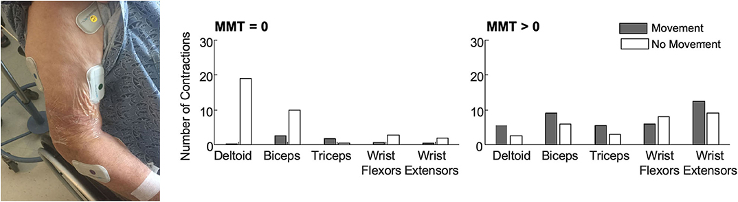
So with similar gains that means telerehabilitation is also a complete fucking failure at 10% full recovery rate.
Why are you accepting such incompetence from your stroke medical professionals?
Evidence-based physiatry: Efficacy of home-based telerehabilitation versus in-clinic therapy for adults after stroke
American Journal of Physical Medicine and Rehabilitation , Volume 99(8) , Pgs. 764-765.
NARIC Accession Number: J84468. What's this?
ISSN: 0894-9115.
Author(s): Tenforde, Adam S. ; Zafonte, Ross ; Hefner, Jaye ; Iaccarino, Mary A. ; Silver, Julie ; Paganoni, Sabrina.
Publication Year: 2020.
Number of Pages: 2.
Abstract: Article reviews a recent report that highlights the potential of telerehabilitation in the management of adults with motor impairments after stroke, as compared to in-clinic rehabilitation. The novel coronavirus pandemic 2019 (COVID-19) has resulted in significant changes in healthcare delivery that may influence management across health systems and conditions, including stroke. To reduce spread of disease, essential medical services are limited for both healthy populations and those with acute, life-threatening conditions, such as stroke. Although studies are inconclusive, patients with risk factors of male sex, older age, cardiovascular disease, hypertension, respiratory disease, and diabetes may be at higher risk for mortality from COVID-19. Notably, most of these risk factors overlap with patients who have experienced stroke. Furthermore, COVID-19 infections cause a hypercoagulable state in some patients that may result in stroke. Limitations in physical distancing, commonly referred to as social distancing, also reduce contact of medical providers with patients. These combined factors create barriers for treatment required to evaluate acute concern of stroke and long-term sequelae. Changes in healthcare delivery including coverage of telehealth visits may address these challenges in delivering effective care. The study results presented suggest that telerehabilitation can have similar gains to in-clinic therapy(So you are suggesting failure to recover is OK because of the failures in the clinic? Trying to get an idea of your reasoning for accepting failure. Have your ever asked your patients if such failure was OK?) for management of upper-limb motor deficits after stroke.
Descriptor Terms: BODY MOVEMENT, IMMUNE SYSTEM DISORDERS, INFECTIONS, LIMBS, MOTOR SKILLS, PHYSICAL MEDICINE, RESPIRATORY DISEASE, STROKE, TELEREHABILITATION.
Can this document be ordered through NARIC's document delivery service*?: Y.
Get this Document: https://journals.lww.com/ajpmr/Fulltext/2020/08000/Evidence_Based_Physiatry__Efficacy_of_Home_Based.17.aspx.
Citation: Tenforde, Adam S. , Zafonte, Ross , Hefner, Jaye , Iaccarino, Mary A. , Silver, Julie , Paganoni, Sabrina. (2020). Evidence-based physiatry: Efficacy of home-based telerehabilitation versus in-clinic therapy for adults after stroke. American Journal of Physical Medicine and Rehabilitation , 99(8), Pgs. 764-765. Retrieved 10/14/2020, from REHABDATA database.
NARIC Accession Number: J84468. What's this?
ISSN: 0894-9115.
Author(s): Tenforde, Adam S. ; Zafonte, Ross ; Hefner, Jaye ; Iaccarino, Mary A. ; Silver, Julie ; Paganoni, Sabrina.
Publication Year: 2020.
Number of Pages: 2.
Abstract: Article reviews a recent report that highlights the potential of telerehabilitation in the management of adults with motor impairments after stroke, as compared to in-clinic rehabilitation. The novel coronavirus pandemic 2019 (COVID-19) has resulted in significant changes in healthcare delivery that may influence management across health systems and conditions, including stroke. To reduce spread of disease, essential medical services are limited for both healthy populations and those with acute, life-threatening conditions, such as stroke. Although studies are inconclusive, patients with risk factors of male sex, older age, cardiovascular disease, hypertension, respiratory disease, and diabetes may be at higher risk for mortality from COVID-19. Notably, most of these risk factors overlap with patients who have experienced stroke. Furthermore, COVID-19 infections cause a hypercoagulable state in some patients that may result in stroke. Limitations in physical distancing, commonly referred to as social distancing, also reduce contact of medical providers with patients. These combined factors create barriers for treatment required to evaluate acute concern of stroke and long-term sequelae. Changes in healthcare delivery including coverage of telehealth visits may address these challenges in delivering effective care. The study results presented suggest that telerehabilitation can have similar gains to in-clinic therapy(So you are suggesting failure to recover is OK because of the failures in the clinic? Trying to get an idea of your reasoning for accepting failure. Have your ever asked your patients if such failure was OK?) for management of upper-limb motor deficits after stroke.
Descriptor Terms: BODY MOVEMENT, IMMUNE SYSTEM DISORDERS, INFECTIONS, LIMBS, MOTOR SKILLS, PHYSICAL MEDICINE, RESPIRATORY DISEASE, STROKE, TELEREHABILITATION.
Can this document be ordered through NARIC's document delivery service*?: Y.
Get this Document: https://journals.lww.com/ajpmr/Fulltext/2020/08000/Evidence_Based_Physiatry__Efficacy_of_Home_Based.17.aspx.
Citation: Tenforde, Adam S. , Zafonte, Ross , Hefner, Jaye , Iaccarino, Mary A. , Silver, Julie , Paganoni, Sabrina. (2020). Evidence-based physiatry: Efficacy of home-based telerehabilitation versus in-clinic therapy for adults after stroke. American Journal of Physical Medicine and Rehabilitation , 99(8), Pgs. 764-765. Retrieved 10/14/2020, from REHABDATA database.


 Katherine M. Steele
 Christina Papazian
 Heather A. Feldner