Changing stroke rehab and research worldwide now.Time is Brain! trillions and trillions of neurons that DIE each day because there are NO effective hyperacute therapies besides tPA(only 12% effective). I have 523 posts on hyperacute therapy, enough for researchers to spend decades proving them out. These are my personal ideas and blog on stroke rehabilitation and stroke research. Do not attempt any of these without checking with your medical provider. Unless you join me in agitating, when you need these therapies they won't be there. Did your doctor somehow miss the recovery protocol that Pedro Bach-y-Rita used to recover fully back in 1958 with only a partial brain?

What this blog is for:

My blog is not to help survivors recover, it is to have the 10 million yearly stroke survivors light fires underneath their doctors, stroke hospitals and stroke researchers to get stroke solved. 100% recovery. The stroke medical world is completely failing at that goal, they don't even have it as a goal. Shortly after getting out of the hospital and getting NO information on the process or protocols of stroke rehabilitation and recovery I started searching on the internet and found that no other survivor received useful information. This is an attempt to cover all stroke rehabilitation information that should be readily available to survivors so they can talk with informed knowledge to their medical staff. It lays out what needs to be done to get stroke survivors closer to 100% recovery. It's quite disgusting that this information is not available from every stroke association and doctors group.

Saturday, October 8, 2022

Feasibility and psychophysical effects of immersive virtual reality-based mirror therapy

 And you somehow think hospitals will buy virtual reality headsets when they can't even provide music for music therapy? If it is just comparable your hospital won't lift a hand. Do they even provide mirrors for your mirror therapy hand rehab?

Feasibility and psychophysical effects of immersive virtual reality-based mirror therapy

Abstract

Background

Virtual reality (VR) has been used as a technological medium to deliver mirror therapy interventions with people after stroke in numerous applications with promising results. The recent emergence of affordable, off-the-shelf head-mounted displays (like the Oculus Rift or HTC Vive) has opened the possibility for novel and cost-effective approaches for immersive mirror therapy interventions. We have developed one such system, ART-VR, which allows people after stroke to carry out a clinically-validated mirror therapy protocol in an immersive virtual environment and within a clinical setting.

Methods

A case cohort of 11 people with upper limb paresis following first time stroke at an in-patient rehabilitation facility received three interventions over a one week period. Participants carried out the BeST mirror therapy protocol using our immersive VR system as an adjunct therapy to their standard rehabilitation program. Our clinical feasibility study investigated intervention outcomes, virtual reality acceptance and user experience.

Results

The results show that the combination of an immersive VR system and mirror therapy protocol is feasible for clinical use. 9 out of 11 participants showed some improvement of their affected hand after the intervention. The vast majority of the participants (9/11) reported experiencing some psycho-physical effects, such as tingling or paraesthesia, in the affected limb during the intervention.

Conclusions

Our findings show that immersive VR-based mirror therapy is feasible and shows effects comparable to those of conventional mirror therapy.

Trial Registration Trial was registered with the ISRCTN Registry (ISRCTN34011164) on December 3, 2021, retrospectively

Implementation of increased physical therapy intensity for improving walking after stroke: Walk ‘n Watch protocol for a multi-site stepped-wedge cluster randomized controlled trial

 Well yeah, higher functioning survivors can walk more, but what are you doing to cure foot drop, spasticity and vertigo for the less able survivors. You're cherry picking here.

Implementation of increased physical therapy intensity for improving walking after stroke: Walk ‘n Watch protocol for a multi-site stepped-wedge cluster randomized controlled trial


View all authors and affiliations
https://doi.org/10.1177/17474930221129982

Abstract

Rationale: 
Clinical practice guidelines support structured, progressive protocols for improving walking after stroke. Yet, practice is slow to change, evidenced by the little amount of walking activity in stroke rehabilitation units. Our recent study (n=75) found that a structured, progressive protocol integrated with typical daily physical therapy improved walking and quality of life measures over usual care. Research therapists progressed the intensity of exercise by using heart rate and step counters worn by the participants with stroke during therapy. To have the greatest impact, our next step is to undertake an implementation trial to change practice across stroke units where we enable the entire unit to use the protocol as part of standard of care.
Aims: 
What is the effect of introducing structured, progressive exercise (termed the Walk ‘n Watch protocol) to standard of care on the primary outcome of walking in adult participants with stroke over the hospital inpatient rehabilitation period? Secondary outcomes will be evaluated and include quality of life.
Methods and sample size estimates: 
This national, multisite clinical trial will randomize 12 sites using a stepped-wedge design where each site will be randomized to deliver Usual Care initially for 4, 8, 12 or 16-months (three sites for each duration). Then, each site will switch to the Walk ‘n Watch phase for the remaining duration of a total 20-month enrolment period. Each participant will be exposed to only one of Usual Care or Walk ‘n Watch. The trial will enrol a total of 195 participants with stroke to achieve a power of 80% with a Type I error rate of 5%, allowing for 20% dropout. Participants will be medically stable adults post-stroke and able to take 5 steps with a maximum physical assistance from one therapist. The Walk ‘n Watch protocol focuses on completing a minimum of 30-minutes of weight-bearing, walking-related activities (at the physical therapists’ discretion) that progressively increases in intensity informed by activity trackers measuring heart rate and step number.
Study outcome(s): 
The primary outcome will be the change in walking endurance, measured by the Six-Minute Walk Test, from Baseline (T1) to 4-weeks (T2). This change will be compared across Usual Care and Walk ‘n Watch phases using a linear mixed-effects model. Additional physical, cognitive, and quality of life outcomes will be measured at T1, T2, and 12-months post-stroke (T3) by a blinded assessor.
Discussion: 
The implementation stepped-wedge cluster-randomized trial enables the protocol to be tested under real-world conditions, involving all clinicians on the unit. It will result in all sites and all clinicians on the unit to gain expertise in protocol delivery. Hence, a deliberate outcome of the trial is facilitating changes in best practice to improve outcomes for participants with stroke in the trial, and for the many participants with stroke admitted after the trial ends.

Get full access to this article

View all access and purchase options for this article.

Friday, October 7, 2022

Stimulation targeting higher motor areas in stroke rehabilitation: A proof-of-concept, randomized, double-blinded placebo- controlled study of effectiveness and underlying mechanisms HHS Public Access

 Didn't do the research completely enough so further research is required, failure in my book. Why choose anodal tDCS rather than these other ones?

Transcranial Brain Stimulation: No Benefit for Stroke Rehab September 2021 

The latest here:

Stimulation targeting higher motor areas in stroke rehabilitation: A proof-of-concept, randomized, double-blinded placebo- controlled study of effectiveness and underlying mechanisms HHS Public Access

Price: EUR 27.50

Comparison of Rehabilitative Interventions That Ameliorate Post-stroke Working Memory Deficit: A Systematic Review

 So you didn't do the research well enough that you called for further research, that is failure in my book, along with not creating any protocols.

Comparison of Rehabilitative Interventions That Ameliorate Post-stroke Working Memory Deficit: A Systematic Review


Abstract

Stroke is one of the most common causes of disability in the world. It has sensory, motor, and cognitive symptoms. Many cognitive domains might get involved in a stroke. This systematic review focuses on working memory domain deficits after stroke and their various rehabilitation methods. This review is based on Preferred Reporting Items for Systematic Reviews and Meta-Analyses(PRISMA) guidelines. For this review, we have searched PubMed, Google Scholar, and Science Direct databases and screened thoroughly with the inclusion criteria of free full-text English papers in the last 10 years that have exclusively studied humans. The articles included in the search are randomized control trials (RCTs), observational studies, meta-analysis studies, systematic reviews, and traditional reviews. Consequent quality assessment was done using the most commonly used tools for each type of study and eight papers were selected. From these papers, full-text articles were studied, analyzed, and tabulated. We found five different rehabilitation methods: transcranial direct-current stimulation, computer-assisted cognitive rehabilitation, physical activity, goal setting, and multimodal rehabilitation. We found that goal setting, computer-assisted cognitive rehabilitation, and multimodal rehabilitation can improve working memory deficits. While transcranial direct current stimulation and physical activity were inconsistent, further studies are needed. The small sample size, no follow-up, the inclusion of only a few studies, the size of the stroke, and comorbid conditions like mild cognitive impairment, dementia, and depression were the main limitations of this study. Future reviews must include a larger number of studies with large sample sizes, including follow-up as an inclusion criterion. We need more clinical trials on these methods for better knowledge.

Introduction & Background

Stroke is the second most common cause of dementia and the third most common cause of morbidity [1]. It can occur due to ischemic (most common) or hemorrhagic causes and can decrease the blood supply of the brain. The most common involved symptoms besides physical deficits (such as complete or partial paralysis, sensory loss, and altered sensations) are dysfunction in learning, memory, and executive functions. This affects nearly 83% of post-stroke patients with cognitive dysfunction, and more than 60% of stroke survivors report cognitive dysfunction, even for up to 10 years [2]. While motor and sensory deficits can affect patients’ quality of life and economic status, memory deficit will affect patients’ daily activities, their profession, and their families. Although the motor and cognitive functions are fundamentally treated as well-defined and separate entities while treating and diagnosing, both entities play an essential role in the post-stroke assessment of behavior and disability [3]. When it comes to cognitive function, working memory has a fundamental role in performing complex behavior [4]. Damage to working memory function causes a drop in the complex cognitive function of the brain to perform everyday activities such as memorization, communication, planning, reading, and writing [5].

What is working memory? It is defined as a multi-component system involved in goal-directed behavior that involves retaining and manipulating information [6]. In simple terms, it is explained as a “sketchpad of conscious thought” [7]. for example, problem-solving in our mind and navigating to our home within our mind. Baddeley and Hitch described it as an essential model with four sub-components arranged hierarchically [6]. They are the phonological loop, visuospatial sketchpad, central executive, and episodic buffer [6]. The central executive supervises the other three subcomponents to store visual, spatial, and phonological information [8]. Working memory reaches its optimal capacity by 20 to 25 years of age. Then each subcomponent declines slowly with further aging at a different rate, which is explained by visual memory declining faster than phonological memory [9]. Many previous imaging studies found that working memory is a part of the lateral prefrontal and parietal cortex of the brain [10]. These areas are supplied by the middle and anterior cerebral artery branches and later drained into cerebral venous sinuses. The stroke affecting these regions will impair the functions. 

Though post-stroke dementia, episodic memory, and long-term memory deficits after stroke have been studied abundantly, working memory deficits after stroke have less information. There is ongoing research on various methods to improve working memory skills and reduce the patient’s morbidity on the efficacy of each rehabilitation. To our knowledge, there is a lack of information on the comparison and efficacies of each intervention; such information is vital for selecting an effective intervention. This review article compares rehabilitative interventions to reduce the working memory deficit after stroke.

Review

Methods

This systematic review was conducted based on the Preferred Reporting Items for Systematic Reviews and Meta-Analyses (PRISMA) 2020 guidelines [11].

First Adaptive Hemorrhagic Stroke Trial Completes Enrollment

 Does your hospital have someone assigned to follow this and create protocols from it if successful? If not, you don't have a functioning stroke hospital. How did they ever get past bloodletting as a stroke intervention if they don't have such a person?

We may as well go back to blood letting as a stroke prescription as discussed in the 1843 book, 'An Essay On The Nature and Treatment of Apoplexy'.

First Adaptive Hemorrhagic Stroke Trial Completes Enrollment

Trial sponsor, NICO Corporation, hopes to show functional recovery for the most deadly, costly stroke

INDIANAPOLIS, Oct. 6, 2022 /PRNewswire/ -- NICO Corporation announced today that enrollment is now complete for the ENRICH (Early MiNimally-invasive Removal of ICH) randomized controlled trial. The trial compares medical and economic outcomes between early surgical intervention of intracerebral hemorrhage (ICH) with minimally invasive parafascicular surgery (MIPS) to current guideline-directed medical management. ENRICH is the first ICH trial using the natural folds of the brain (trans-sulcal) to access and surgically address hemorrhages within 24 hours of onset. It is also the first trial to adaptively enroll patients based on ICH location to maximize the likelihood of patient benefit.

(PRNewsfoto/NICO Corporation)

ENRICH is the first ICH trial using natural folds of brain to access & surgically address hemorrhages within 24 hrs.

ENRICH utilizes a MIPS approach that uniquely integrates advanced imaging technologies with the NICO BrainPath® for non-disruptive navigable access using a parafascicular route to the ICH and the automated Myriad® to pursue the goal of maximum clot evacuation. The goal of the trans-sulcal MIPS approach is to allow surgeons to access and remove the hemorrhage more quickly – providing an opportunity for improved recovery.

"Historically, supratentorial intracerebral hemorrhage is a disease where all earlier surgical trials have failed to meet their primary endpoints, these studies, however, paved the way for future successes by providing specific metrics needed to impact functional outcomes," said Dr. Gustavo Pradilla of Emory University, co-principal investigator of the ENRICH trial.

Intracerebral hemorrhage, or hemorrhagic stroke, is the deadliest, most costly, and most debilitating form of stroke. As many as half of ICH patients die within 30 days, and there is no current proven therapy to treat the disease. ICH costs the U.S. more than $12 billion annually. More than 120,000 people are diagnosed with an ICH annually in the U.S. and 2.1 million people worldwide. The incidence of ICH is expected to rise due to the aging population and increased use of anticoagulants.

The ENRICH multi-center trial is led by the Emory Stroke Center of Emory University hospitals and Marcus Stroke & Neuroscience Center of Grady Memorial Hospital in Atlanta. The trial leverages multi-disciplinary teams, including stroke neurology, neurosurgery, and neuro-critical care at 33 sites that include major medical and academic centers, as well as large community hospitals. NICO has invested $10 million and more than five years in the trial, initiated in January 2017. Results are expected to be announced in Q1 2023 after the final 180-day patient follow-up period concludes.

In May, the American Heart Association-American Stroke Association announced changes to the stroke guidelines to include minimally invasive surgery approaches like MIPS when treating ICH. "We are thrilled that ENRICH is now fully enrolled and want to express our sincere gratitude to all patients and healthcare providers involved in this groundbreaking adaptive trial," said Jim Pearson, president and CEO of NICO Corporation, sponsor of the ENRICH trial. "We are hopeful the trial results will reinforce the recent changes to ICH guidelines and further define the role of MIPS in treating patients who suffer from ICH."

NICO currently holds over 250 issued or pending patents, with 60 focusing solely on safe and repeatable non-disruptive trans-sulcal access through eloquent areas of the brain with BrainPath. NICO's patents also cover efficient removal of clots formed by brain bleeds down a small corridor using the automated Myriad. It is the first and only company in the world to develop and patent technologies to create an entirely new minimally invasive surgical market in neurosurgery for subcortical and skull base lesions, including ICH. Its technologies have been featured in more than 180 peer-reviewed published papers with over 550 unique authors from major academic centers. For more information, visit NICOneuro.com, and follow the latest news on LinkedIn and Twitter.

For more information about the ENRICH trial or patient selection criteria, visit ClinicalTrials.gov or EnrichTrial.com.

Contact: Sue Goin
sue.goin@sapphire-com.com
317.402.869

Cision
Cision

View original content to download multimedia:https://www.prnewswire.com/news-releases/first-adaptive-hemorrhagic-stroke-trial-completes-enrollment-301643074.html


Poststroke pain may be controlled by targeting neuropathic symptoms

 This is after the fact, WHOM will be doing the research that will prevent post stroke pain?

Poststroke pain may be controlled by targeting neuropathic symptoms

LAS VEGAS — Central neuropathic pain can be challenging to treat, but several pharmacologic and nonpharmacologic options are available, according to a presenter at BRAINWeek 2022.

“Central neuropathic pain is one of the most difficult pain syndromes to treat,” Michael Bottros, MD, associate professor of anesthesiology and clinical operations and medical director of pain services at Keck School of Medicine the University of Southern California, Los Angeles, said during his presentation. “There are a number of reasons for developing central neuropathic pain syndromes, and stroke is just one of them.”

Person in pain with fibromyalgia
Source: Adobe Stock.

Bottros explained that pain is among the most common complications of stroke, with an estimated prevalence between 39% and 55%, and tends to appear in central locations like the head, shoulders and upper extremities. Central poststroke pain (CPSP) is neuropathic in origin and affects sensory pathways, accounting for roughly 25% of poststroke pain cases, he said.

Onset of poststroke pain, Bottros stated, can develop immediately following stroke in some patients, 3 to 6 months after stroke in others and up to several years in some. However, CPSP onset within a few months is most common, with later onset a possible sign of stroke recurrence.

“Pain is the most common complication,” Bottros said. “The majority of pain described by people who have a stroke is musculoskeletal.”

To make an accurate pain assessment, clinicians should work around emotional response in their patients, he noted, and find an accurate and objective alternative to pain scales, particularly in cases of CPSP.

Bottros cited the Quantitative Sensory Testing (QST) method as the most objective, as CPSP encompasses a range of pain sensations, locations and emotions within patients that can interfere with diagnosis.

First-line treatment for CPSP is pharmacologic, Bottros said, and includes antidepressants such as amitriptyline, while anticonvulsants like gabapentin and pregabalin are good second-line choices. Lamotrigine monotherapy also is moderately effective and generally well tolerated, although this treatment comes with more serious potential side effects. And while opioids are considered generally ineffective in treating CPSP, Bottros reported that IV ketamine provided relatively rapid pain relief, which lasted 2 to 3 hours, in a double-blind, placebo-controlled study.

In the realm of nonpharmacologic treatment, various forms of neurostimulation (motor cortex, transcranial magnetic stimulation, deep brain and vestibular caloric) demonstrated beneficial pain reducing effects.

Regional blocking of nerves, Bottros added, resulted in fairly rapid pain reduction as a result of signals from the brain being cut off before reaching the locus of pain. This, he said, may be an indicator that pain may not be generated and perceived in the central nervous system.

“CPSP has variable timetable to onset following stroke,” Bottros said. “Sensory afferent input could play an important role in patients hiding the effects of poststroke pain.”

First pass effect in posterior circulation occlusions: Analysis from the CICAT registry

When you enter the emergency room you'll have to demand your doctor accomplish the thrombectomy in one pass or they better have followup protocols that restore functionality to the same that would have occurred in one pass.

First pass effect in posterior circulation occlusions: Analysis from the CICAT registry

Abstract

Background:

The first pass effect (FPE) is an independent predictor of functional independence in patients with large vessel occlusion in anterior circulation ischemic strokes. However, whether it predicts outcome in posterior circulation large vessel occlusion (PC-LVO) is uncertain. We aimed to study the frequency, characteristics, and predictors of FPE and its association with clinical outcomes in PC-LVO.

Method:

We performed an analysis from the prospective CICAT Registry. All patients with PC-LVO who underwent endovascular therapy between January 2016 and January 2020 were included. A centrally assessed clinical follow-up was performed at 3 months by blinded investigators. FPE was defined as the achievement of modified Thrombolysis In Cerebral Infarction 3 in a single pass of the endovascular thrombectomy device, and multi-pass effect (MPE) if it was achieved in more than one pass. A multivariable analysis was performed to identify whether FPE is an independent predictor of functional independence defined as a modified Rankin Score of 0–2.

Results:

We analyzed data from 265 patients in who FPE was achieved in 105 (39.6%). Patients with FPE were more likely to achieve functional independence compared to the non-FPE group (52.4% vs 25.1%, p < .001) and the MPE group (52.4% vs 26.7%, p < .001). FPE was independently associated with functional independence (adjusted odds ratio (aOR): 2.10, 95% confidence interval (CI) 1.01-4.37) but MPE was not (aOR: 0.92, 95% CI 0.40-2.13). Independent predictors of FPE were the use of direct aspiration, embolic mechanism of stroke, and the absence of general anesthesia (GA) use.

Conclusions:

FPE is an independent predictor of functional independence in PC-LVO and was associated with a significantly better outcome than MPE.

Get full access to this article

View all access and purchase options for this article.

References

1. Rha JH, Saver JL. The impact of recanalization on ischemic stroke outcome: a meta-analysis. Stroke 2007; 38(3): 967–973.

Thursday, October 6, 2022

New technology providing good vibrations for stroke patients

I think this is the research they are referring to: 

You'll have to ask your doctor how to implement subthreshold vibration.

 Neuroplasticity after upper extremity rehabilitation therapy with sensory stimulation in chronic stroke survivors

The latest here: Your doctor can tell you the name of that device.

Identifying how inflammation affects stroke recovery

 Instead of looking for useless biomarkers why not do the research that prevents this inflammation?

Identifying how inflammation affects stroke recovery

A new research grant will enable University of Cincinnati researchers to learn more about how inflammation affects patient recovery after certain kinds of strokes.

Kyle Walsh, MD, is the principal investigator of the CAPSTONE study, and his team received a five-year, $2.5 million grant from the National Institute of Neurological Disorders and Stroke (NINDS).

Research foundation

Kyle Walsh Emergency Medicine in White Coat

Kyle Walsh, MD. Photo/University of Cincinnati.

The name of the study, CAPSTONE, is an acronym for Central And Peripheral STrOke inflammatioN with Exosomes. The research focuses on patient recovery after intracerebral hemorrhage (ICH), a particular type of stroke caused by a blood vessel in the brain rupturing. 

ICH strokes account for about 20% of all strokes, but are often deadly and cause high disability. They also occur in Black and Hispanic populations twice as often and an average of 10 years earlier in life compared to their white counterparts.

Walsh explained that the brain suffers injury from the bleeding itself during an ICH stroke, called the “primary injury,” but following this a number of inflammatory processes occur both inside the brain and in the circulating blood. Previous research has suggested that while some of these inflammatory processes help repair the damage, others are harmful and contribute to a “secondary injury.”

Daniel Woo, MD, co-investigator of the study, said sometimes inflammation can persist and become chronic in the brain long after it has done its job, which is believed to lead to neurodegeneration. Nearly 40% of patients who survive an ICH stroke develop progressive cognitive decline, comparable to dementia, within a few years after the stroke occurred, he said.

Research methodology

In the CAPSTONE study, the research team will analyze different inflammatory biomarkers to see which are associated with positive outcomes and which are tied to poor outcomes in patients. 

“So we’ll be using the blood and plasma samples from those patients to look at a number of different mediators of inflammation,” said Walsh, associate professor in the Department of Emergency Medicine in UC’s College of Medicine and a UC Health attending physician in the Emergency Department, Neurosciences Intensive Care Unit and with the UC Stroke Team. 

These mediators include three main categories: gene expression (messenger RNA or mRNA), specific types of RNA called microRNA and exosomes, which are essentially small pieces of cells that travel between or “talk” from one cell to another. 

The patient samples will be obtained from Woo’s ROSELAWN grant that is recruiting 500 patient cases of ICH and following 250 patients long term.

I’m hopeful for the future about the potential of having an actual treatment for ICH based on these inflammatory biomarkers.

Kyle Walsh, MD

“Many studies may only follow patients for three months or six months after stroke. ROSELAWN actually follows patients much longer, up to a few years after the ICH, to look for clinical outcomes as well as long-term cognitive decline,” Walsh said. 

Woo noted that in the past, it was difficult to identify exactly what inflammatory processes were occurring inside the brain during stroke recovery, as researchers were limited by only being able to draw blood and see what was going on in the larger bloodstream. 

With new technology, however, testing this same circulating blood provides an opportunity to identify biomarkers from inside the brain. Walsh said this is because exosomes that are released from brain cells into the circulating bloodstream can be tagged and isolated, and the microRNA inside the exosomes can be characterized.

“We can look at the inflammation that’s occurring in a living human just by looking at these exosomes,” said Woo, vice chair of research in UC’s Department of Neurology and a UC Health physician. “This study will do this very fancy exosome testing, looking at the information that occurs not just at baseline, but also to see if it’s persisting years later, and what kind of inflammation that is.”

Potential treatments

Daniel Woo, MD,
Professor,
Vice Chair of Clinical Research,
Neurology and Rehabilitation Medicine, College of Medicine

Daniel Woo, MD. Photo/Andrew Higley/UC Marketing + Brand.

The ultimate goal of the study is to identify which inflammatory biomarkers lead to worse outcomes in the hopes of targeting treatments to improve patient outcomes, including to prevent long-term degeneration in the brain. Some of these biomarkers may already have treatments available that could be tested in patients.

“CAPSTONE is critically important in helping us break down inflammation into very specific parts of which cells are doing it, which actual proteins or what we call cytokines are actually causing it, and it looks very deeply into the molecular mechanisms,” Woo said.

There will likely be multiple biomarkers that are found to cause worse outcomes, so the team will also aim to prioritize treatments for what is found to have the strongest effects. But in the future, there may be the potential to personalize treatments based on which group of biomarkers each patient has.

“I’m very excited to see what markers we’re able to identify on this larger scale,” Walsh said. “I’m hopeful for the future about the potential of having an actual treatment for ICH based on these inflammatory biomarkers.”

Support and mentorship

Walsh said he is grateful for pilot grant funding from UC’s Department of Emergency Medicine, Department of Neurosurgery and the UC Gardner Neuroscience Institute that led first to a larger American Heart Association grant and now to the NINDS funding. He also credited Woo and former UC faculty member Opeolu Adeoye as mentors who have supported him as he advances as a researcher.

“It’s just such a testament to the importance of mentoring and of mentors who are willing to dedicate the time and their expertise and resources. In the case of CAPSTONE, of course the mentoring from Dr. Woo over several years has been invaluable, but it’s also the opportunity to utilize some of the resources from one of his grants,” Walsh said. “I would say it’s really almost impossible to succeed in this type of research without strong mentors, for many different reasons.”

Woo said often young researchers will think that mentors will simply tell them the trick to get funding for their research and progress in their careers, but he encourages his mentees simply to do the best science they can and ask questions about what can do the most good for the most people in the area they are interested in.

“Kyle [Walsh] has been fantastic to work with and an outstanding mentee. He has been great at taking that constant thought — that idea that, ‘I’m not here to get funding, I’m here to do the best science I can and to think about what’s the most good that I can do,’” Woo said. “I want Kyle to become a generational leader teaching others in the emergency department and other areas to have that same philosophy.”

Other co-investigators for the CAPSTONE study include UC’s Scott Langevin, PhD, and Leyla Esfandiari, PhD, and Carl Langefeld, PhD, of Wake Forest University.

Featured photo at top courtesy of Unsplash.

Next Lives Here

The University of Cincinnati is classified as a Research 1 institution by the Carnegie Commission and is ranked in the National Science Foundation's Top-35 public research universities. UC's graduate students and faculty investigate problems and innovate solutions with real-world impact. Next Lives Here.

NYU researchers partner with FDA to identify biomarkers for stroke rehabilitation

What a pile of shit! Do you not understand that biomarkers DO ABSOLUTELY NOTHING to get survivors recovered. A valid question: Are you a blithering idiot or not?

NYU researchers partner with FDA to identify biomarkers for stroke rehabilitation


A unique collaboration between the engineering sector (NYU Tandon), the clinical sector (NYU Langone), and the regulatory sector (the U.S. Food and Drug Administration) will bring biomarker data to bear on neurorehabilitation technology.

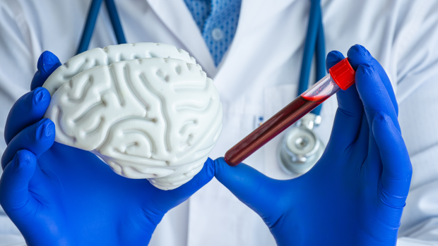
Person in doctors coat holds a model brain next to a red vial.

BROOKLYN, New York, Wednesday, September 28, 2022 — Stroke is the leading cause of age-related motor disabilities and is becoming more prevalent in younger populations as well. But while there is a burgeoning marketplace for rehabilitation devices that claim to accelerate recovery, including robotic rehabilitation systems, recommendations for how and when to use them are based mostly on subjective evaluation of the sensorimotor capacities of patients who use them. (What you really need is an objective diagnosis which leads directly to EXACT PROTOCOLS THAT DELIVER RECOVERY! Call it by the correct term; objective diagnosis.)

S. Farokh Atashzar, assistant professor of Electrical & Computer Engineering and Mechanical & Aerospace Engineering at the NYU Tandon School of Engineering and the director of the Medical Robotics and Interactive Intelligent Technologies (MERIIT) Lab; in collaboration with John-Ross Rizzo, associate professor of Biomedical Engineering at NYU Tandon and Ilse Melamid Associate Professor of rehabilitation medicine at the NYU School of Medicine, are working with Dr. Ramin Bighamian from the U.S. Food and Drug Administration (FDA) to design a regulatory science tool (RST) based on data from biomarkers in order to improve the review processes for such devices and how best to use them. 

The team will design and validate a robust recovery biomarker enabling a first-ever stroke rehabilitation RST based on exchanges between regions of the central and peripheral nervous systems. 

“There is currently no approved RST for assessing the efficacy of rehabilitative devices for post-stroke motor recovery,” explained Atashzar. “This unique collaboration will shed light on how neurological markers can demystify the complex patterns of neural communication, realizing an objective ‘neurophysiological window to degradation in the interaction between anatomy, activation, and pathway in post-stroke patients.”

The research team will also promote FDA regulatory science on next-generation brain-machine interfaces. Additionally, the project will include seminars and undergraduate research to promote STEM education with a focus on engaging students from underrepresented groups.

The project is supported by the U.S. National Science Foundation.  

“This project exemplifies how, as we build our expertise in areas like neural engineering, we are cultivating a highly collaborative research atmosphere — across disciplines such as data science and medicine, between Tandon and other schools at NYU, and spanning the lab and the classroom — all with the greater good in the mind of helping patients,” said Jelena Kovačević, Dean of NYU Tandon. “The importance of this work with the NYU School of Medicine speaks for itself, but it is also heartening to see validation of it from both the FDA and NSF.”