Changing stroke rehab and research worldwide now.Time is Brain! trillions and trillions of neurons that DIE each day because there are NO effective hyperacute therapies besides tPA(only 12% effective). I have 523 posts on hyperacute therapy, enough for researchers to spend decades proving them out. These are my personal ideas and blog on stroke rehabilitation and stroke research. Do not attempt any of these without checking with your medical provider. Unless you join me in agitating, when you need these therapies they won't be there. Did your doctor somehow miss the recovery protocol that Pedro Bach-y-Rita used to recover fully back in 1958 with only a partial brain?

What this blog is for:

My blog is not to help survivors recover, it is to have the 10 million yearly stroke survivors light fires underneath their doctors, stroke hospitals and stroke researchers to get stroke solved. 100% recovery. The stroke medical world is completely failing at that goal, they don't even have it as a goal. Shortly after getting out of the hospital and getting NO information on the process or protocols of stroke rehabilitation and recovery I started searching on the internet and found that no other survivor received useful information. This is an attempt to cover all stroke rehabilitation information that should be readily available to survivors so they can talk with informed knowledge to their medical staff. It lays out what needs to be done to get stroke survivors closer to 100% recovery. It's quite disgusting that this information is not available from every stroke association and doctors group.

Monday, December 1, 2025

Treatment of apathy in stroke patients: a systematic review

 Apathy is COMPLETELY YOUR DOCTOR'S FAULT!  EXACT 100% RECOVERY PROTOCOLS and your patient will gladly do the millions of reps needed because they are looking forward to recovery! If you can't see that, you are incredibly blitheringly stupid!

 Treatment of apathy in stroke patients: a systematic review


  • 1Stroke Unit, Department of Neurology, Torrecárdenas University Hospital, Almería, Spain
  • 2Fundation for Biosanitary Research of Eastern Adnalusia (FIBAO), Torrecárdenas University Hospital, Almería, Spain
  • 3Department of Psychology, Faculty of Psychology, CTS-280 Clinical and Experimental Neuroscience Research Group and Research Center CiBiS, University of Almeria, Almeria, Spain
  • 4Department of Nursing, Physiotherapy and Medicine, Faculty of Health Science, Health Research Center (CEINSA), University of Almería, Almeria, Spain

Introduction: Post-stroke apathy is a prevalent yet frequently underdiagnosed neuropsychiatric syndrome, reported in up to one-third of stroke survivors, and is consistently associated with poorer functional recovery and cognitive decline. We aimed to review the current evidence on available pharmacological and non-pharmacological treatments for post-stroke apathy, and to evaluate their efficacy and safety.

Methods: A systematic review was conducted following PRISMA guidelines and registered in the PROSPERO database (CRD42022332559). We searched PubMed, Web of Science, and Scopus for randomized and non-randomized clinical trials published until November 2024. Eligible studies included adults with ischemic or hemorrhagic stroke and a defined diagnosis of apathy. Interventions included pharmacological treatments and non-pharmacological strategies, such as neuromodulation techniques. Data extraction and risk of bias assessment were independently performed by two reviewers using the RoB-2 tool.

Results: Ten clinical trials involving 2,359 patients were included. Pharmacological interventions with escitalopram and donepezil (alone or combined with intensive language action therapy) showed potential benefits. Nefiracetam yielded mixed results depending on dose and coexisting depression. Non-pharmacological approaches such as problem-solving therapy, motor relearning programs, strategy training, and complex rehabilitation programs demonstrated significant improvement in apathy scores. High-frequency repetitive transcranial magnetic stimulation also showed efficacy. However, heterogeneity in study design and apathy assessment scales limited direct comparisons.

Conclusion: Several interventions, including escitalopram, donepezil, motor relearning programs, strategy training, and rTMS, have demonstrated potential effectiveness in treating post-stroke apathy. Nevertheless, evidence remains scarce and heterogeneous, underscoring the need for larger, high-quality randomized controlled trials to establish definitive treatment guidelines.

Systematic review registration: https://www.crd.york.ac.uk/PROSPERO/view/CRD42022332559.

Low-dose statins combined with repetitive transcranial magnetic stimulation reduce post-stroke depression

 Why is there discussion on this? Didn't this earlier research prove the benefit of immediate statin therapy? Or do you INCOMPETENTLY NOT KNOW ABOUT IT?

Association Between Acute Statin Therapy, Survival, and Improved Functional Outcome After Ischemic Stroke: The North Dublin Population Stroke Study March 2011

Or don't you have a functioning stroke doctor who incompetently missed this research from 2011?

1. Statins.

tested in rats from 2003

http://Statins induce angiogenesis, neurogenesis, and synaptogenesis after stroke Statins induce angiogenesis, neurogenesis, and synaptogenesis after stroke  

Simvastatin Attenuates Stroke-induced Splenic Atrophy and Lung Susceptibility to Spontaneous Bacterial Infection in Mice

Or,

Simvastatin attenuates axonal injury after experimental traumatic brain injury and promotes neurite outgrowth of primary cortical neurons 

October 2012

tested in humans, March 2011

http://www.medwirenews.com/39/91658/Stroke/Acute_statin_therapy_improves_survival_after_ischemic_stroke.html

And now lost even to the Wayback Machine

So, I think this below is the actual research.

Association Between Acute Statin Therapy, Survival, and Improved Functional Outcome After Ischemic Stroke April 2011 

The latest here:

 Low-dose statins combined with repetitive transcranial magnetic stimulation reduce post-stroke depression


Chaohua Cui
&#x;Chaohua Cui1*†Chuhua Cai&#x;Chuhua Cai2†Qiulian YinQiulian Yin1Haoye GuanHaoye Guan1Tonghua LongTonghua Long1
  • 1Life Science and Clinical Medicine Research Center, Affiliated Hospital of Youjiang Medical University for Nationalities, Baise, Guangxi, China
  • 2Drug Clinical Trial Management Office, The Second Affiliated Hospital Of Guangxi University of Science and Technology, Liuzhou, Guangxi, China

Background: The effectiveness of statins in preventing post-stroke depression (PSD) remains controversial. Lower testosterone levels caused by statins could potentially increase the risk of PSD. Low-dose statins could offer better benefits due to their lesser impact on testosterone levels. Repetitive transcranial magnetic stimulation (rTMS) has been shown to reduce PSD in the acute stage. The combination of low-dose statins and rTMS may provide a promising therapeutic option for patients with PSD.

Methods: Data were collected from ischemic stroke patients. Unsupervised machine learning methods were employed to explore the risk factor of PSD. Our prospective cohort study collected data from ischemic stroke patients over 3 years. The patients’ conditions determined the prescription of statins. The outcomes measured included the incidence of PSD, favorable functional outcome (FFO) at 6- and 12-months post-onset.

Results: A total of 545 patients were included in the study. The mean age was 60.99 ± 12.768 years, and 31.9% of the participants were female. The combination of low-dose statins and rTMS subgroup had the lowest incidence of PSD at both 6 and 12 months (p < 0.001). In multivariable logistic regression, the combination of low-dose statins and rTMS was inversely related to PSD at 6 months (OR = 0.370, p = 0.003) and 12 months (OR = 0.386, p = 0.004).

Conclusion: The combination of statins and the rTMS group related the lower incidence of PSD in stroke patients. Combining low-dose statins and rTMS could reduce PSD and further improve the prognosis of ischemic stroke patients.

Introduction

Post-stroke depression (PSD) is a common complication among ischemic stroke patients. These patients have worse functional outcomes and higher mortality rates than those without PSD (1). Antidepressants are the primary treatment for PSD patients. However, longer treatment cycles and more side effects affect the compliance of PSD patients (2). Additionally, an increasing number of PSD patients develop tolerance to antidepressants (2). These PSD patients require a safer and simpler treatment.

Statins could affect the incidence of PSD of stroke patients (3–6). Different studies have reached distinct conclusions about the effect. One Asian study suggests that statins increase the incidence rate of PSD (4). Other studies have shown the opposite result, indicating that statins decrease the incidence rate of PSD (5, 6). The various effects of statins and different types of studies could partly explain these contradictory conclusions (7). Several studies have demonstrated that reduced testosterone levels may elevate the risk of PSD (8, 9). Therefore, low-dose statins may offer greater benefits to PSD patients, as they exert a less pronounced effect on testosterone levels (8, 9).

Statins may have varying effects on PSD depending on the stage of stroke. The use of statins before a stroke may reduce the risk of PSD, whereas post-stroke use could potentially increase it (10, 11). Thus, identifying an effective treatment strategy for patients in the acute phase of stroke with PSD is essential. Our analysis of a retrospective cohort, utilizing unsupervised machine learning methods, revealed that stroke patients receiving statins during the acute phase in conjunction with rTMS exhibited a lower incidence of PSD. Several studies have indicated that high-frequency rTMS is an effective treatment for PSD, particularly for patients in the acute phase of stroke (12–14). rTMS may counteract the adverse effects of statins in acute PSD patients, while statins could provide longer-term benefits for those in the chronic phase of PSD. Consequently, combining statins with rTMS may provide a comprehensive solution to these challenges.

This study aims to explore whether the combination of statins and rTMS can decrease PSD in ischemic stroke patients. Additionally, we aim to analyze further whether the combination of low-dose statins and rTMS better affects PSD patients.

Methods

Cohort 1: retrospective explored cohort

Patients

The study cohort was a retrospective observational cohort. The cohort comprised consecutive patients with ischemic stroke. Patients were recruited from the Neurology and Rehabilitation Departments of the Affiliated Hospital of Youjiang Medical University for Nationalities between January 1, 2018, and May 30, 2019.

The inclusion criteria included ischemic stroke patients aged 18 years or older who had undergone head CT or MRI examinations, met the WHO ischemic stroke diagnostic criteria and received conventional medicine and rehabilitation therapy post-admission. The exclusion criteria were as follows: (1) patients with a recent history of depression before onset; (2) patients unable to be evaluated for depression due to aphasia, disturbance of consciousness, cognitive disorder, or other conditions; (3) patients who taken other lipid-lowering drugs such as fenofibrate; (4) patients with intracerebral hemorrhage, subarachnoid hemorrhage, or severe systemic disease; (5) Patients who are intolerant to statins or rTMS treatment; (6) patients who withdrew from the study or could not provide outcome events.

Data collected

Baseline patient data were collected from electronic medical records, including demographic information, vital signs, and laboratory data. The data collected at admission included age, gender, heart rate, and blood pressure. Critical laboratory data collected at admission included PLT (platelet), INR (international normalized ratio), ALT (alanine aminotransferase), AST (aspartate aminotransferase), CRP (C-reactive protein), among others. Blood lipid levels, including TC (total cholesterol), TG (triglycerides), HDL-C (high-density lipoprotein cholesterol), and LDL-C (low-density lipoprotein cholesterol), were recorded at admission and discharge. Comorbidities, such as renal insufficiency, epilepsy, and pneumonia, were also documented. Medical histories and medication profiles were obtained using structured questionnaires completed by patients or their relatives. Two experienced neurologists, blinded to the patient’s conditions and outcomes, collected and evaluated these data.

Post-stroke depression

The diagnosis of post-stroke depression (PSD) was identified using ICD-9-CM codes 296.2, 296.3, 300.4, or 311. The Mini International Neuropsychiatric Interview (MINI) (6–8), a structured diagnostic psychiatric interview based on the Diagnostic and Statistical Manual of Mental Disorders, 4th Edition (DSM-IV), was also used. According to these criteria, patients were diagnosed with major depression if they presented one core symptom and at least four additional depressive symptoms. Alternatively, patients were diagnosed with minor depression if they exhibited at least one core symptom and between two and four additional symptoms. PSD was defined as encompassing both major and minor depressive disorders.

Classify data by unsupervised machine learning

We utilized Python 3.8 for our analysis. For cluster analysis, we first standardized all data using the StandardScaler module from the sklearn library, and then classified data using Hierarchical Clustering methods (AgglomerativeClustering module, sklearn library). The heatmap illustrated the distinct characteristics among the classified patient groups. We compared outcome events across the different identified groups. Grouping can yield clinical significance by clarifying differences in PSD among patient groups. Subsequently, we applied chi-square tests and t-tests to further analyze the factors within the selected groups. Significant differences were found in statin use and rTMS across the Hierarchical Clustering groups. We subsequently validated the impact of statin use and rTMS on the incidence of PSD.

Cohort 2: prospective validated cohort

Study subjects

This study was a prospective cohort study. Ischemic stroke patients were recruited from the neurology and rehabilitation departments of affiliated hospitals of Youjiang Medical University for Nationalities. Patients were enrolled from January 2020 to December 2022 and followed up until December 2023.

Patients who took statins after admission were assigned to the statin group and the remaining patients were assigned to the control group. The study included only patients who did not take statins before admission. Patients were prescribed statins and specific types based on the guidelines for the management of stroke (15).

The inclusion criteria and exclusion criteria were similar as cohort 1.

Baseline data

Baseline patient data were similar as cohort 1.

We evaluated patients’ NIHSS (National Institutes of Health Stroke Scale) scores and mRS (Modified Rankin Scale) scores at admission. Two experienced neurologists, blinded to patients’ conditions and outcomes, collected and evaluated these clinical scale scores.

Statin use

We collected data on patients’ statin use during hospitalization and follow-up. Low-dose and medium-high-dose statins were defined according to the ACC/AHA guidelines on statin intensity (16). The types of statins used in our cohort included atorvastatin, rosuvastatin, simvastatin, and others. Statins were classified into lipophilic (atorvastatin, simvastatin, pitavastatin, lovastatin) and hydrophilic (rosuvastatin) categories. For patients whose statin type or dosage was adjusted during their course of treatment due to changes in their condition, we typically record the statin type or dosage used at the first administration during hospitalization. If the duration of statin use after the change in type or dosage exceeds 70% of the total usage time, the changed type or dosage will be recorded. Lower statin compliance was defined as using statins continuously for less than 40% of the follow-up period. The cohort did not include patients with lower statin compliance. When patients experience significant side effects from statin use and need to discontinue the medication, they are withdrawn from the cohort.

rTMS

Multiple rTMS protocols have been proposed for treating post-stroke depression; this study adopted the treatment approaches from several well-powered studies with robust conclusions (12–14). All patients underwent rTMS therapy for 4 weeks, with five sessions per week, totaling 20 sessions. rTMS was administered to the left dorsolateral prefrontal cortex (DLPFC) at 10 Hz, with magnetic stimulation strength set at 80–100% of the motor threshold level. Each daily treatment included 20 sequences (20 min), each consisting of 4 s of continuous stimulation followed by a 56-s interval (12–14). When patients experience intolerable side effects from transcranial magnetic stimulation therapy or are deemed unsuitable for further treatment by a physician, they will be withdrawn from the cohort.

Outcome events

The primary outcome was the incidence rate of PSD at 6- and 12-months post-onset. The secondary outcomes included favorable functional outcomes (FFO) at 6- and 12-months post-onset. A favorable functional outcome was a modified Rankin Scale (mRS) score of less than 3. Outcome events were collected post-discharge through face-to-face interviews.

Statistical analysis

Statistical analyses were conducted using SPSS 23.0 for Windows. The threshold for statistical significance was set at p < 0.05.

Baseline character

Continuous variables (e.g., blood pressure, laboratory measures) normally distributed between two groups were analyzed using a t-test and expressed as mean ± standard deviation (SD). Continuous variables (e.g., NIHSS) that were abnormally distributed between two groups were analyzed using the Mann–Whitney U test and expressed as the median and interquartile range (IQR). Categorical variables (e.g., gender, history of disease) and ordinal variables (e.g., mRS) were analyzed using the chi-square test and expressed as numbers and percentages.

Outcome variables

A chi-square test was employed to compare the incidence rates of PSD, optimism, and FFO between the two groups. Additionally, we compared the incidence of post-stroke depression across various subgroups. These subgroups were categorized by different doses, type of statins and whether patients underwent repetitive transcranial magnetic stimulation treatment.

We employed multivariate logistic regression analysis to control for confounding factors. In cases where the extreme data exhibited significant imbalance, subgroup analysis and propensity score matching were considered for data processing. However, since the validation cohort did not show a notable imbalance in extreme risk factors, only multivariate logistic regression was used for data analysis.

Logistic regression was used to analyze the relationship between the combination of low-dose statins and rTMS, or other risk factors, and the incidence rate of PSD at different time points. Eligible factors for multivariable regression were screened as follows: (1) a p-value of less than 0.05 in univariable logistic regression; (2) factors selected using LASSO regression; (3) factors meeting criteria (1) and (2). The eligible data were then analyzed using multivariable logistic regression. Odds ratios (ORs), 95% confidence intervals (CIs), and p-values were calculated using logistic regression.

Results

Cohort 1

The exploratory cohort included 117 patients. Thirty-two were female (27.4%), and the mean age was 63.15 ± 11.002 years.

The heatmap suggested that two groups (HCgroup1 and HCgroup2) were optimal for hierarchical clustering methods (Figure 1). The heatmap also indicated significant distinctions between statins and rTMS (Figure 1). Comparing the incidence of PSD between the two groups, HCgroup1 (3.4%) had a significantly lower rate of PSD (p < 0.001) than HCgroup2 (17.7%).


Study clarifies how spermine can guard against Alzheimer’s and Parkinson’s

Your incompetent? doctor and hospital won't get human testing going, will they?

 Study clarifies how spermine can guard against Alzheimer’s and Parkinson’s

Researchers at the Paul Scherrer Institute PSI have clarified how spermine - a small molecule that regulates many processes in the body's cells - can guard against diseases such as Alzheimer's and Parkinson's: it renders certain proteins harmless by acting a bit like cheese on noodles, making them clump together. This discovery could help combat such diseases. The study has now been published in the journal Nature Communications. 

Our life expectancy keeps rising - and as it does, age-related illnesses, including neurodegenerative diseases such as Alzheimer's and Parkinson's, become increasingly common. These diseases are caused by accumulations in the brain of harmful protein structures consisting of incorrectly folded amyloid proteins. Their shape is reminiscent of fibres or spaghetti. To date, there is no effective therapy to prevent or eliminate such accumulations.

Yet a naturally occurring molecule in the body called spermine offers hope. In experiments, researchers led by study leader Jinghui Luo, in the Center for Life Sciences at the Paul Scherrer Institute PSI, have discovered that this substance is capable of extending the life span of small nematode worms, improving their mobility in old age, and strengthening the powerhouses of their cells - the mitochondria. Specifically, the researchers observed how spermine helps the body's immune system eliminate nerve-damaging accumulations of amyloid proteins.

The new findings could serve as a basis for developing novel therapies for such diseases.

A central mediator of cellular processes

Spermine is a vital substance for the organism. It belongs to the so-called polyamines, which are relatively small organic molecules. Spermine, first discovered more than 150 years ago, is named after the seminal fluid, as it is found in particularly high concentrations there. But it also occurs in many other cells of the body - especially those that are active and capable of dividing.

Spermine promotes cell mobility and activity and controls numerous processes. Above all, it interacts with the nucleic acids of the genome, regulating the expression of genes and their conversion into proteins. This ensures that cells can properly grow and divide and ultimately die. Spermine is also central to an important cellular process called biomolecular condensation: In this process, certain macromolecules, such as proteins and nucleic acids, segregate and collect within the cell in a droplet-like form, so that important reactions can take place there.

In connection with neurodegenerative diseases such as Alzheimer's or Parkinson's, there has previously been evidence that spermine can protect nerve cells and alleviate age-related memory loss. Lacking until now, however, has been a more precise understanding of how spermine intervenes in nerve-damaging processes - understanding that might make it possible to derive medical benefits from it.

Assisting cellular waste removal

Jinghui Luo's group has now investigated this in more detail. In addition to optical microscopy, the researchers also used the SAXS scattering technique at PSI's Swiss Light Source SLS to shed light on the molecular dynamics of these processes. The investigations were conducted both in a glass capillary (in vitro) and in a living organism (in vivo). The nematode C. elegans served as a model organism.

It was shown that spermine causes the harmful proteins to gather and, in a sense, clump together through biomolecular condensation. This facilitates a process called autophagy, which occurs routinely in our cells: Damaged or unnecessary proteins are wrapped up in small membrane vesicles and safely degraded with enzymes - a natural recycling process, in effect.

Autophagy is more effective at handling larger protein clumps. And spermine is, so to speak, the binding agent that brings the strands together. There are only weakly attractive electrical forces between the molecules, and these organise them but do not firmly bind them together."

Jinghui Luo, study leader 

Administration of eptifibatide as rescue therapy during mechanical thrombectomy in acute ischemic stroke

Your competent? doctor has known of eptifibatide use for over a decade, right?


  • Eptifibatide (5 posts to February 2013)
  •  Administration of eptifibatide as rescue therapy during mechanical thrombectomy in acute ischemic stroke

    David Cernik,David Cernik1,2*Jiri NeumannJiri Neumann3Jarmila NeradovaJarmila Neradova2Veronika HlinenaVeronika Hlinena2Pavol Viglas,Pavol Viglas2,5David CihlarDavid Cihlar5Filip CihlarFilip Cihlar6
    • 1Department of Neurology, Univerzita Palackeho v Olomouci Lekarska fakulta, Olomouc, Czechia
    • 2Department of Neurology, Masarykova nemocnice Usti nad Labem oz, Ústí nad Labem, Czechia
    • 3Department of Neurology, Krajska zdravotni as Nemocnice Chomutov oz, Chomutov, Czechia
    • 4Department of Physical Education and Sport, Faculty of Education, Univerzita Jana Evangelisty Purkyne v Usti nad Labem, Ústí nad Labem, Czechia
    • 5Department of Radiology, Faculty of Medicine in Hradec Kralove, Charles University, Hradec Kralove, Czechia
    • 6Department of Radiology, J. E. Purkinje University, Masaryk Hospital Krajska zdravotni as, Ústí nad Labem, Czechia

    Background: The success of the technical implementation of mechanical thrombectomy (MT) depends on many factors. Considering the experience of the interventional radiologist and the anatomical vascular differences of individual patients, it has technical limits. In our group, we focused on verifying the effectiveness and safety of eptifibatide intra-arterial administration when a sufficient degree of recanalization was not achieved with standard instruments.

    Methods: The study included 1,350 patients who underwent MT between 2008 and 2022. Neurological deficit was assessed with National Institutes of Health Stroke Scale (NIHSS) and clinical outcome with modified Rankin scale (mRS). Presence of symptomatic intracerebral hemorrhage (SICH) was assessed according to the SITS-MOST criteria. In some patients, eptifibatide was administered in the case of insufficient recanalization or in the case of an acute periprocedural tendency to reocclusion. Achieved recanalization using the Thrombolysis in Cerebral Infarction scale (TICI).

    Results: Eptifibatide was administered during the procedure to 181 patients (age 67.8 ± 12.2 years, 57.5% men). In the eptifibatide group, successful recanalization (≥TICI2b) was achieved in 80.1%, the incidence of ICH was 21.6% and SICH 3.9%. In the group without eptifibatide, successful recanalization was achieved in 80.5%, ICH 19.9% and SICH 5.2%.

    Conclusion: The use of eptifibatide is an effective and safe procedure in technically more difficult cases of MT. It enables a similar degree of recanalization to be achieved without increasing the risk of SICH.

    Introduction

    Mechanical thrombectomy (MT) is a standard, highly effective and safe treatment for symptomatic cerebral artery occlusion in patients with acute ischemic stroke (AIS). Unfortunately, not all patients achieve a good clinical outcome. One of the most important factors is successful recanalization. Currently, a wide range of instruments are available, which differ in size, design and principle of use. The choice of instrument or technique can have a major impact on the outcome of the intervention. In positive randomized studies, stent-retrievers were used exclusively, which are currently completely dominant worldwide. Another technique is aspiration thrombectomy. In 2017, a randomized study ASTER (The Contact Aspiration vs. Stent Retriever for Successful Revascularization) was published, which did not demonstrate a significant difference in the number of achieved recanalizations between stent-retrievers and aspiration, however, patients treated with aspiration more often needed additional treatment compared to stent-retrievers (1). The combination of aspiration and stent-retriever after failure of the primary thrombectomy method can increase the success rate of recanalization, so some centers primarily use various combinations of aspiration thrombectomy and stent-retriever, e.g., the ARTS (Aspiration-Retriever Technique for Stroke, ARTS) method (2). Despite the use of various techniques, a significant proportion of procedures still fail to achieve successful recanalization of the occluded artery. Currently, the ADAPT (A Direct Aspiration first-Pass Technique) technique is increasingly used, where the primary recanalization technique is the use of aspiration (6). Despite various techniques and instruments, a significant proportion of patients still fail to achieve a sufficient level of recanalization. Eptifibatide is a glycoprotein IIB/IIIA receptor antagonist that is used in combination with a fibrinolytic agent to treat myocardial infarction. Efforts to improve recanalization rates by administering eptifibatide (or other glycoprotein IIb/IIIa inhibitors) in endovascular procedures have generally not yielded conclusive results. The approach in our work is different. Eptifibatide is administered only in the case of primary endovascular failure. The aim of the work is to show that this rescue technique can achieve a significant degree of recanalization in previously almost hopeless cases.

    Methods

    Patients with acute ischemic stroke due to large vessel occlusion who underwent mechanical thrombectomy were included into a monocentric retrospective study. Intravenous thrombolysis was administered to most patients.

    Initial neurological deficit was assessed using the National Institutes of Health Stroke Scale (NIHSS). The achieved recanalization was evaluated using the Thrombolysis in Cerebral Infarction scale (TICI). Eptifibatide (Integrilin, Glaxo Operations UK Ltd.) was applied as rescue therapy after careful consideration of the risks in case of unsuccessful recanalization (TICI <2b) or a clear tendency for immediate reocclusion already during the endovascular procedure.

    The indication criteria for the use of eptifibatide were intracranial artery occlusion intractable to mechanical thrombectomy, periprocedural rethrombosis of atherosclerotic stenosis of intracranial arteries, implantation of an intracranial stent with a clear periprocedural tendency to reocclusion, acute stent thrombosis of carotid or vertebral arteries. In the case of rethrombosis of stenosis and occlusion of the intracranial artery, eptifibatide was administered intraarterially according to the effect of the treatment. The maximum dose for an i.v. bolus (180 micrograms/kg) was not exceeded. Continuous administration was followed by continuous infusion at a dose of 2 micrograms/kg/min if the bolus intra-arterial administration was ineffective until the patient was switched to dual antiplatelet therapy (but not longer than 8 h). Before starting eptifibatide administration, cone beam CT was performed since 2015 to exclude periprocedural hemorrhage.

    We evaluated the safety of the chosen procedure (occurrence of bleeding), the resulting degree of recanalization at the end of the procedure and the clinical effect.

    Symptomatic intracerebral hemorrhage (SICH) on follow-up CT scan 24 h later was assessed according to the SITS-MOST criteria. The presence of intracerebral hemorrhage (ICH) was strictly defined as any new hyperdensity on follow-up CT. As part of secondary prevention, a statin and antiplatelet therapy were used (preferably dual antiplatelet therapy in the form of acetylsalicylic acid and clopidogrel, if ICH was not present on the control CT). In the case of ICH, antiplatelet monotherapy (acetylsalicylic acid) was used at a safe interval according to the size of the ICH. In the case of cardioembolization etiology of AIS, secondary anticoagulant prevention was preferred. Clinical outcome was assessed using the modified Rankin Scale (mRS).

    The aim of the study was to determine efficacy, qualified by achieving successful recanalization of TICI2b ≥2b and achieving self-sufficiency at a three-month interval, assessed by the mRS scale ≤2. The second, equally important endpoint was the assessment of safety, qualified by the rate of hemorrhagic complications during rescue therapy.

    STATISTICA 13.0 (Tibco Software Inc., 2018, United States) was used for statistical data processing. The Shapiro–Wilk test was used to determine the normality of the data. Due to the non-normal distribution of frequencies, non-parametric statistical procedures were used. The Mann Whitney test was used to determine any differences and the chi-square test for possible connections. For both tests, statistical significance was defined as p < 0.05.

    Results

    Patients who underwent mechanical thrombectomy from 2008 to 2022 were included in the study.

    Of the 1,350 MTs, 409 patients (30.3% of all MTs performed) failed to achieve successful recanalization. 181 patients were given eptifibatide (44.3% of MTs with primary inadequate recanalization) and 145 of them subsequently achieved successful recanalization (35.5% of MTs with primary inadequate recanalization, 80.1% of MTs in which eptifibatide was administered).

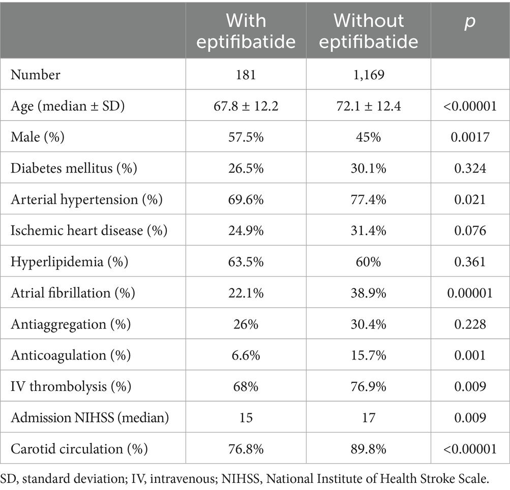
    Patients with eptifibatide were younger (67.8 vs. 73.7%, p < 0.00001), more often male (57.5 vs. 45%, p = 0.002), with a milder baseline clinical condition (15 vs. 17 points in NIHSS, p = 0.009). Patients also suffered from fewer comorbidities. The incidence of arterial hypertension was lower (69.6 vs. 77.4%, p = 0.021), the incidence of atrial fibrillation was also lower (22.1 vs. 38.9%, p = 0.00001) and occlusion in the carotid basin was significantly less common (76.8 vs. 89.8%, p < 0.00001). There were also significant differences in previous use of anticoagulant therapy (6.6 vs. 15.7, p = 0.001) and administration of intravenous thrombolysis before endovascular procedure (68 vs. 76.9%, p = 0.009). The basic and demographic characteristics of the group are summarized in Table 1.

    Table 1
    www.frontiersin.org

    Table 1. Demographic and baseline clinical characteristics of enrolled patients.

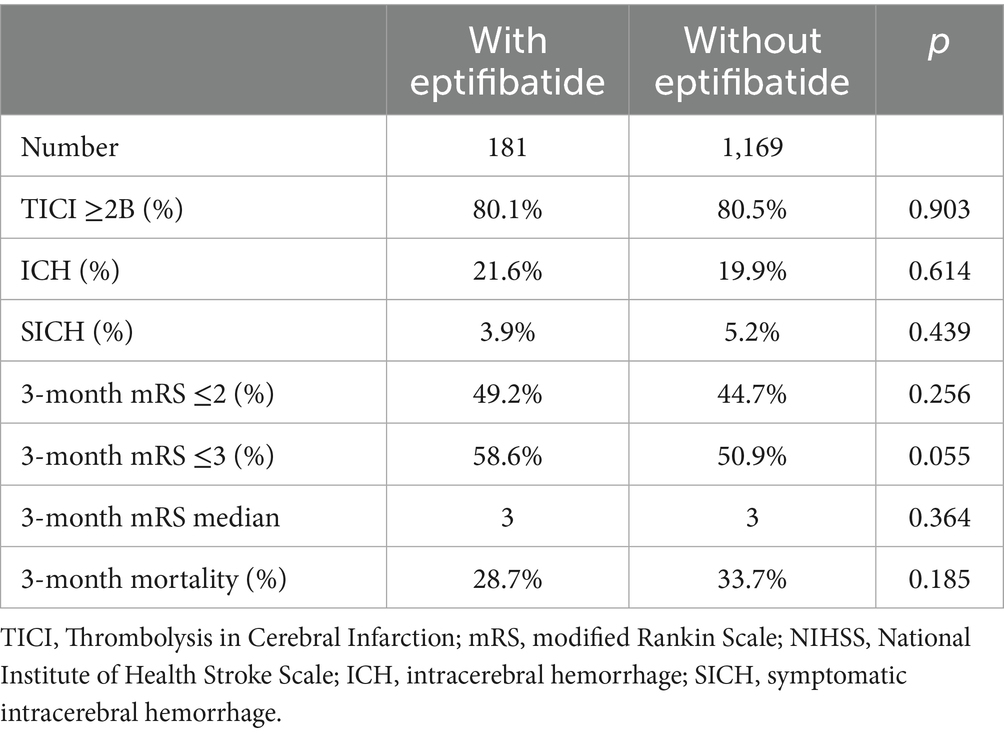
    In the group initially treated with endovascular failure, successful recanalization was achieved in 80.1% (TICI ≥2b) thanks to the administration of eptifibatide. No statistically significant difference was found compared to the group without eptifibatide (80.5%, p = 0.903).

    The incidence of complicating intracerebral hemorrhage was statistically insignificantly higher in the eptifibatide group (21.6 vs. 19.9%, p = 0.614). However, the incidence of symptomatic intracerebral hemorrhage was statistically insignificantly lower (3.9 vs. 5.2%, p = 0.439).

    The clinical outcome tended to be better in the eptifibatide group, but no statistically significant difference was found. Good clinical status was achieved by 49.2 vs. 44.7% (mRS ≤2, p = 0.256) at 3 months. Relatively good outcome (mRS ≤3) was achieved by 58.6 vs. 50.9%, when the difference was already on the verge of statistical significance (p = 0.055). Three-month mortality was also lower (28.7 vs. 33.7%, p = 0.185). The results are summarized in Table 2.

    Table 2
    www.frontiersin.org

    Table 2. Results—comparison between the patients with eptifibatide and standard treatment.

    Discussion

    In our study, no difference in the recanalization rate was found between the eptifibatide group and the group without eptifibatide. However, this result is excellent with regard to our basic criterion for administering eptifibatide (primary failure of the basic endovascular procedure). Without salvage therapy, successful recanalization would not have occurred in this group. With the use of eptifibatide, the rate of successful recanalization reached 80%. The clinical outcome is significantly dependent on the success of recanalization. Full self-sufficiency was achieved with the use of salvage therapy in 49% of patients (without successful recanalization, the chance of a good clinical outcome is at the level of a few percent according to common clinical experience). The safety profile of the selected treatment was favorable. There was no significantly higher incidence of ICH and SICH. The number of ICHs was numerically slightly higher in the eptifibatide group, however, the difference was not statistically significant. Such a small difference may also be significantly influenced by the asymmetry of the two groups (there are numerically one order of magnitude fewer patients with eptifibatide).

    Ma et al. (3) in a matched control analysis show the effectiveness of eptifibatide administration within the framework of endovascular procedure (162 patients, 81 with eptifibatide and 81 controls, successful recanalization 91.3% versus 81.5%). No significant difference in ICH or SICH was found. Latacz et al. (4) in a cohort with AIS treatment for tandem lesions (115 patients with eptifibatide) also did not find an increased incidence of ICH or SICH. Rana et al. (5) (54 patients with eptifibatide for MT) also report a favorable safety profile with the use of eptifibatide (no significant difference in the incidence of ICH or SICH compared to the two control groups). The mentioned studies are consistent with our safety results. They also show efficacy similar to our case. However, there is a fundamental difference in the approach to administering eptifibatide. In our case, it is only in the case of primary failure of mechanical thrombectomy. For the same reason, we cannot compare the clinical results with these studies. Here again, we can state that the patients who were administered eptifibatide largely benefited.

    In our study, patients with eptifibatide tended to have a better clinical outcome (58.6 vs. 50.9%, assessed for mRS ≤3, p = 0.055). However, this group was younger and had fewer significant comorbidities (see Table 1).

    The limitations of our study are primarily its retrospective nature and the absence of an appropriate control group. The administration of eptifibatide was entirely at the discretion of the interventional radiologist. As shown by the basic characteristics of the group, there is a certain tendency to treat younger patients more intensively. Another limitation of the study is that clinical status was monitored only after 3 months. However, clinical outcome is traditionally dependent on the achievement of recanalization, not on the method by which it is achieved. Due to the safety profile of the treatment, longer follow-up is irrelevant. Given the indication for eptifibatide administration only as salvage therapy, based on our data we cannot clearly comment on the general benefit of eptifibatide as adjuvant therapy to MT, nor was this our goal. However, our data show that if eptifibatide is rationally administered, it does not carry a statistically significant risk of hemorrhagic complications and, on the contrary, provides a very significant chance of successful recanalization.

    Conclusion

    Despite advances in endovascular treatment, there are still patients in whom this procedure fails. In our study, eptifibatide was used as salvage therapy in primary failure of endovascular treatment. In 80% of patients primarily without the possibility of recanalization with standard endovascular techniques, this procedure led to successful recanalization. Given the above limitations, we draw two conclusions from our data. The administration of eptifibatide, after careful individual consideration, can be a highly effective salvage therapy in primary failure of mechanical thrombectomy (the last therapeutic option). The second conclusion is the favorable safety profile of the treatment in this indication, which is consistent with the general results of previously published studies using eptifibatide as adjuvant therapy to mechanical thrombectomy. A larger randomized trial would be needed to verify the results.

    Data availability statement

    The raw data supporting the conclusions of this article will be made available by the authors, without undue reservation.