Does your doctor have ANY PROTOCOLS TO ENSURE YOUR CERBRAL BLOOD FLOW IS CORRECT? I thought not, so incompetence is standard for your doctor!
Do you prefer your doctor, hospital and board of director's incompetence NOT KNOWING? OR NOT DOING? Your choice; let them be incompetent or demand action!
Alzheimer’s may begin with a silent drop in brain blood flow
- February 24, 2026
- Keck School of Medicine of USC
- Subtle changes in brain blood flow and oxygen use are closely linked to hallmark signs of Alzheimer’s, including amyloid plaques and memory-related brain shrinkage. Simple, noninvasive scans may one day help spot risk earlier—by looking at the brain’s vascular health, not just its plaques.
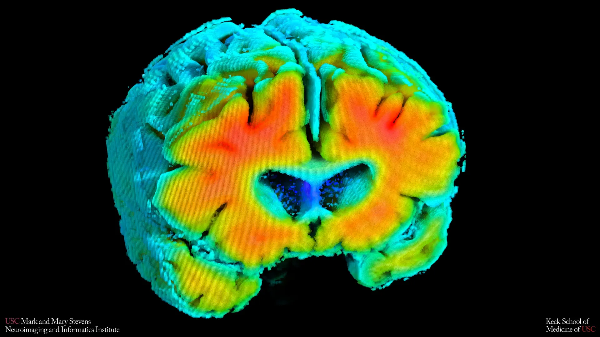 Amyloid PET imaging was used in the study to measure plaque buildup in the brain, a hallmark of Alzheimer's disease. Warmer colors indicate higher amyloid levels. Participants with healthier patterns of brain blood flow and oxygen regulation had lower amyloid burden, reinforcing the idea that vascular function may be linked to Alzheimer's-related changes. Credit: Stevens INI
Small shifts in how blood moves through the brain and how brain cells receive oxygen may be closely connected to the risk of Alzheimer's disease. That is the conclusion of new research from the Mark and Mary Stevens Neuroimaging and Informatics Institute (Stevens INI) at the Keck School of Medicine of USC.
The study, published in Alzheimer's and Dementia: The Journal of the Alzheimer's Association, examined older adults both with and without cognitive impairment. Researchers found that simple, noninvasive measures of brain blood flow and oxygen levels were linked to well known signs of Alzheimer's, including amyloid plaque buildup and shrinkage of the hippocampus, the part of the brain that plays a central role in memory. The results suggest that the health of the brain's blood vessels may influence the disease process early on and could help flag people at risk before noticeable symptoms develop.
"Amyloid and tau are often considered the primary players in Alzheimer's disease, but blood flow and oxygen delivery are also critical," said Amaryllis A. Tsiknia, lead author of the study and USC PhD candidate. "Our results show that when the brain's vascular system functions more like it does in healthy aging, we also see brain features that are linked to better cognitive health."
Noninvasive Tools to Measure Brain Circulation
To study these changes, the team relied on two painless techniques that can be used while a person rests quietly. Transcranial Doppler ultrasound tracks how quickly blood travels through the brain's major arteries. Near infrared spectroscopy evaluates how effectively oxygen reaches brain tissue near the surface of the cortex.
Researchers then applied advanced mathematical modeling to combine these readings into overall indicators of cerebrovascular function. These indicators reflect how well the brain adjusts blood flow and oxygen delivery in response to natural fluctuations in blood pressure and carbon dioxide.
Vascular Health Linked to Amyloid and Memory Centers
Participants whose vascular indicators more closely resembled those of cognitively healthy adults tended to have lower amyloid levels and a larger hippocampus. Both features are associated with reduced Alzheimer's risk.
"These vascular measures are capturing something meaningful about brain health," said Meredith N. Braskie, PhD, senior author of the study and assistant professor of neurology at the Keck School of Medicine. "They appear to align with what we see on MRI and PET scans that are commonly used to study Alzheimer's disease, providing important information about how vascular health and standard brain measures of Alzheimer's disease risk may be related."
The researchers also observed that people diagnosed with mild cognitive impairment or dementia showed weaker vascular function compared to cognitively normal participants. This finding supports the view that declining blood vessel health in the brain is part of the broader Alzheimer's disease continuum.
"These findings add to growing evidence that Alzheimer's involves meaningful vascular contributions in addition to classic neurodegenerative changes," said Arthur W. Toga, PhD, director of the Stevens INI. "Understanding how blood flow and oxygen regulation interact with amyloid and brain structure opens new doors for early detection and potentially prevention."
Potential for Earlier and Broader Screening
Compared with MRI and PET imaging, these methods are less costly and easier to perform. They do not involve injections, radiation exposure, or demanding tasks for patients. That simplicity could make them useful for large scale screening or for individuals who are unable to undergo more intensive brain imaging.
The authors caution that the findings represent a single snapshot in time and do not establish cause and effect. Ongoing long term studies are tracking participants to see whether shifts in these vascular measures can predict future cognitive decline or response to treatment.
"If we can track these signals over time, we may be able to identify people at higher risk earlier and test whether improving vascular health can slow or reduce Alzheimer's-related brain changes," Tsiknia said.
About the Study
In addition to Tsiknia and Braskie, the study's other authors are Peter S. Conti, Rebecca J. Lepping, Brendan J. Kelley, Rong Zhang, Sandra A. Billinger, Helena C. Chui and Vasilis Z. Marmarelis.
This work was supported by the Office of The Director, National Institutes of Health, under Award Number S10OD032285, and by the National Institute on Aging [R01AG058162].
No comments:
Post a Comment